
Unlock crystallographic & microstructural secrets of your samples
Diffraction Contrast Tomography in your laboratory
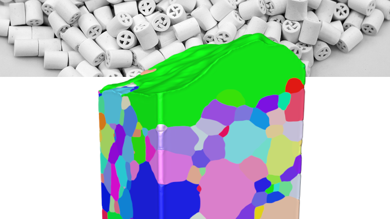
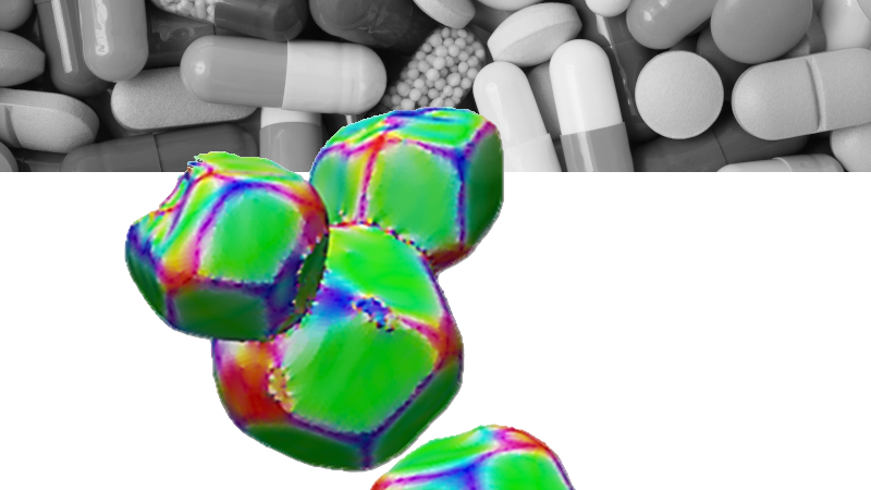
Do you want to perform non-destructive mapping of grain morphology in 3D to characterize materials like metals, alloys or ceramics? Discover the first commercially available lab-based diffraction contrast tomography (DCT) technique for complete three-dimensional imaging of grains in your sample. Two powerful solutions — LabDCT Pro and CrystalCT — allow you to directly visualize 3D crystallographic grain orientation. Powered by the advanced GrainMapper3D software, it opens new ways to investigate a variety of polycrystalline materials.


Get superior sample representivity from advanced diffraction scan modes
Introducing ZEISS Xradia CrystalCT and LabDCT Pro

ZEISS Xradia Versa LabDCT Pro and Xradia CrystalCT
ZEISS Xradia Versa LabDCT Pro and Xradia CrystalCT advance materials characterization, modeling, and discovery through ground-breaking diffraction scanning modes.
- Provides unprecedented sample representivity
- Enables scanning larger sample volumes
- Simplifies sample prep, and handling of irregular / realistic sample shapes
- Increases speed
- Addresses sample specificity
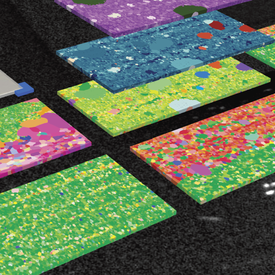
Inspired by nature’s golden angle, advanced scanning modes deliver helical phyllotaxis schema to manage a wide range of sample shapes and sizes, and overcomes some of the previous challenges of conventional DCT:
- Helical phyllotaxis when your sample is tall and narrow
- Helical phyllotaxis raster when your sample is large and wide
- Helical phyllotaxis with high aspect ratio tomography (HART) for flat samples.
The technology behind LabDCT Pro and CrystalCT
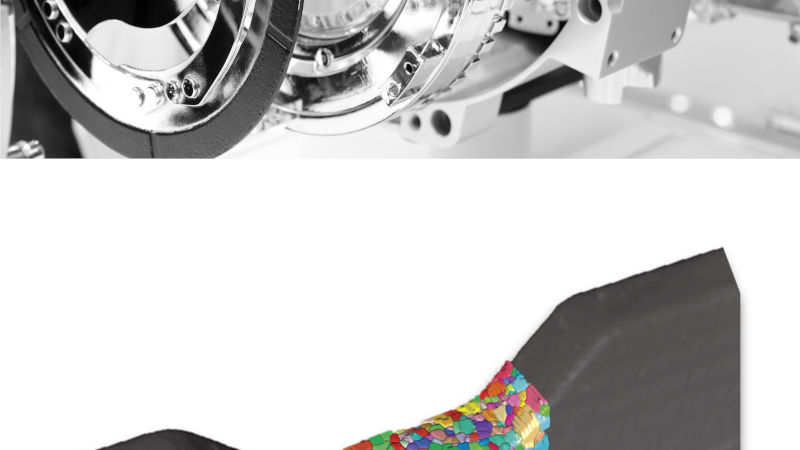
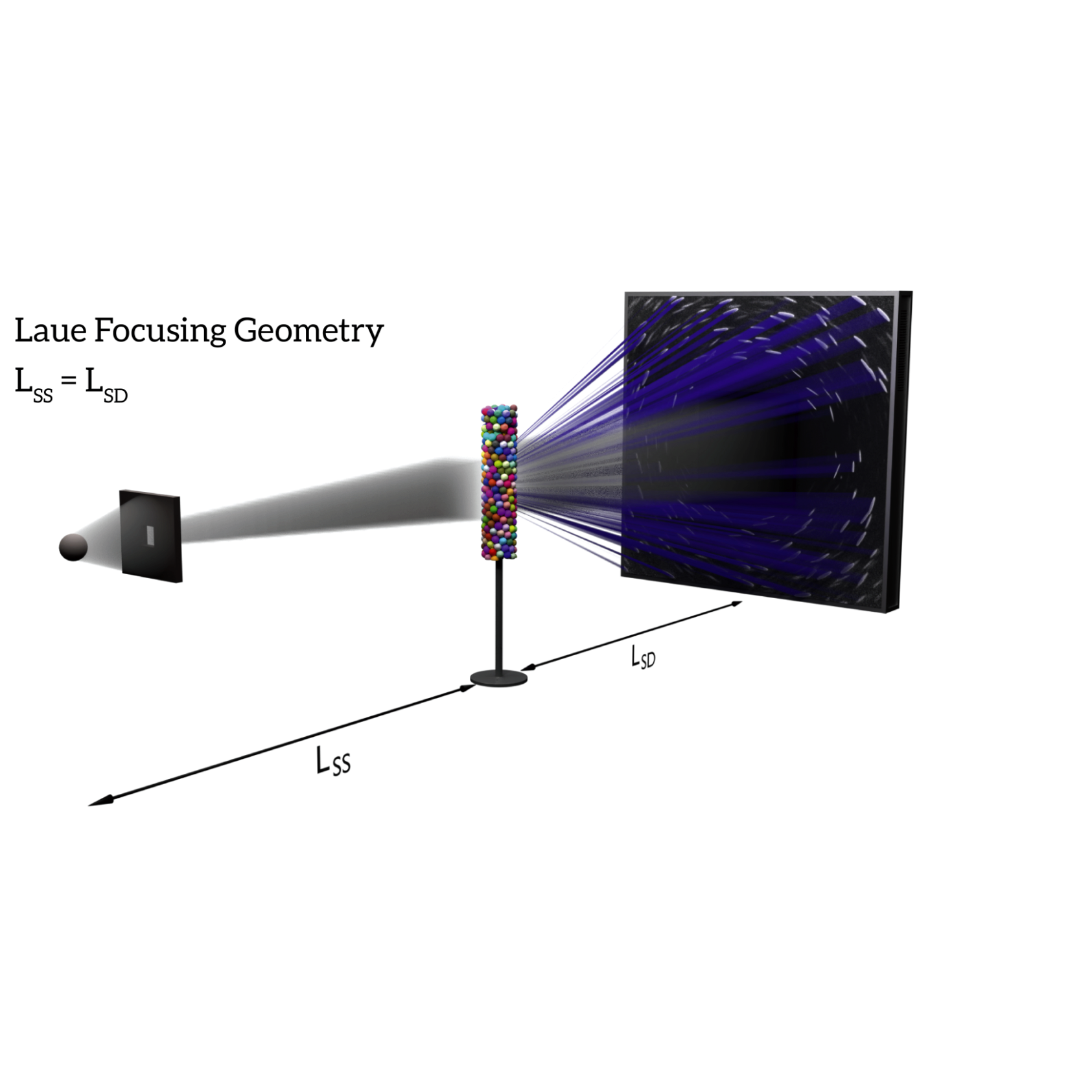
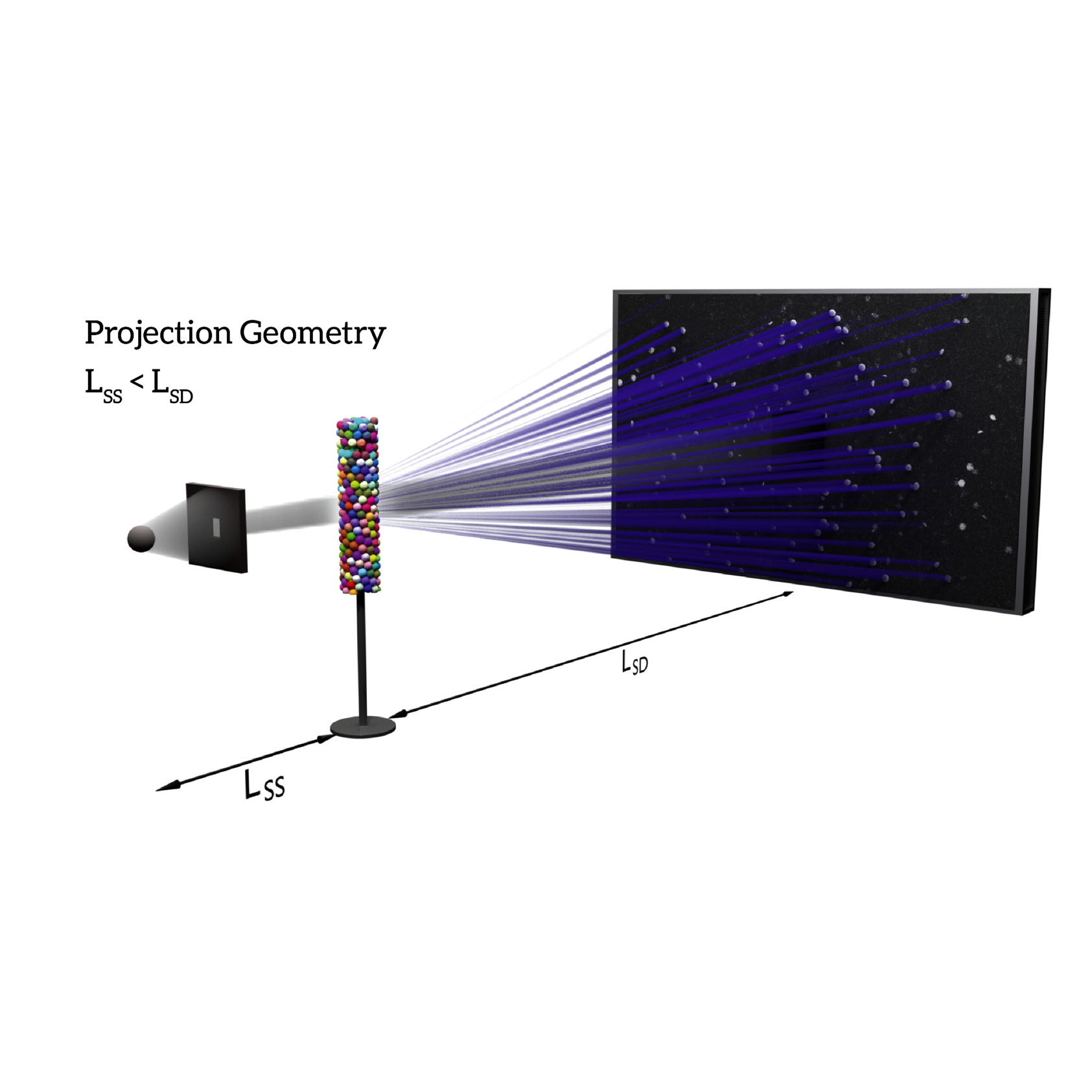
DCT projection images produce distinct diffraction patterns on the detector based on their focusing geometries. In Laue focusing mode, grains produce diffraction spots that are sharp lines on the scintillator-coupled objective-based detector. Projection geometry uses the flat panel detector positioned in the magnifying or defocused position to collect diffraction spots that appear as projected shape profiles of the corresponding diffracting grains. The LabDCT Pro module on the ZEISS Xradia 620 Versa enables grain mapping on both the dedicated 4X DCT objective and on the optional flat panel detector providing the flexibility to choose between high resolution or high throughput large area grain mapping, whereas ZEISS Xradia CrystalCT uses its own dedicated flat panel detector in projection mode.
-


LabDCT Pro: Laue focusing geometry with the 4X DCT objective
Download image (541 KB) -

LabDCT Pro or CrystalCT: Projection or Laue focusing geometry onto a flat panel detector
Download image (755 KB)


3D Crystallographic Grain Mapping

3D Grain Reconstruction: Index grain data precise, fast & automated
After the initial acquisition as the first step in your workflow you then start to reconstruct. Load your absorption tomography and your diffraction data into GrainMapper3D. Let it identify potential candidates for grain orientations of a given polycrystal by using back and forward projections.
An automated, iterative search for grains in the sample volume is your next step. Grain reconstruction results are stored as stacks of slices or volume datasets that contain the full description of the indexed grains. Eventually, share 3D LabDCT Pro results with your collaborators or customers using the standalone GrainMapper3D Viewer application.
GrainMapper 3D is a 3D crystallographic grain reconstruction software developed by ZEISS collaboration partners Xnovo Technology ApS, Denmark
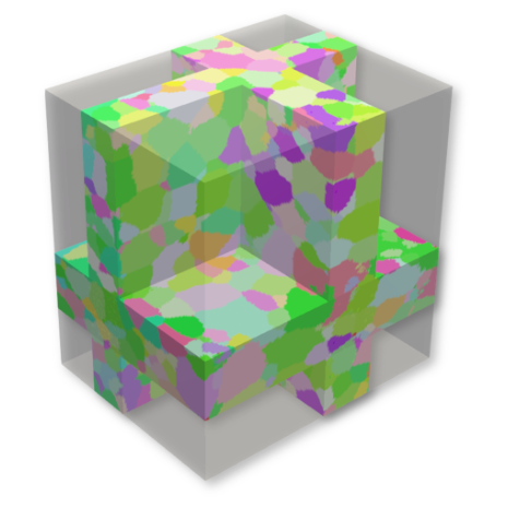

3D Grain Mapping: Get all information in one file
Your final step is to get out all the information you need in one single file. Shape, orientation and spatial locations of all grains in the sample volume are exported into an open data format.
Finish your experiment with subsequent analyses using customized software or simulation tools. The advanced indexation routines now support the more complex lower symmetry crystal systems.