

ZEISS Xradia Versa X-ray Microscopes
Discover More with Non-destructive 3D X-ray Imaging at Submicron Resolution
Extremely versatile ZEISS Xradia Versa 3D X-ray microscopes (XRM) provide superior 3D image quality and data for a wide range of materials and working environments. Xradia Versa XRM feature dual-stage magnification based on synchrotron-caliber optics and revolutionary RaaD™ (Resolution at a Distance) technology for high resolution even at large working distances, a vast improvement over traditional micro-computed tomography. Non-destructive imaging preserves and extends the use of your valuable sample, enabling 4D and in situ studies.
-
Xradia 630 Versa


ZEISS Xradia 630 Versa 3D expands the horizon of what researchers are able to achieve with their X-ray microscope. The system delivers breakthrough resolution performance, takes accessibility to the next level with an intuitive user experience, and accelerates productivity with faster time-to-results.
-
Xradia 620 Versa

Xradia 620 Versa unlocks new degrees of versatility for your research with non-destructive imaging at maximum flexibility to accelerate your research. Move from exploration to discovery in a seamless workflow. 600-family technical specs with a wider range of imaging options.
-
Xradia 610 Versa

Xradia 610 Versa extends the limits of your exploration with innovative source and optics technology. Higher X-ray flux delivers faster tomography scans with industry-leading resolution and contrast.
-
Xradia 510 Versa

Xradia 510 Versa breaks the one-micron resolution barrier for 3D imaging and in situ / 4D investigations. ZEISS Xradia 510 Versa makes synchrotron-caliber research even more practical for mid-sized imaging centers and industrial laboratories.


Xradia 630 Versa
ZEISS Xradia 630 Versa, with higher energy capabilities of the exclusive 40X Prime objective, enables you to push the limits of submicron imaging like never before.
The system achieves unparalleled resolution performance of 450-500 nm across the full range of energy, from 30 kV to 160 kV, unlocking entirely new capabilities for your research. NavX guides users through automated workflows with intelligent system insights to deliver results easily and efficiently. AI-based DeepScout changes the game for understanding your sample with throughput boosts 100 times faster.
Xradia 630 Versa
ZEISS Xradia 630 Versa, with higher energy capabilities of the exclusive 40X Prime objective, enables you to push the limits of submicron imaging like never before. The system achieves unparalleled resolution performance of 450-500 nm across the full range of energy, from 30 kV to 160 kV, unlocking entirely new capabilities for your research. NavX guides users through automated workflows with intelligent system insights to deliver results easily and efficiently. AI-based DeepScout changes the game for understanding your sample with throughput boosts 100 times faster.


Head of objectives with the 40X-P
Breakthrough Resolution Performance to Expand Your Research Horizons
The ZEISS 40X-Prime Objective
With more X-ray photons available on ZEISS Xradia 600-series Versa, you can now achieve even faster time to results for varied samples without compromising resolution. Unique to ZEISS Xradia 630 Versa is the 40X-Prime (40X-P) objective lens.
ZEISS Xradia 630 Versa XRM, with the higher energy capabilities of the exclusive 40X-Prime (40X-P) objective, enables you to push the limits of submicron imaging like never before. Known for their ability to achieve resolution at a Distance (RaaD™), ZEISS Xradia Versa platforms allow high resolution imaging of a wide array of sample types and sizes over a long range of length scales.
With 40X-P, the system achieves unparalleled resolution performance of 450-500 nm across the full range of source voltage, from 30 kV to 160 kV, defining RaaD 2.0. Unlocking entirely new application capabilities for researchers, the ZEISS 40X-P objective enables ZEISS Xradia 630 Versa to push industry standards of submicron imaging resolution.
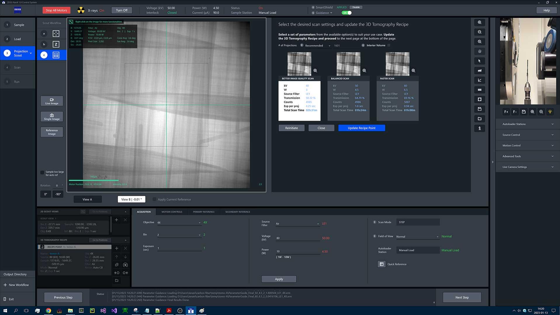

NavX User Interface
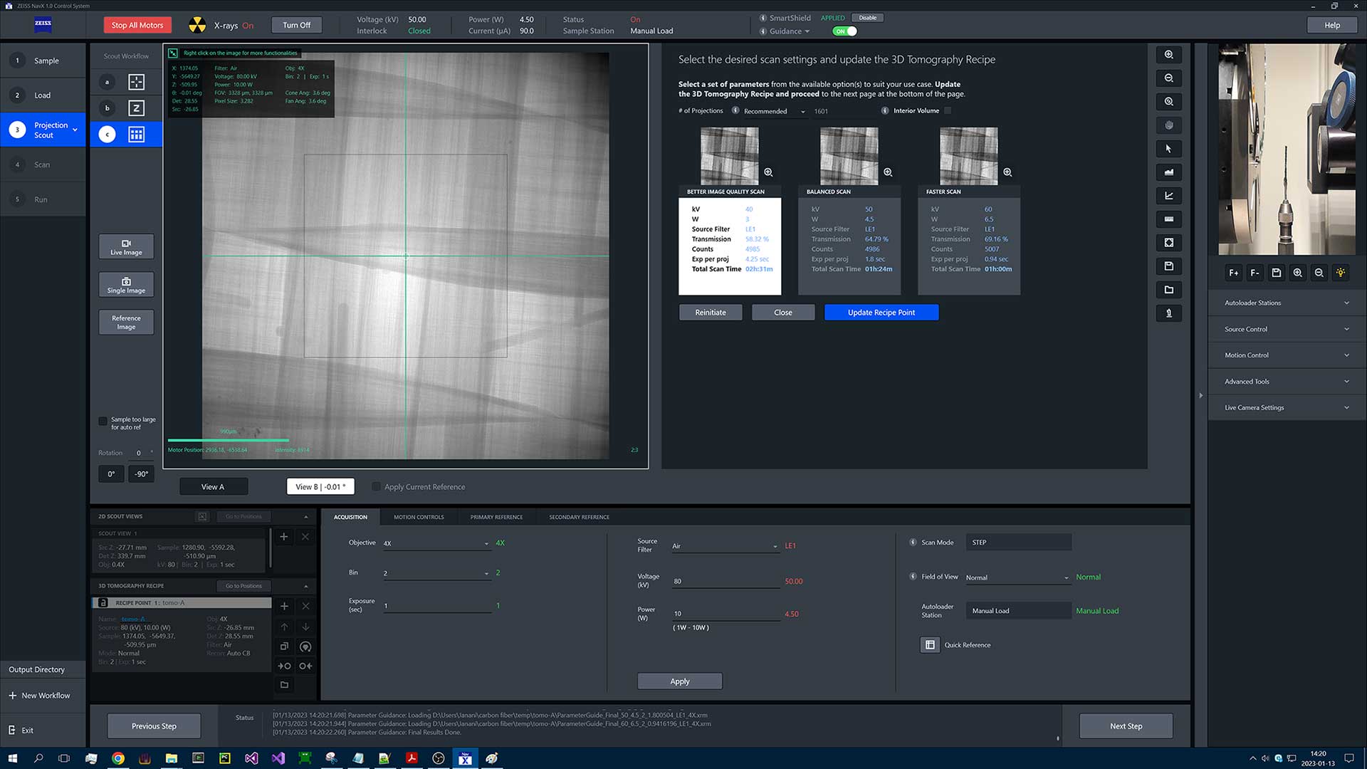

NavX User Interface
NavX User Interface
NavX User Interface
The physics of X-ray imaging can be complex, so ZEISS XRM researchers studied user habits, dove into their challenges, and employed human-centered design (HCD) principles to enable even the newest user in a busy environment to be immediately productive. NavX™, the new user interface for ZEISS Xradia 630 Versa, guides users through automated workflows with intelligent system insights and delivers experimental results more easily and efficiently while also allowing experienced users to explore the full versatility of the platform.
NavX enables you to automate your workflow and provides guidance on the impact the parameters you've chosen will have on your setup. That guidance is directly embedded in the software, taking you through choices in a natural and familiar way.
Additionally, the NavX File Transfer Utility (FTU) takes the data that is being produced by the microscope and automatically transfers it to other locations so that users have their data where they need it, when they need it. These advancements make NavX much more capable for remote operation, advancing user productivity.
NavX intuitive navigation follows the evolution of the XRM user base and revolutionizes X-ray navigation and control with seamless and integrated workflows complementing the planning and execution of advanced correlative workflows..


Flat Panel Extension
The flat panel extension (FPX), standard on your ZEISS Xradia 630 Versa X-ray microscope, further increases the versatility of the system, directly supporting the Advanced Reconstruction Toolbox’s AI-based DeepScout for deep learning and neural network training. Leverage FPX to perform a low resolution, large field of view, "scout” scans, and identify interior regions for higher resolution “zoom” scans on a variety of different sample types. The Volume Scout workflow streamlines this process within NavX.


Non-destructive three-dimensional grain map of an Armco iron sample with illustrations of the various grain analysis that can be performed on a typical LabDCT Pro dataset.


LabDCT Pro for diffraction contrast tomography (DCT)
Unlocking Crystallographic Information
LabDCT Pro for diffraction contrast tomography (DCT), available exclusively on Xradia 630 and 620 Versa, enables non-destructive mapping of grain orientation and microstructure in 3D. Direct visualization of 3D crystallographic grain orientation opens up a new dimension in the characterization of polycrystalline materials like metal alloys, geomaterials, ceramics, or pharmaceuticals.
- LabDCT Pro supports specimens with crystal structures from cubic symmetry to systems with lower symmetry such as monoclinic materials
- Acquire high resolution crystallographic information using the dedicated 4X DCT objective. For even larger samples, use large area mapping and increase your throughput with the Flat Panel Extension (FPX).
- Obtain comprehensive 3D microstructure analysis from larger representative volumes and wide-ranging sample geometries
- Investigate microstructural evolution with 4D imaging experiments.
- Combine 3D crystallographic information with 3D microstructural features.
- Combine modalities to understand structure-property relationships.


Xradia 620 Versa
Boost the performance of your Xradia 620 and 630 Versa and explore more with their advanced capabilities. Enhance absorption contrast for low-Z or similar-Z materials with the Dual Scan Contrast Visualizer (DSCoVer). Unlock 3D crystallographic information with laboratory-based Diffraction Contrast Tomography (LabDCT). Improve scan speed and accuracy of large or irregular samples with advanced acquisition techniques such as High Aspect Ratio Tomography (HART).
A reconstruction of the grain microstructure of Armco Iron, acquired with LabDCT. The grains are colored by crystallographic orientation, and reconstruction reveals the true grain shape. The background shows an example of a diffraction pattern that is collected during the LabDCT acquisition.


Achieve New Degrees of Freedom
With more X-ray photons available on ZEISS Xradia 600-series Versa, you can now achieve even faster time to results for varied sample sizes without compromising resolution. Xradia 620 Versa offers additional unique features and imaging capabilities.
- Improve scan speed and accuracy of large or irregular samples with advanced acquisition techniques such as High Aspect Ratio Tomography (HART)
- Automated Filter Changer (AFC) enables seamless filter changing without manual intervention, and your selection can be programmed and recorded for each recipe
- Unlock crystallographic information in your own lab with the optional LabDCT


Gain an Edge in Contrast
Dual Scan Contrast Visualizer (DSCoVer), exclusive to Xradia 620 Versa, extends the detail captured in a single energy absorption image by combining information from tomographies taken at two different X-ray energies. DSCoVer takes advantage of how X-rays interact with matter based on effective atomic number and density. This provides you with a unique capability for distinguishing, for example, mineralogical differences within rocks as well as among difficult-to-discern materials such as silicon and aluminum.


Investment Protection
As your imaging needs evolve, so should your instrument. Unlike traditional microCT systems, the ZEISS Xradia Versa family is built on an established ZEISS 3D X-ray microscope platform that is upgradeable, expandable and reliable, paving the way for future enhancements and protecting your investment. Select the system that is right for you today and expand as your needs require.
- Protect your investment by upgrading your system at any time with the latest capabilities and innovations
- Constant development means that you can add advanced capabilities such as in situ sample environments, unique imaging modalities, and productivity enhancing modules
- Field conversion from basic systems up to most advanced systems in most instances


Non-destructive three-dimensional grain map of an Armco iron sample with illustrations of the various grain analysis that can be performed on a typical LabDCT Pro dataset.
LabDCT Pro for diffraction contrast tomography (DCT)
Unlocking Crystallographic Information
LabDCT Pro for diffraction contrast tomography (DCT), available exclusively on Xradia 630 and 620 Versa, enables non-destructive mapping of grain orientation and microstructure in 3D. Direct visualization of 3D crystallographic grain orientation opens up a new dimension in the characterization of polycrystalline materials like metal alloys, geomaterials, ceramics, or pharmaceuticals.
✔ LabDCT Pro supports specimens with crystal structures from cubic symmetry to systems with lower symmetry such as monoclinic materials
✔ Acquire high resolution crystallographic information using the dedicated 4X DCT objective. For even larger samples, use large area mapping and increase your throughput with the Flat Panel Extension (FPX).
✔ Obtain comprehensive 3D microstructure analysis from larger representative volumes and wide-ranging sample geometries
✔ Investigate microstructural evolution with 4D imaging experiments.
✔ Combine 3D crystallographic information with 3D microstructural features.
✔ Combine modalities to understand structure-property relationships.


When investigating a smartphone camera lens module in the assembled state, the assessment of geometrical properties requires a non-contact, non-destructive measurement method to quantify relational parameters. MTX allows the accuracy-verified measurement of properties like thickness of annular wedges, centration interlock diameters, gaps between wedges, lens- tolens tilt, or apex heights and centration. These parameters are important for functional inspection and the enhancement of manufacturing designs and processes, to enable production of versatile cell phone cameras.
Metrology Extension
Adding Measurement Accuracy to X-ray Microscopy
With the Metrology Extension (MTX) you turn your Xradia 620 Versa into a verified measurement accuracy system far beyond the limits of conventional CT technology. This is essential for academic and industrial labs where miniaturization and integration of components drive a growing demand for high-resolution metrology. Benefit from high resolution X-ray imaging combined with high-precision metrology.
✔ Leading CT metrology accuracy: Calibrated with MTX, ZEISS Xradia Versa provides a market-leading maximum permissible error value of MPESD = (1.9 + L/100) μm for measurements in small-scale volumes, where L is the measured length in mm.
✔ Small volumes at high resolution: MTX enables measurements with high dimensional accuracy within small reconstructed volumes of 125 mm3.
✔ Simple calibration workflow: The MTX package provides an integrated user-guided calibration workflow.
✔ Once the calibration routine has been executed, you perform precise measurements and make the data available to standard metrology software for further processing.


Xradia 610 Versa
By leveraging RaaD capability, Xradia Versa 600-series maintain the highest resolution across large working distances, accommodating samples contained within environmental chambers and high precision in situ load rigs. 600-series XRM offer even higher resolution and throughput over previous generations. Xradia Versa seamlessly integrate with other ZEISS microscopes to solve your multi-scale correlative challenges.
Cement paste sample blended with resin, enabling the paste to acquire higher porosity resulting in better freeze-thaw behavior. Courtesy Nanjing University of Science and Technology, China


True spatial resolution of 0.5 µm demonstrated on JIMA resolution target.
Highest Resolution without Compromise
ZEISS Xradia Versa use a two-stage magnification architecture to enable submicron resolution imaging at large working distances—RaaD—for a diverse set of sample sizes and types. With more X-ray photons available, the ZEISS Xradia 600 Series Versa provide faster time to results for the widest range of sample sizes and types, without compromising resolution.
✔ Deliver image quality without compromising throughput
✔ Image larger, denser objects including intact components and devices, in 3D
✔ ZEISS Xradia 600-series Versa obtain true spatial resolution of 500 nm with a minimum achievable voxel size of 40 nm


Higher X-ray Flux
ZEISS Xradia 600 Series Versa leverage 25 W X-ray sealed source technology, delivering high X-ray flux that pushes the boundaries of performance. It provides excellent thermal management, with increased flux capacity and high throughput while maintaining stringent world-class Versa spot size performance. An innovative source control system ensures source responsiveness, enabling a faster scan setup for an easy and engaging user experience
✔ Faster tomography scans—up to 2X higher output—enable more sample runs and more regions of interest to explore
✔ Higher flux offers higher contrast-to-noise ratio and exceptional performance at high energy (kV), without compromising resolution
✔ Sealed sources mean higher vacuum and longer filament life


Premier In Situ and 4D
ZEISS Xradia 600 Series Versa can non-destructively characterize the 3D microstructure of materials under controlled variations (in situ) and enable you to observe the evolution of structures over time (4D). By leveraging RaaD, Xradia Versa XRM maintain the highest resolution across large working distances, accommodating sample, environmental chamber, and high precision in situ load rigs without sacrificing resolution.
✔ Characterize and quantify your sample under varying conditions and native-like environments in situ and over time
✔ Seamlessly integrate with other ZEISS microscopes to solve multi-scale correlative imaging challenges


Xradia 510 Versa
Benefit from the two-stage magnification technique offered by ZEISS Xradia Versa to uniquely achieve RaaD, which enables you to effectively study the widest range of sample sizes. Intuitive Scout-and-Scan control software enables a broad range of user skillsets in your busy lab.
Polymer with urethane backbone. Imaging after in situ experiments. Simulation of fluid flow demonstrates permeability. Courtesy National Chemical Laboratory, India


A Class Above MicroCT
Extend scientific research beyond the limits of projection-based microCT systems at submicron resolution with ZEISS Xradia 510 Versa XRM. Traditional computed tomography relies on a single stage of geometric magnification and maintaining high resolution for larger samples is impossible due to the longer working distances required. ZEISS Xradia Versa XRM feature a unique two-stage process based on synchrotron caliber optics. Multilength-scale capabilities enable you to image the same sample across a wide range of magnifications. An added benefit: ZEISS Xradia 510 Versa is easy to use by everyone in your busy lab.
✔ Reduce dependence on geometric magnification and maintain submicron resolution even at large working distances
✔ Experience versatility for wide range of materials, including soft and low Z, with unique contrast solutions that overcome the limitations of traditional computed tomography
✔ Characterize the microstructure of materials in their native-like environments in situ and study the evolution of properties over time (4D)


True Spatial Resolution
ZEISS XRM systems are specified on true spatial resolution, the most meaningful measurement of your microscope’s performance. Spatial resolution refers to the minimum separation at which two features can be resolved by an imaging system. You would typically measure it by imaging a standardized resolution target with progressively smaller line-space pairs. Spatial resolution accounts for critical characteristics such as X-ray source spot size, detector resolution, magnification geometry, and vibrational, electrical and thermal stability.
✔ 0.7 μm true spatial resolution with a minimum achievable voxel size of 70 nm
✔ Energy-tuned detectors enable highest resolution across broad ranges of sample types and densities
✔ Source operates across the entire application space (30-160 kV) with a wide range of detectors, eliminating the need for manual hardware reconfigurations


Reveal Hidden Details
Your imaging requires superior contrast capabilities to reveal details necessary to accurately visualize and quantify features. ZEISS Xradia Versa systems deliver flexible, high contrast imaging for even your most challenging materials–low atomic number (low Z) materials, soft tissue, polymers, fossilized organisms encased in amber, and other materials of low contrast.
- Our comprehensive approach employs proprietary enhanced absorption contrast detectors that provide you with superior contrast by maximizing collection of low energy photons while minimizing collection of contrast-reducing high energy photons
- Tunable propagation phase contrast measures the refraction of X-ray photons at material transitions to allow you to visualize features displaying little or no contrast during absorption imaging
Breakthrough Imaging with ZEISS Xradia Versa XRM
View highlights of ZEISS Xradia Versa 3D X-ray microscopes: non-destructive imaging, higher resolution with higher throughput.
The Technology Behind Xradia Versa X-ray Microscopes
-


The Versatile Advantage of RaaD
The Versatile Advantage of RaaD
The two-stage magnification technique offered by ZEISS Xradia Versa uniquely achieves Resolution at a Distance, or RaaD, which enables you to effectively study the widest range of sample sizes, including those within in situ chambers.
Images are initially magnified via geometric projection as with conventional microCT. The projected image is cast onto a scintillator, converting X-rays to a visible light image which is then optically magnified by microscope optics before acquisition by a CCD detector.
Reducing dependence on geometric magnification enables ZEISS Xradia Versa solutions to maintain submicron spatial resolution down to 500 nm at large working distances.
-

Tensile testing of laser welded steel under increasing load.
Push the Limits of Scientific Advancement
ZEISS Xradia X-ray systems provide the industry's premier 3D imaging solution for the widest variety of in situ rigs, from high-pressure flow cells to tension, compression, and thermal stages. Moving beyond the three dimensions of space, leverage the non-destructive nature of X-ray investigation to extend your studies into the dimension of time with 4D experiments.
These studies require samples to be further away from the X-ray source to accommodate various types of in situ rigs. On traditional microCT systems, this significantly limits the resolution achievable for your samples. ZEISS XRM are uniquely equipped with dual-stage magnification architecture with RaaD technology that enable the highest resolution for in situ imaging.
ZEISS Xradia XRM platforms can accommodate a variety of in situ rigs, from high-pressure flow cells to tension, compression, and thermal stages, to user-customized designs. You can add the optional in situ Interface Kit to your ZEISS Xradia XRM, which includes a mechanical integration kit, a robust cabling guide, and other facilities (feed-throughs) along with recipe-based software that simplifies your control from within the Versa Scout-and-Scan user interface. When your needs require pushing the resolution limits of your in situ experiments, convert your ZEISS Xradia microCT or XRM to an Xradia 620 Versa X-ray microscope and leverage RaaD technology for the maximum performance tomographic imaging of samples within in situ chambers or rigs.
-
Begin Your Multi-Scale, Multi-Modal, Multi-Dimensional Microscopy with Non-destructive 3D Imaging
Because of the non-destructive nature of X-rays and the versatile array of sample types and sizes they are able to image, correlative microscopy often begins with, or is enabled by, ZEISS Xradia Versa XRM.
Using the Scout-and-Zoom capability of Versa, you are able to clearly define your region of interest (ROI) before sacrificing your sample to premature cutting or other sample prep. Rapidly scout at low resolution with a large field of view, and then zoom to the ROI at higher resolution, whether using the range of Versa objectives (up to 40X), nanoscale Xradia Ultra XRM, or ZEISS light, electron, or FIB-SEM microscopes. This prevents premature sample destruction and allows for maximum workflow efficiency while combining full sample context with key sample information.
Additionally, the ability to perform interior tomography, or to clearly see inside your sample in 3D, further reduces the risk of losing sight of your ROI. Achieve greater efficiency by pinpointing a specific “address” to which to navigate for accurate and efficient next steps for interrogating your sample.
Finally, examine your sample under varying conditions and over time with in situ and 4D studies before performing further analysis — chemical, surface, etc.— with other ZEISS modalities.
Leverage the widest array of microscopy solutions available — exclusively from ZEISS — to perform multi-modal, multi-lengthscale, multi-dimensional analyses, by starting with non-destructive 3D X-ray microscopy.

Full correlative sample workflow for the project. Initial XRM scans highlight key areas for higher resolution imaging and target locations for thin section orientation within the volume. Subsequent 2D analysis includes electron and light microscopy, leading to correlation with in situ microanalytical data.


Features That Make Every Versa Platform More Powerful
-
Wide Field Mode
Wide Field Mode (WFM) can be used to image across an extended lateral field of view. The wide lateral field of view can provide 3x larger 3D volume for large samples, or give a higher voxel density for a standard field of view. All Xradia Versa systems are capable of WFM with the 0.4x objective. In combination with Vertical Stitching, WFM enables you to image larger samples at exceptional resolution.


, and Dr. J. König (Jozef Stefan Institute)")
, and Dr. J. König (Jozef Stefan Institute)")


.")
.")


.")
.")


")
")
")
")
")
")


")
")
")
")
")
")


Accessories
Upgrade your microscope with additional accessories to enhance its capabilities


Autoloader
Maximize Your Instrument’s Utilization
Maximize use and minimize user intervention with the optional ZEISS Autoloader. Reduce the frequency of user interaction and increase productivity by enabling multiple jobs to run. Load up to 14 sample stations, which can support up to 70 samples, queue, and allow to run all day, or off-shift.


In Situ Interface Kit
Push the Limits of Scientific
ZEISS Xradia platforms can accommodate a variety of in situ rigs, from high-pressure flow cells to tension, compression, and thermal stages, to user-customized designs. Moving beyond the three dimensions of space, leverage the non-destructive nature of X-ray investigation to extend your studies into the dimension of time with 4D experiments.


Lithium ion battery
Visualization and Analysis
ZEISS Recommends Dragonfly Pro
An advanced analysis and visualization software solution for your 3D data acquired by a variety of technologies including X-ray, FIB-SEM, SEM and helium ion microscopy. Available exclusively through ZEISS, ORS Dragonfly Pro offers an intuitive, complete, and customizable toolkit for visualization and analysis of large 3D grayscale data. Dragonfly Pro allows for navigation, annotation, creation of media files, including video production, of your 3D data. Perform image processing, segmentation, and object analysis to quantify your results.


Related Applications
Downloads
-
-

3D Imaging Systems
Your Guide to the Widest Selection of Optical Sectioning, Electron Microscopy and X-ray Microscopy Techniques.
File size: 5 MB -

ZEISS Xradia 610 and 620 Versa
Your 3D X-ray Microscopes for Faster Sub-Micron Imaging of Intact Samples
File size: 11 MB -

ZEISS Xradia 630 Versa X-ray Microscope
Expanded Accessibility. Improved Productivity. Extended Capabilities.
File size: 30 MB -

3D X-ray Microscope Field Conversion and Upgrade Options
File size: 2 MB -

40×-Prime Objective from ZEISS
Enhance Resolution and Image Quality On ZEISS Xradia 630 Versa
File size: 3 MB -

Extending the Frontiers of Semiconductor Failure Analysis
ZEISS Xradia 630 Versa 3D X-ray Microscopy
File size: 1 MB -

Flyer: ZEISS DeepRecon Pro for Electronics and Failure Analysis
Faster 3D X-ray data acquisition and superior imaging quality for electronics failure analysis
File size: 1 MB -

Identify, Access, Prepare, Analyze Your Sample with Precise Navigational Guidance
File size: 651 KB -

Metrology Extensionfor ZEISS Xradia Versa
Adding measurement accuracy to X-ray microscopy.
File size: 812 KB -

ZEISS DeepRecon
Faster throughput, superior image qualityfor industry
File size: 1 MB -

ZEISS Mineralogic 3D
The next dimension in automated mineralogy
File size: 1 MB -

ZEISS Mineralogic 3D for Mining - Flyer
Your geometallurgy goals realized with maximum efficiency
File size: 677 KB -

ZEISS ORS Dragonfly
Outstanding 3D visualization with best-in-class graphics
File size: 689 KB -

ZEISS PhaseEvolve
Reveal contrast that has never been seen before
File size: 2 MB -

ZEISS Xradia Versa with FPX
Larger samples, higher throughput
File size: 1 MB -

ZEISS ZEN AI Toolkit
Segmentation and Classification by Machine Learning
File size: 1 MB
-
-
-

Diffraction Contrast Tomography
Unlocking Crystallographic Information from Laboratory X-ray Microscopy
File size: 1 MB -

Originally Published at ISTFA 2022
A Correlative Microscopic Workflow for Nanoscale Failure Analysis and Characterization of Advanced Electronics Packages
File size: 5 MB -

Resolution of a 3D X-ray Microscope
Defining Meaningful Resolution Parameters
File size: 932 KB -

X-ray Nanotomography in the Laboratory
with ZEISS Xradia Ultra 3D X-ray Microscopes
File size: 6 MB -

ZEISS Xradia 510 Versa
Submicron X-ray Imaging: Maintain High Resolution Even at Large Working Distances
File size: 13 MB -

3D X-ray Imaging in Life Science Research
An Introduction to Capturing the 3D Structure of Biological Specimens Using X-rays
File size: 3 MB -

4D Study of Silicon Anode Volumetric Changes in a Coin Cell Battery using X-ray Microscopy
File size: 1 MB -

ZEISS Microscopy Solutions for Geoscience
Understanding the fundamental processes that shape the universe expressed at the smallest of scales
File size: 15 MB -

ZEISS Microscopy Solutions for Oil & Gas
Understanding reservoir behavior with pore scale analysis
File size: 7 MB -

ZEISS Xradia Versa X-ray microscopes
3D Quantitative Histology of Zebraish
File size: 1 MB
-
-
-

ZEISS Xradia 610 & 620 Versa
탐구의 한계를 확장해 보세요.
File size: 26 MB -

ZEISS Xradia 630 Versa 3D 엑스레이 현미경
반도체 고장 분석의 지평을 넓히다
File size: 2 MB
-


