The Future of Diabetic Retinopathy Management
As ophthalmic imaging technologies continue to expand their capabilities, eye care practitioners have an increasing abundance of data points necessary for managing diabetic eye disease over time. The increasingly robust data continuity available to clinicians has proven to decrease diagnostic error while optimizing treatment for patients. This is also true for complicated cases of diabetic retinopathy (DR) with proliferation or edema. However, this is less true for patients with moderate and severe stages of DR when our power of prediction of the evolution of more advanced stages of diabetic retinopathy hasn’t improved and probably even has decreased as it still relies on longitudinal data from last century when diabetes control was not as good as today.
To improve prediction the future management and treatment of DR will have to rely increasingly on longitudinal clinical data via various imaging technologies and also artificial intelligence (AI) platforms that allow integrating this increasing amount of data. The implementation of AI and machine learning in the eye care world will provide auxiliary data points that factor into the classification/risk assessment of a patient’s current level of DR. Data points that are not commonly used to assess DR will become increasingly important for DR risk stratification (eg. trends in blood pressure, HBA1c, medication use).
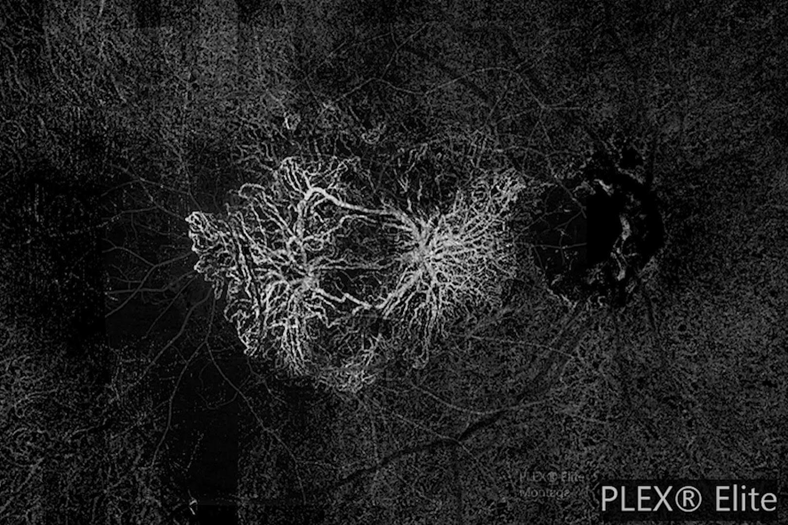
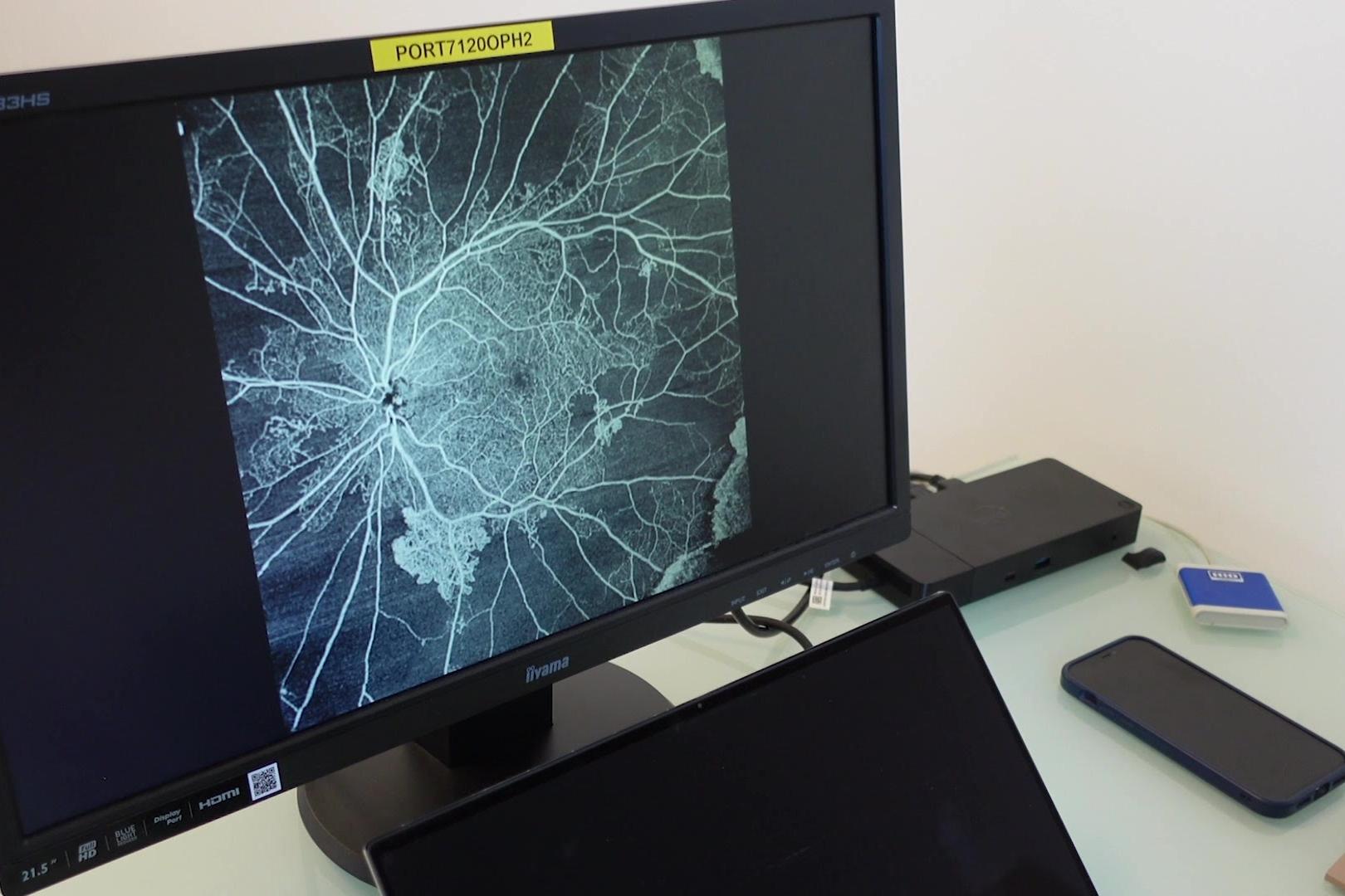
Optical coherence tomography angiography (OCT-A), a non-invasive imaging method used to assess retinal vascular integrity and perfusion may also impact the classification and management of DR. OCT-A provides a superior assessment of retinal vessels as compared to fundus photography alone in a field that today is comparable to the 7-field ETDRS fundus photos. Unlike fundus photography, classifying DR severity based on OCT-A is not widely used as it is still finding its place in clinical practice and research. When it comes to future technologies that will shape and change the way we manage diabetic retinopathy, OCT-A seems very promising.
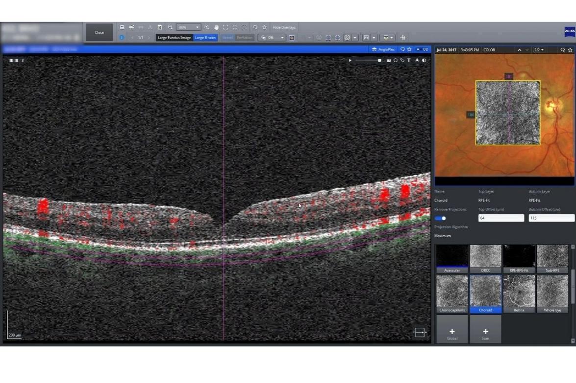
Over the past several decades, spectral domain OCT has also revolutionized the diagnosis and management of diabetic macular edema (DME). It continues to play an integral role in clinical practice, research, and the development of new AI prediction models.
The Future of Diabetic Retinopathy Management with Artificial Intelligence & Diagnostic Imaging

Shifting the Paradigm in Diabetic Retinopathy Classification
Over 50 years ago, the Early Treatment Diabetic Retinopathy Study (ETDRS) Research Group developed an objective grading scale for DR severity. This grading scale enabled clinicians to estimate the risk of a patient progressing to proliferative retinopathy based on 7-field fundus photos. At the time when the ETDRS scale was developed, clinicians had little more than fundus photos to supplement their clinical evaluation when managing diabetic retinopathy. With the plethora of better noninvasive imaging technologies available today, the ETDRS grading scale no longer should play as significant a role as it once did. With more precise imaging, with wider field imaging and new possibilities to integrate non ocular data we should be able to achieve a more predictive scale for DR.
With the advent of artificial intelligence (AI) and new innovations in ophthalmic imaging technology, the ETDRS grading scale may soon become obsolete. Currently, the Intelligent Evaluation of Diabetic Retinopathy (EviRed) project in France aims to update and optimize the outdated ETDRS grading scale. With nationwide involvement, this project aims to combine AI, clinical data of patients, and ophthalmic imaging to create a new system for stratifying DR risk assessment.
EviRed uses myriad ophthalmic imaging technologies and other clinical data points to feed its algorithm. These include OCT, OCT-A, and ultra-widefield imaging, as well as other non-imaging variables harvested from the patient’s medical history (eg. blood pressure, blood sugar, weight, age, and gender). This data is all compiled in one step and has a real potential to have superior diagnostic accuracy when compared to the older ETDRS grading system. Previously, less emphasis was placed on these auxiliary biomarkers when treating patients for DR; however, this data may be more important than previously thought. Through harnessing the predictive accuracy of AI, EviRed aims to optimize DR treatment algorithms, open the way to future preventive treatments and decrease the margin of error for treatment outcomes.
Validating the EviRed Algorithm
A clinical trial is currently underway in France that aims to train and validate the EviRed prediction model for DR risk. The trial will recruit approximately 5,000 diabetic patients who will be followed for 24 months on average. Over this time, our research group hopes to collect enough data from ultrawidefield photography, OCT, and OCT-A, as well as various other biomarkers to help refine and validate the algorithm. The primary endpoint of the study will be prediction of the progression severe DR. Close observation will be made to determine the model’s accuracy in assessing DR severity and risk of progression. Its performance will also be compared to predictions made by ophthalmologists using the ETDRS classification or similar models.

How Diagnostic Imaging & AI will Impact Workflow for EyeCare Professionals
AI and advanced diagnostic imaging offer tremendous value in improving clinical decision making and optimizing workflow efficiency. With more clinical data readily available in one place, eye care practitioners are better prepared to manage complex patient cases while spending less time sifting through previous data points.
AI platforms like EviRed aim to collect, as much automatically as allowed, clinical data points over just several years, and help clinicians to predict with better accuracy which patients are at risk for progressing to complicated DR. To obtain the diagnostic accuracy of EviRed, by classical methods without AI, other predictive models would have previously taken several decades to determine. This will optimize patient care, improve treatment outcomes, and help to better prevent blindness related to diabetes.

What Advancements of Key Diagnostic Tools Mean for Diabetic Retinopathy Management
For the eye care practitioner managing patients with diabetic retinopathy, the advancements made in key diagnostic tools like fundus imaging and OCT are paramount. Tools like the ZEISS CLARUS and ZEISS CIRRUS offer the ability to very quickly visualize the development of ocular disease, crucial for all disease management but especially crucial in cases of diabetic macular edema. While few studies exist on the effects of these new technologies on disease management, there is clear potential for these tools to improve diagnostic efficiency and management decisions.
With OCT-A advancements, ophthalmologists have the ability to see vessels better than with any other system. While OCT-A does not show as wide a field of view as ultra-widefield imaging, it still shows more details on vessels than any other such device, allowing doctors to validate their management choices. In particular, OCT-A is useful for monitoring patients undergoing anti-VEGF treatments; for these patients, while their ETDRS score may improve, their perfusion perhaps has not, and OCT-A can be a critical tool for assessing any changes in risk related to that.
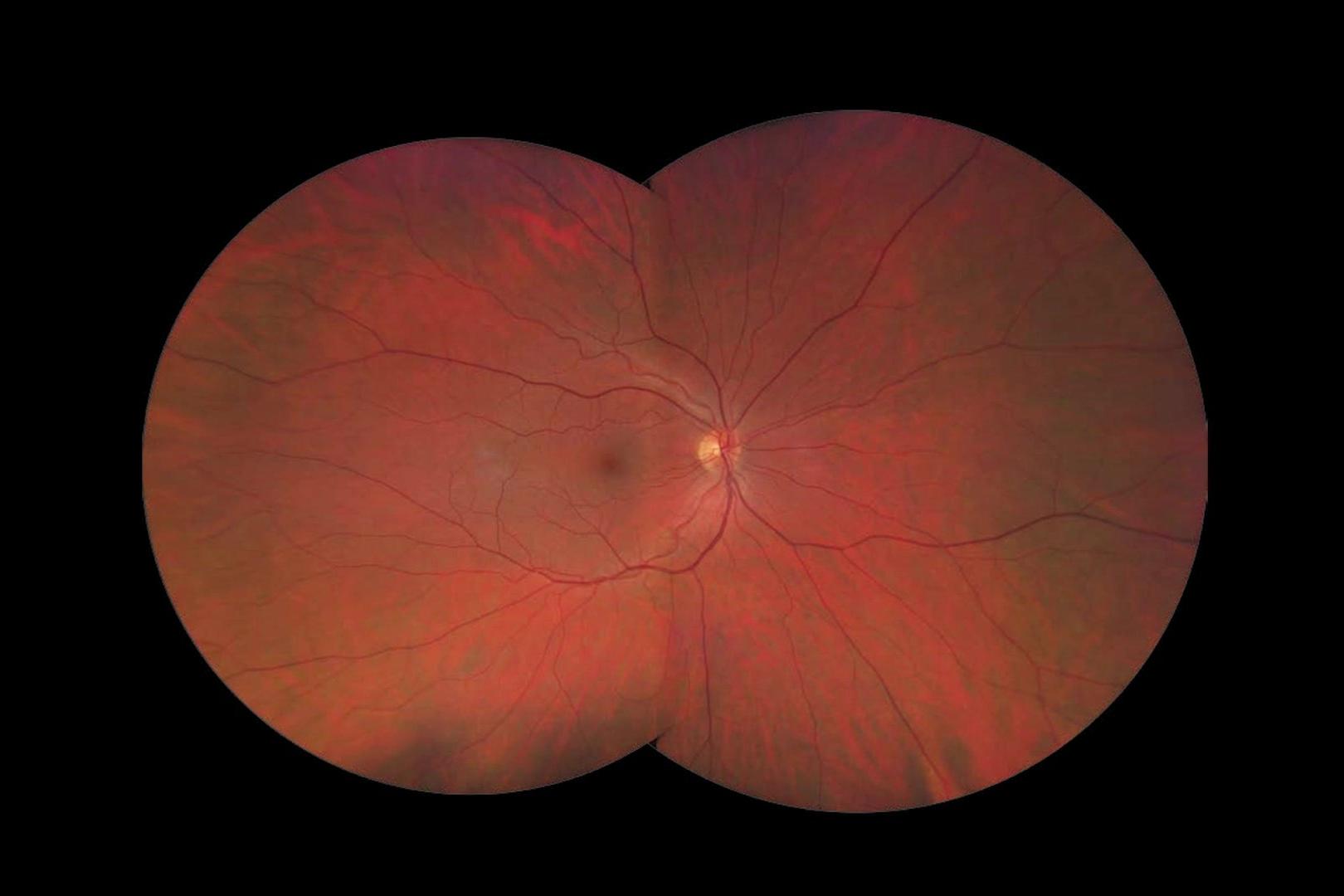

As ultra-widefield imaging becomes more prevalent in optimizing workflow for managing DR, it also enables clinicians to identify and document retinal lesions outside of the conventional 7-field area typically used with fundus photography. For instance, the risk of progression for two patients with early proliferative DR may be very different if one has also neovascularization outside the 7-field area and the other does not. In other cases, neovascularization can be only outside the 7-field area and missed if ultra-widefield imaging is not used. Advances in widefield fundus imaging and OCT-A have given practitioners the tools to enhance diagnostic accuracy while improving risk evaluation and treatment outcomes for patients with DR. Research has shown that treatments are modified in up to 15% of diabetic retinopathy patients based on the additional fundus coverage depicted in ultrawidefield imaging.
Advanced Diagnostic Imaging, Machine Learning, and the Future of Eye Care
Advanced diagnostic imaging and AI will also help to promote resources like telemedicine in the eye care world, allowing for lower risk patients to be seen remotely while higher risk patients continue being seen in person for their exams and treatment. Diabetic patients with no history of retinopathy or a stable history of mild retinopathy findings are typically ideal candidates for telemedicine consults. When it comes to managing patients with diabetic retinopathy, AI and machine learning, in addition to OCT, OCT-A and ultrawide field imaging are here to stay. The development of AI platforms such as EviRed will provide tremendous value for managing diabetic retinopathy and enhance treatment algorithms for eye care providers.