A Whole New Level
The ZEISS ELYRA 7 isn’t just any old microscope system. It can be used to take high-res photos of living cells at maximum speed – without damaging the samples too much.
ZEISS was well aware that it was bringing an exceptional new product to market at the end of last year. But no one expected it to be so well-received. At the annual meeting of the American Society for Cell Biology, the top US trade fair for cell biologists, the first live demos of the ZEISS ELYRA 7 microscope system blew the scientific community away. “They had such a warm reception and every demo was packed,” says product manager Dr. Klaus Weisshart.
Despite no single demo being tailored to a particular customer, one system was sold at the booth – even though purchasing a sophisticated system like this normally has to be budgeted for in advance. “Everything is just right: the customer can reap the benefits right after the first experiment. But I’ve never seen a customer won over so quickly,” says Weisshart.
The number of visitors to the website testifies to the high level of interest in the new product. While the site used to get around 60 visitors per day, this figure skyrocketed during the launch of the ZEISS ELYRA 7 to around 8,000 visits. Simply astonishing. That’s because the microscope system is not suitable for the mass market – and it never will be. After all, a system like this costs about as much as a home. The ZEISS ELYRA 7 enables users to achieve superresolution – it’s microscopy on a whole new level. In the area of optical high resolution, great efforts are therefore being made because innovative microscope systems like these guarantee high market visibility. Given the system’s leading position, there’s hope that other products will also benefit from this increased awareness.
The target group that ZEISS is addressing is highly unique: microscope systems are predominantly used by biological research institutes, aka imaging or core facilities, which make them accessible to scientists from a host of different labs. “These service providers are among our top multipliers,” says Weisshart.

A target group with clear needs
Other than for cell biologists, who are the most important users, there are also several areas of application that could be of great interest to developmental biologists. The product manager believes that the market’s requirements are clear: “Everyone wants to show more sensitivity, so they inspect their samples more quickly and at a higher resolution.” For a long time, improving one of the three parameters – sensitivity, speed and resolution – was detrimental to the other two (see diagram). For instance, in the past, if you wanted to keep an eye on processes in the millisecond range, it was not possible to depict this in high resolution. Or you had to increase the light intensity to a degree that either damages the cell or significantly impacts the process. “In order to prevent this, you have to disrupt system dependencies. That’s exactly what we were able to achieve by integrating Lattice SIM into the ZEISS ELYRA 7,” says Weisshart.
Everyone involved was determined to get the system to market without delay
Dr. Ingo Kleppe, Project Manager for ZEISS ELYRA 7
 © Sebastian Reuter

The two groundbreaking benefits of the ZEISS ELYRA 7
The new microscope system allows you to work with much less laser light. If the phototoxicity, i.e. the destructive impact of light on the cell sample, is reduced, processes can be observed for a bit longer. This way, you can achieve high-res images with less damage to the sample. This works because a checkered pattern is used instead of a grid pattern. In the past you had to rotate the sample in order to show the frequency signal – thanks to Lattice SIM, this is no longer necessary. Suddenly, you see something you’ve never noticed before. This means you can observe the processes at different levels, which makes the microscope system incredibly fast.
ZEISS has been working on developing new microscope systems for many years together with Professor Eric Betzig, the US physicist and recipient of the Nobel Prize in Chemistry (see interview on p. 35). The idea to use Lattice SIM for structured illumination came from ZEISS. “Everyone involved was determined to get the system to market without delay,” says project manager Dr. Ingo Kleppe. Development was completed in record time: it took less than a year to go from the initial idea to the market launch. “It’s a tremendous help for us in the market when a key opinion leader acts as an ambassador for our brand”, says Weisshart.
Betzig holds several patents for superresolution, photo-activated localization microscopy (PALM). Scientists are now in a position to view processes in living cells and tissues down to the molecular level. His work has led to the development of the first commercially available super-resolution microscope based on PALM technology. In 2007, ZEISS obtained the exclusive rights to market PALM, and this served as the basis for the development of the ZEISS ELYRA microscope system in 2012. The Sales staff was impressed by the ZEISS ELYRA 7 from the get-go. Additional customer demos have already been held. The system continues to be very well-received.
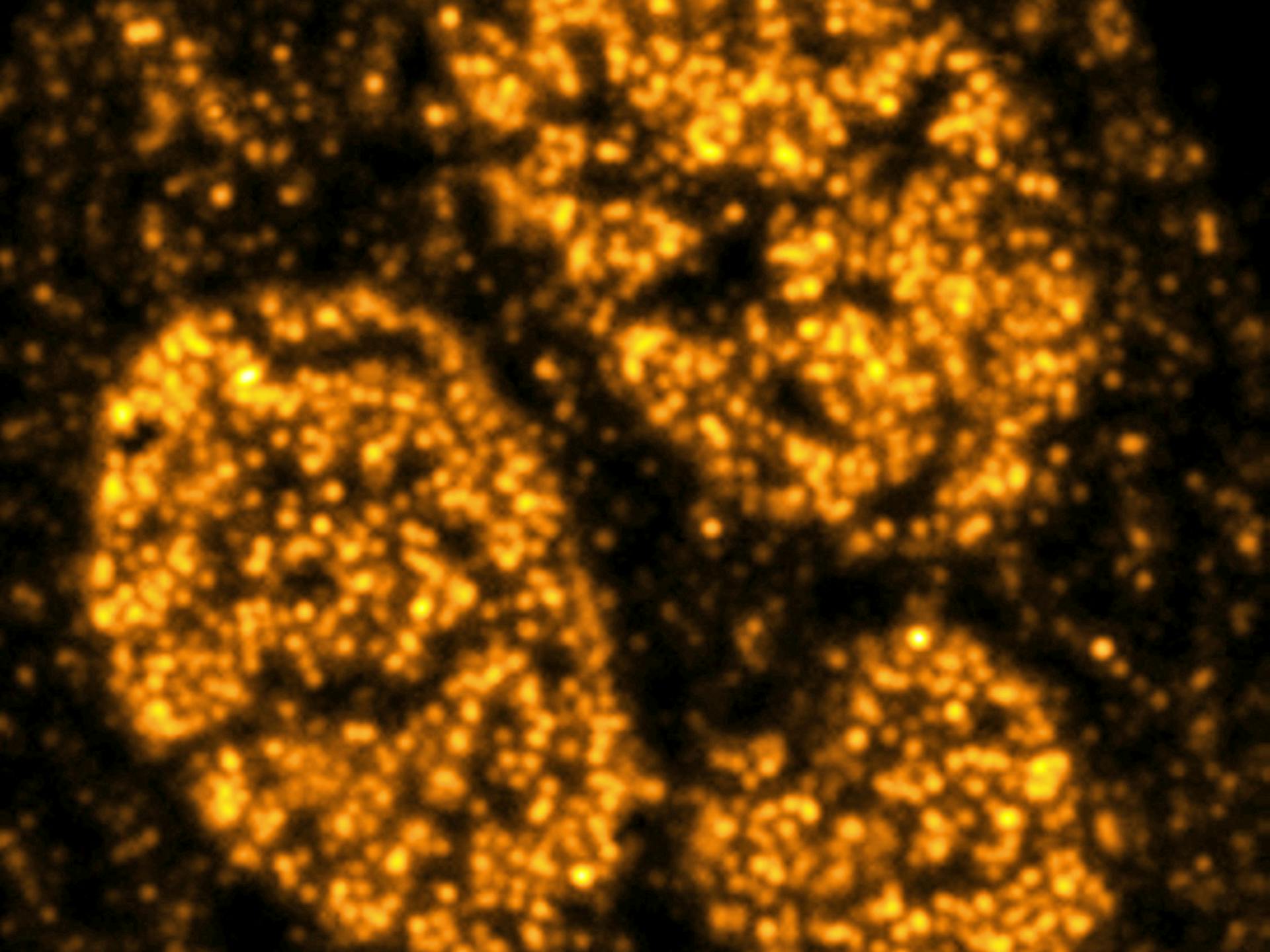
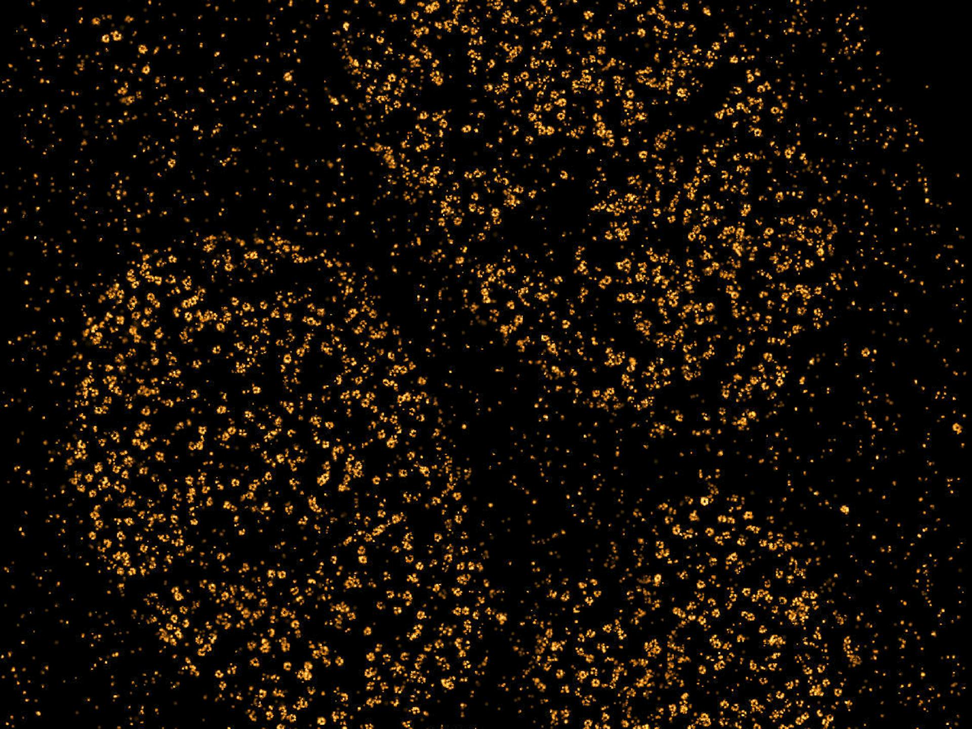
RESOLVING MOLECULAR STRUCTURES SMLM (single molecule localization microscopy) enables individual proteins to be localized and mapped with great precision. Eightfold symmetry of the nuclear pore complex in A6 cells. Gp210 was labeled with Alexa Fluor 647. Widefield image, SMLM and magnified image section.
The term LATTICE SIM
SIM stands for “structured illumination microscopy.” This technology enables users to achieve a resolution that was previously unthinkable for a light microscope, but which was made possible by dividing spatial and temporal illumination. Lattice refers to the checkered pattern used in the ZEISS ELYRA 7 microscope system that tremendously accelerates the imaging process.