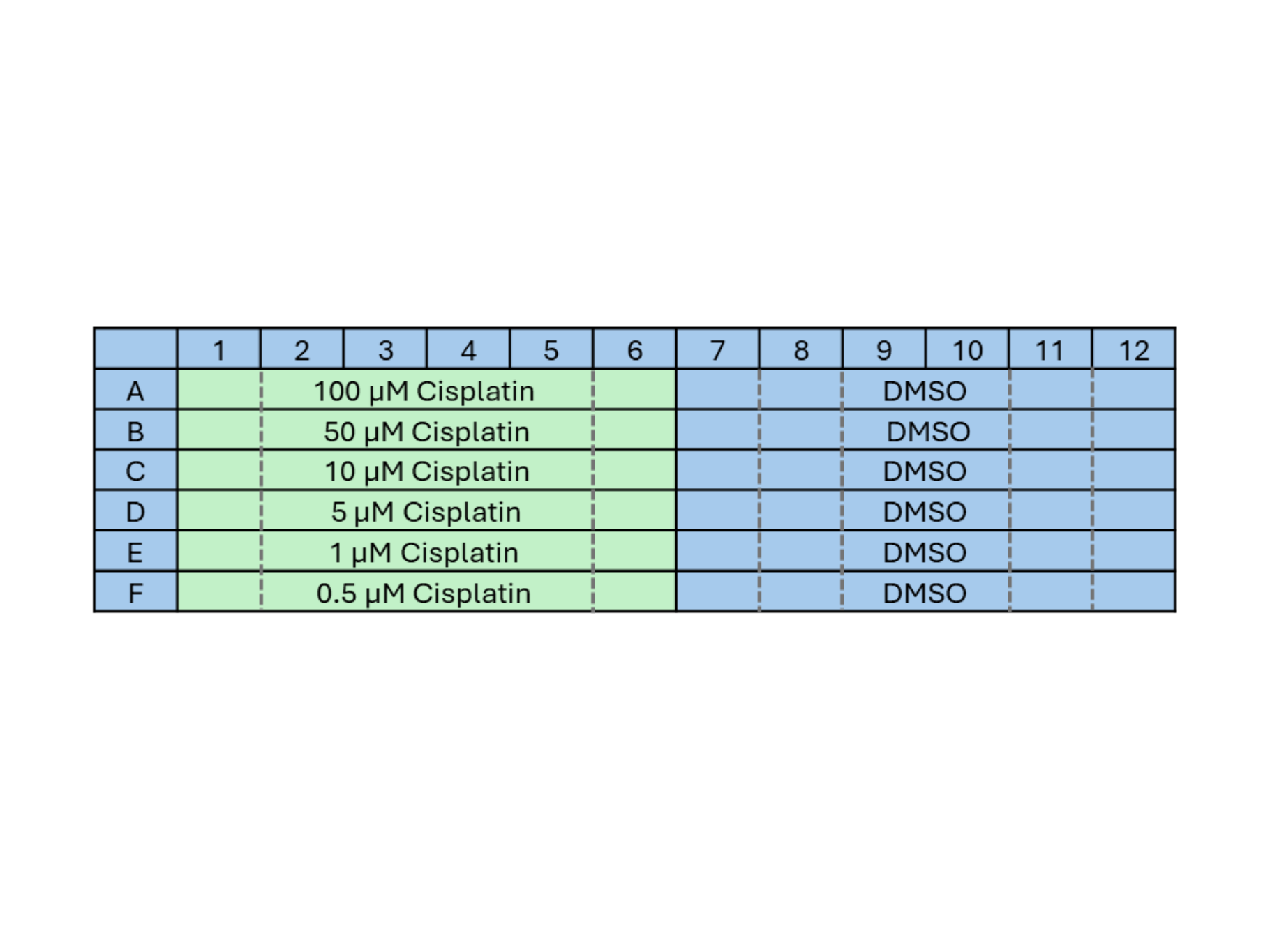
HeLa cells were initially cultured in DMEM media supplemented with 10% fetal bovine serum and 1% penicillin-streptomycin to promote optimal growth and prevent contamination. Once the cells reached 70-80% confluence in T-75 flasks, they were trypsinized and resuspended in fresh media to create a single-cell suspension. The cell density was adjusted 3,000 cells per well, and the cells were carefully seeded into a 96-well plate, allowed to adhere for 24 hours in a CO2 incubator at 37°C, and then treated with varying concentrations of cisplatin, along with appropriate controls. All drug doses and controls were added to FluoBrite cell culture media, for optimal imaging.
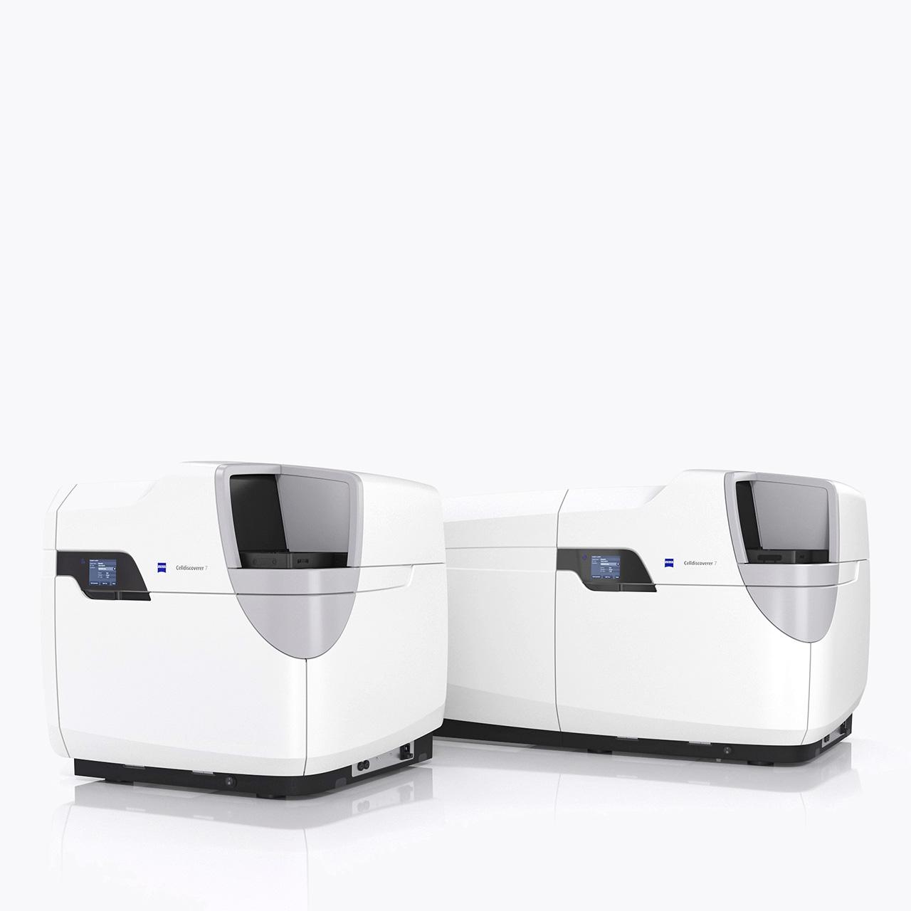
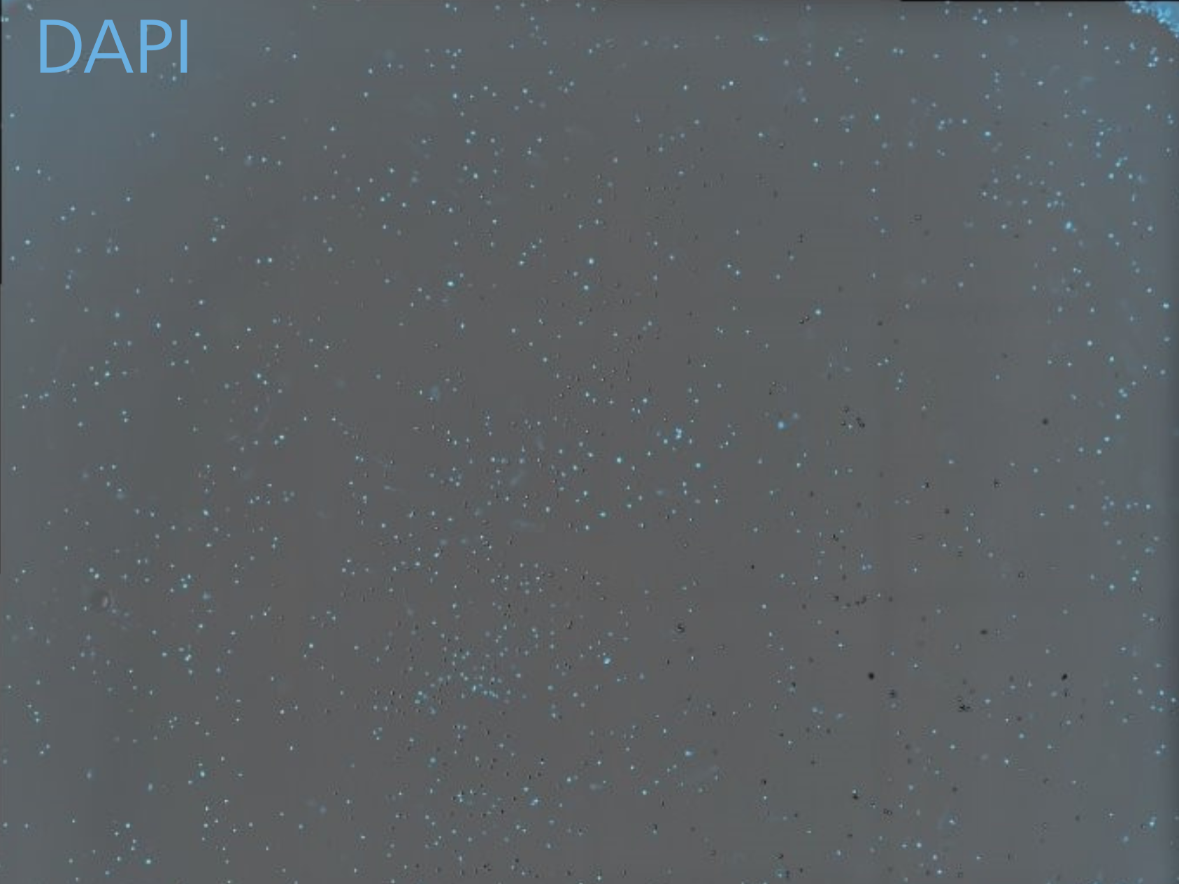
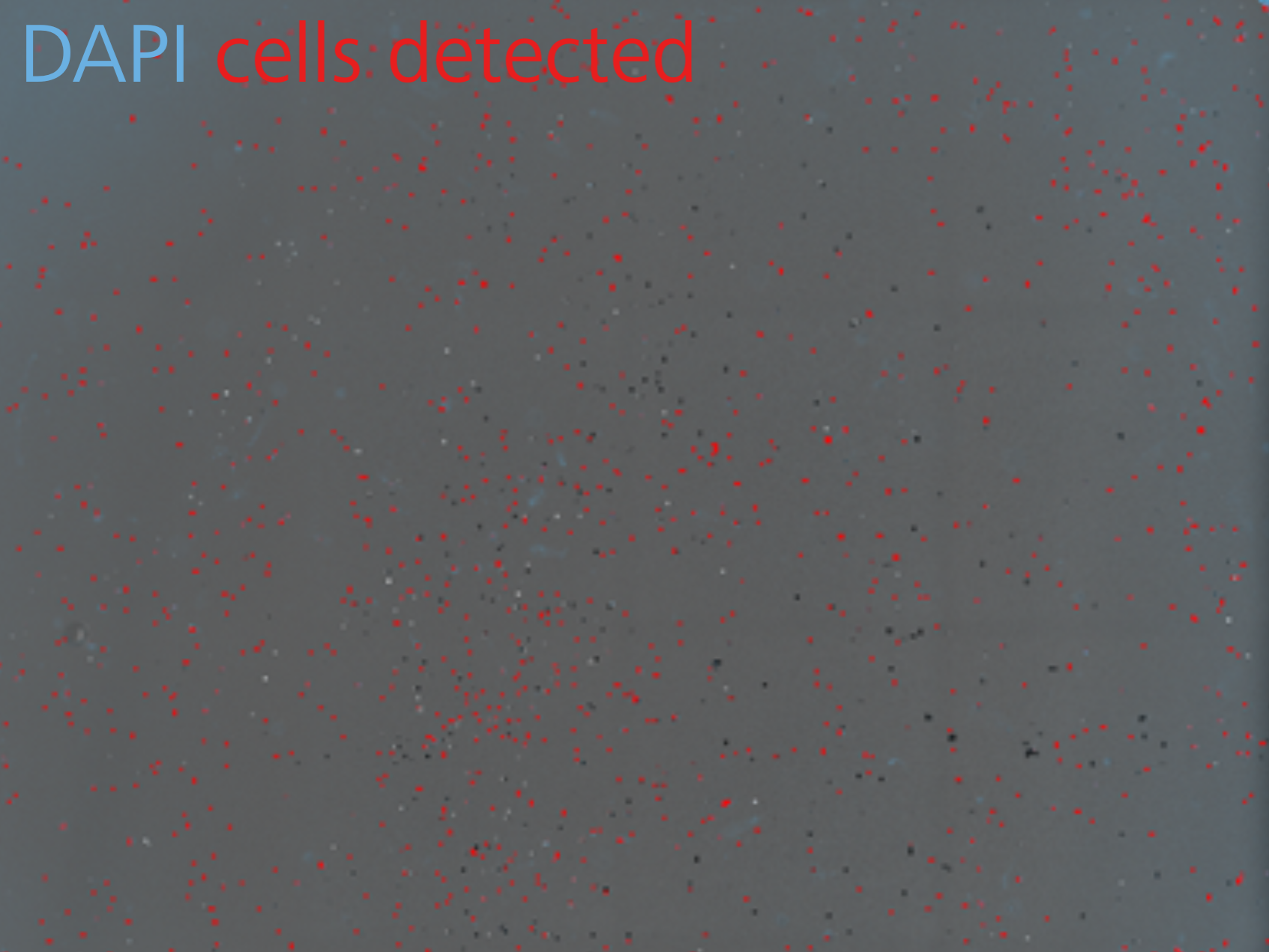
Following cisplatin treatment, the cells were stained with DAPI to visualize the nuclei, incubating for an additional 5 minutes in the dark. The plate was then transferred to the Celldiscoverer 7. Utilizing the automated HCS workflow the type of plate is automatically recognized, and imaging regions are placed within the wells. Smart automation features such as autofocusing and automatic exposure setting streamline the process. The complete environmental control offered by Celldiscoverer 7, allows the sample to be continuously imaged for 48 hours. The cells were imaged in brightfield with Oblique illumination which angles the light onto the sample, significantly enhancing the contrast and creating a pseudo 3D appearance. Additionally, the DAPI label was imaged using fluorescence widefield microscopy, with excitation 385 nm LED. An area of 75% of each well was imaged allowing to ensure a statistically relevant sample size. Sample focus was maintained with automated SW features, ensuring the cells remain in focus of the entirety of the experiment.
:721-7) imaged every 15 minutes for 72 hours using autoimmersion")
:721-7) imaged every 15 minutes for 72 hours using autoimmersion")