You explore to understand and imagine what’s next.
With our solutions you challenge the status quo.
Everyday – in science and in industry.
Together we shape what matters most – for generations to come.
From curiosity, to knowledge, to progress.
From curiosity to lasting impact.
… I owe a large part of my success, which I had achieved in the name of science, to your excellent microscopes.
Zeissians are very meticulous. Customers have certain expectations when they buy a ZEISS system. It is important for us to meet these – and that is also my personal goal.
Addressing global megatrends
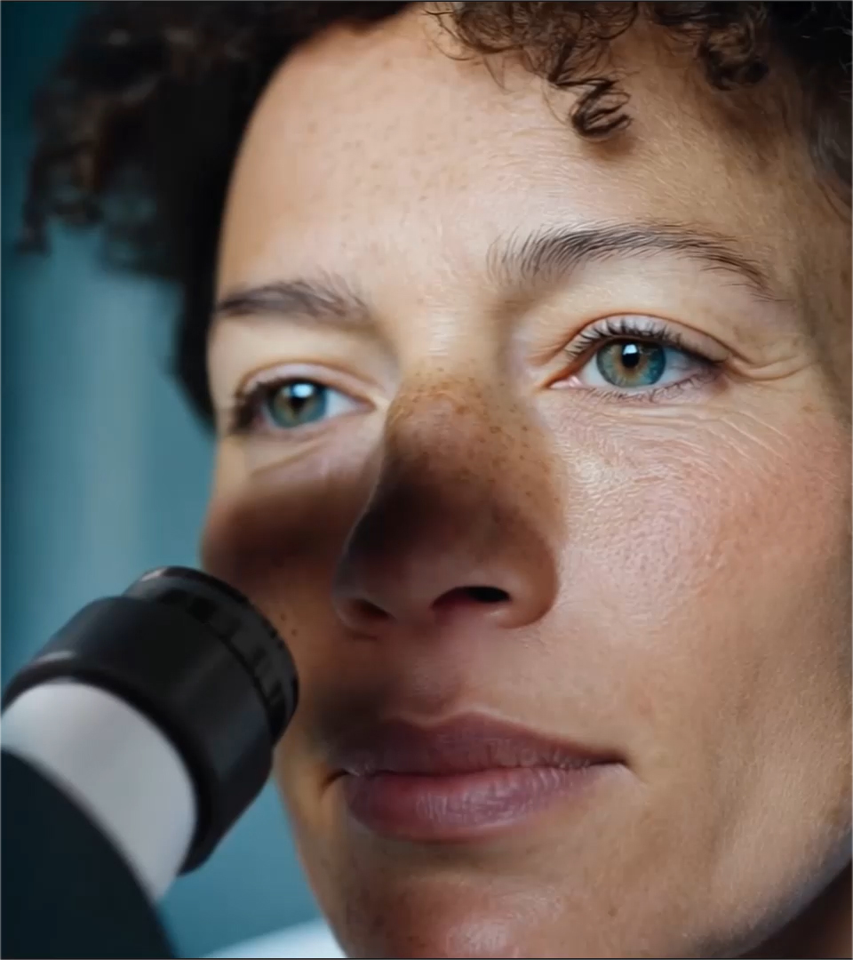
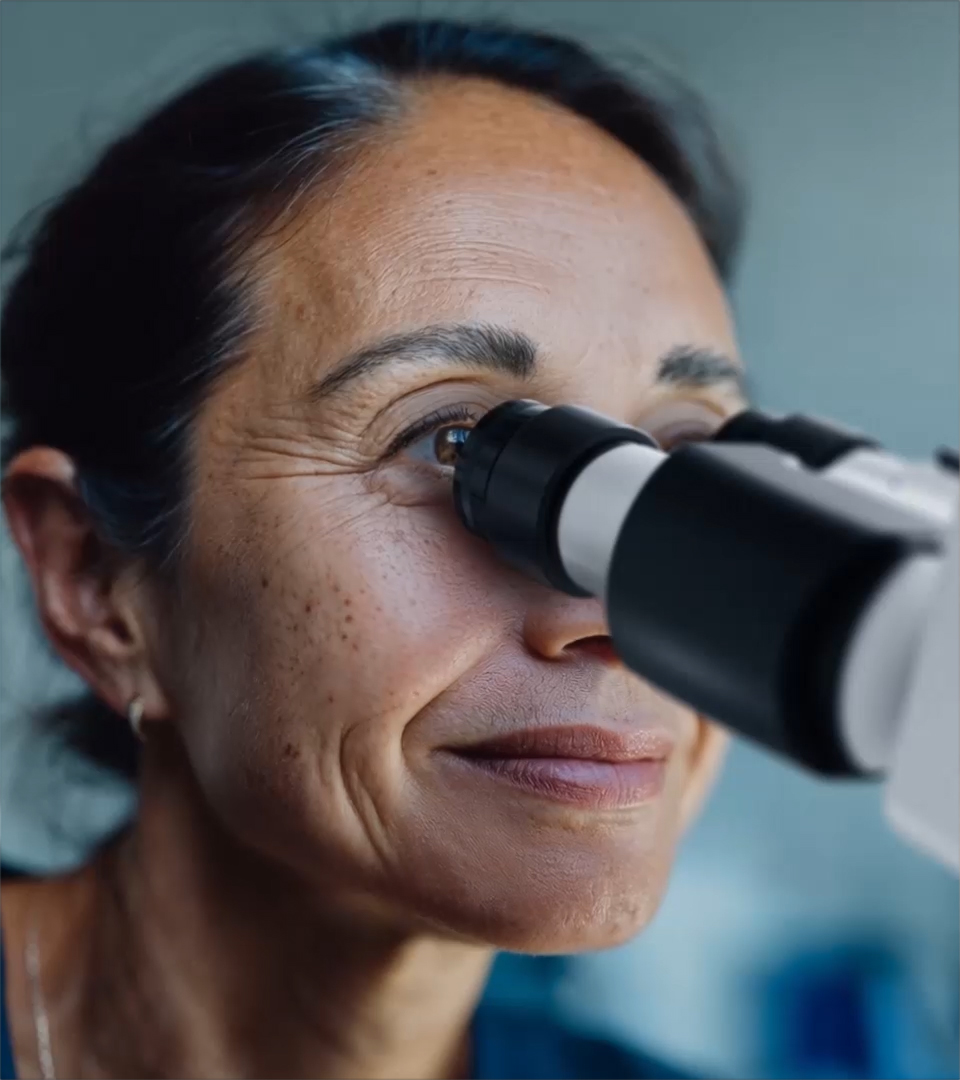
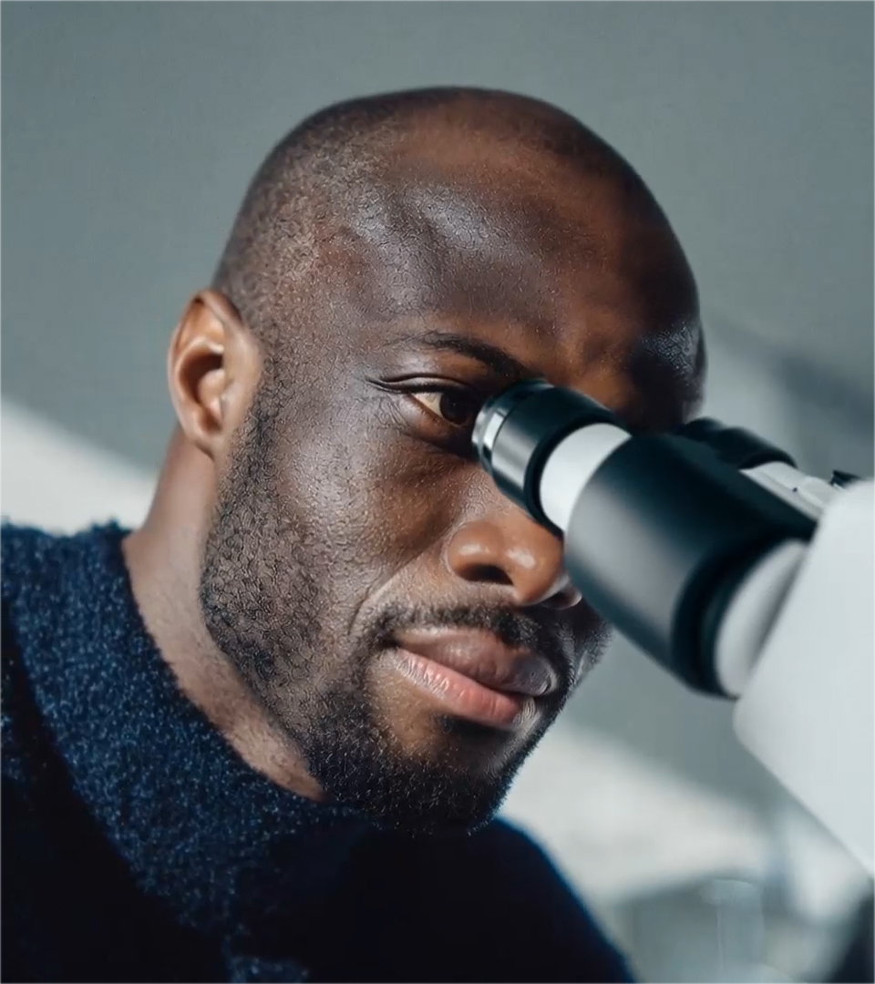
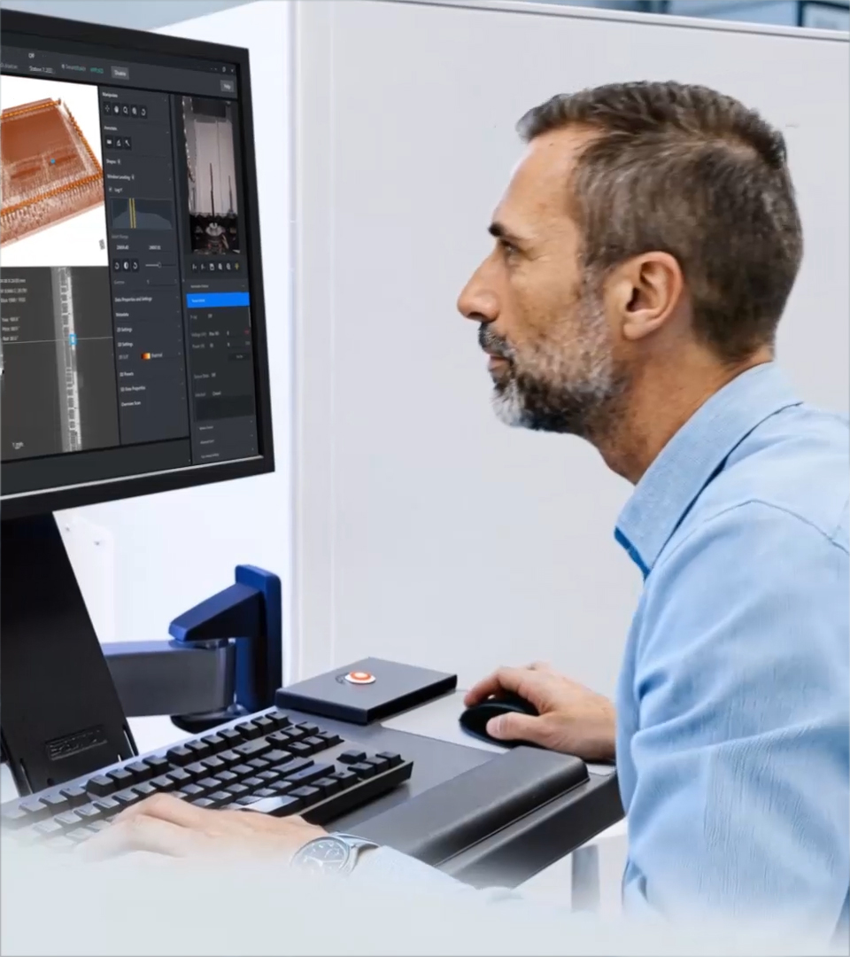
Our customers lead the way in scientific and industrial advancements by leveraging our cutting-edge microscopy solutions to transform complex questions into pioneering discoveries.
Silver society and growing population
From rising diseases like cancer or Alzheimer's and infertility challenges to increased food demands driven by population growth, microscopy is crucial for uncovering causes and developing effective treatments and sustainable solutions.
Digitalization & urbanization
From 6G communications and healthcare to new materials, microscopy uncovers microstructures to detect failures and optimize performance, while innovative teaching technologies powered by microscopy ignite scientific curiosity.
Green energy and neo-ecology
From transitioning to sustainable energy to preserving natural environments, microscopy enhances the analysis of batteries and underground formations, optimizing efficiency and sustainability.