Virology is an important field within microbiology and is concerned with the study of viruses and viral diseases. The impact of viruses on the population is enormous, as they can cause severe endemic or even pandemic diseases, such as the Spanish flu (H1N1 influenza virus), Aids (Human immunodeficiency virus HIV), dengue fever (dengue virus DENV), or most recently Covid-19 (Coronavirus SARS-CoV-2).
Studying viruses, their interaction with host cells, and the immune response to the virus helps to develop vaccines and treatments against viral pathogens. Therefore, virology is often strongly connected to immunology.
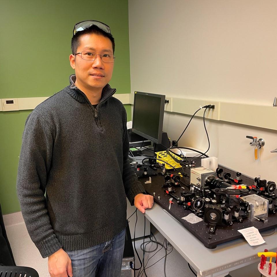
Allen Liu is an Associate Professor of Mechanical Engineering at the University of Michigan, USA.
User Story
Analyzing Influenza A Virus entry through Lattice Lightsheet microscopy
Researchers at the University of Michigan used ZEISS Lattice Lightsheet 7 to watch influenza A virus enter living cells in real time. By fluorescently labeling the virus, clathrin, and the membrane-bending protein epsin, they captured high-resolution 3D movies showing how the virus exploits epsin to bend the cell membrane and trigger endocytosis. The gentle lattice light sheet illumination minimized photobleaching, allowing long, continuous imaging and revealing detailed virus–host interactions that traditional confocal methods often miss.
Unlike traditional microscopy, lattice light sheet technique allowed us to visualize the viral entry on the entire cell surface. Furthermore, we were able to track IAVs before and after they attach to the cell membrane.
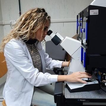
Dr. Massimo Zollo (left) with his collaborator, Dr. Veronica Ferrucci, at their ZEISS Elyra 7 super-resolution system.
User Story
Antiviral actions of polyphosphates against SARS-CoV-2 visualized with super-resolution microscopy
At CEINGE in Italy, researchers used ZEISS Elyra 7 super-resolution microscopy to visualize how long-chain polyphosphates inhibit SARS-CoV-2 infection. By immunostaining viral spike proteins, ACE2, and viral RNA in HEK293-ACE2 cells, they directly observed that polyphosphates disrupt syncytia formation and reduce viral replication. The high spatial resolution of Elyra 7 allowed precise colocalization studies in subcellular compartments, strengthening mechanistic insights into antiviral actions at the molecular level.
Super-resolution microscopy using ZEISS Elyra 7 turned out to be a powerful tool to visualize SARS-CoV-2 viral proteins (through immunofluorescence) and/or RNA (via HuluFISH) in specific cellular compartments, such as membrane or cytosol.
Microscopy Solutions for Virology
Microscope Requirements
For observation and maintenance purposes in cell culture, inverted light microscopes with a small footprint, LED fluorescence option, good ergonomics and high-quality optics for reliable digital documentation are essential tools. Immunofluorescence opens up the rapid detection of viral agents with direct (DFA) or indirect fluorescence antibody (IFA) tests, including antibody test kits against herpes simplex (HSV), Influenza A, other respiratory viruses and enteroviruses.
Automated boxed microscopes with integrated calibration, environmental control and fluorescence options are ideal for lab environments with high throughput demands, enabling fully automated 2D and 3D screening of cell cultures and tissues. Confocal microscopes give the virologist the option to investigate the details of cellular invasion in greater detail and prepare the respective sample for further investigation with immune electron microscopy.
Recent developments in scanning electron microscopy (SEM) have shown to meet the resolution and image quality requirements for virus studies. The large field of view imaging mode in combination with correlative light microscopy and automated workflows saves valuable time in finding relevant viral spots and provides fast results, even in 3D.
Recommended Products for Virology
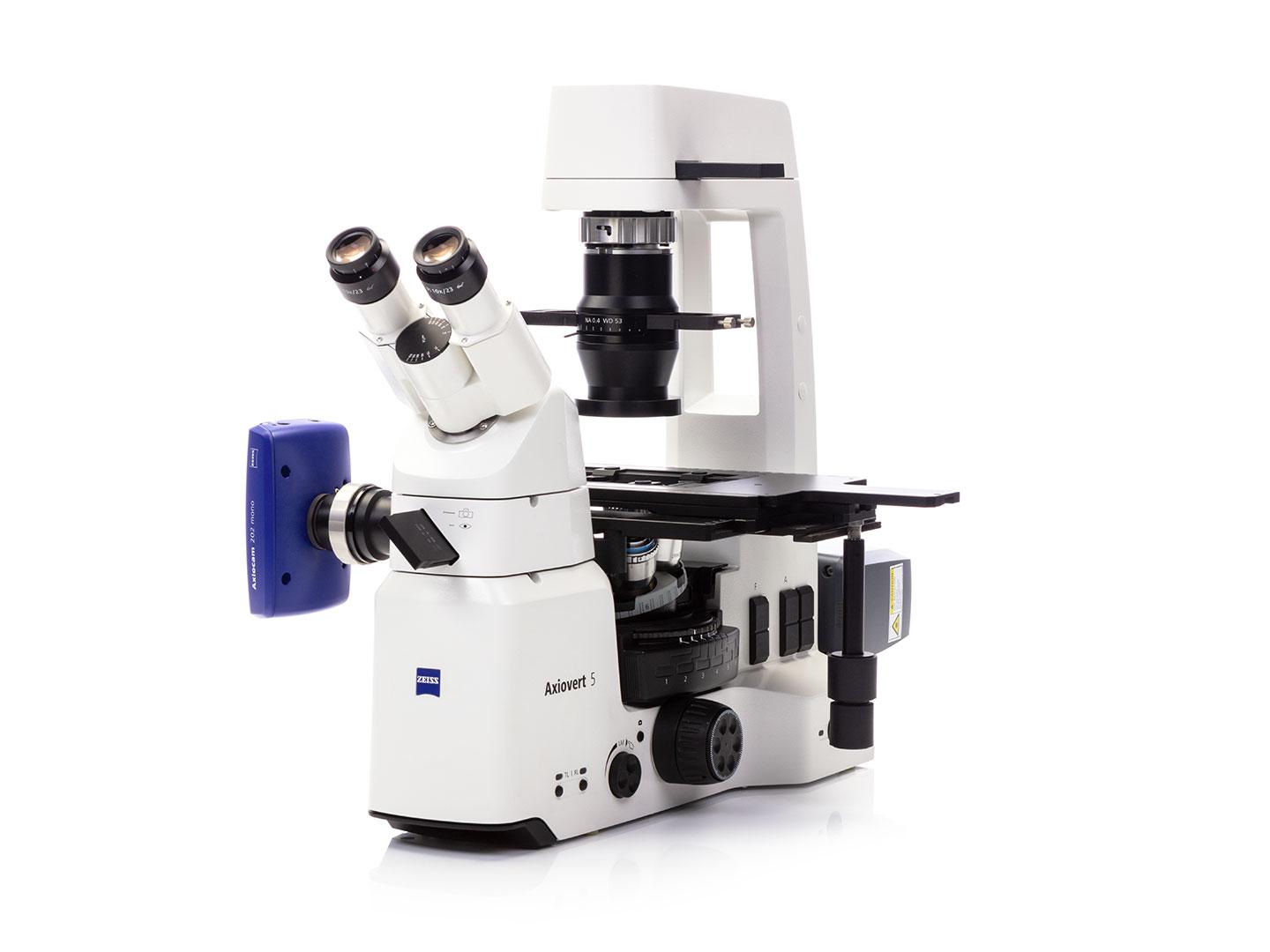
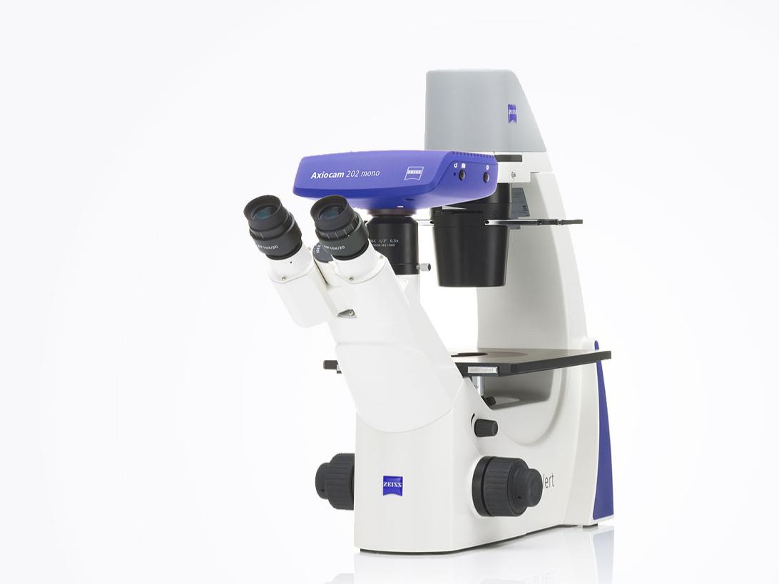
Your Smart Microscope for Cell Culture and Research
ZEISS Axiovert 5
Axiovert 5 brings smart microscopy into your cell culture lab. All you need to do is focus on your samples and workflow. Simply push Snap to get crisp images for documentation.
Examine and Assess your Living Cells Quickly and Easily
ZEISS Primovert
Place ZEISS Primovert right inside your Laminar Flow Box. Examine unstained cells in phase contrast and GFP-labeled cells in fluorescence contrast quickly and efficiently.
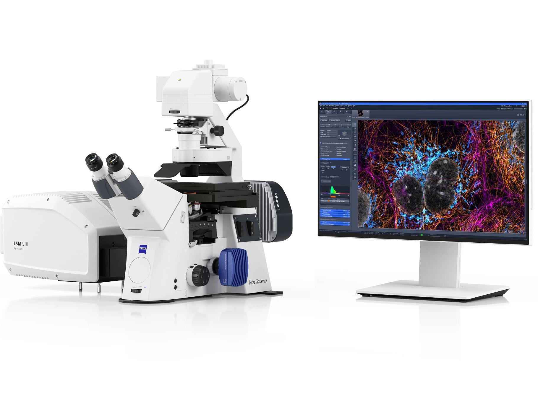
Your Compact Confocal Microscope for Innovative Imaging and Smart Analysis
ZEISS LSM 910
ZEISS LSM 910 combines high-quality confocal imaging with innovative possibilities for your next research endeavors. Conduct multi-color, live experiments with spectral precision. Gain more information with gentle super-resolution imaging or study the dynamics of life in 3D at impressively high speed.
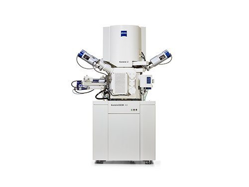
FE-SEM For Highest Demands in Sub-nanometer Imaging, Analytics and Sample Flexibility
ZEISS GeminiSEM
ZEISS GeminiSEM stands for effortless imaging with sub-nanometer resolution. These FE-SEMs (field emission scanning electron microscope) combine excellence in imaging and analytics.
ZEISS Primostar 1 is a durable and user-friendly microscope designed for educational applications, featuring fixed Koehler illumination that simplifies setup and ease of use.
Axiovert 5 digital brings AI into your cell lab to ease your daily work. It will make your processes more efficient and your results more reproducible. Stay relaxed, even when there is a lot going on around you.
Your Microscope for Digital Teaching and Routine Lab
ZEISS Primostar 3
No matter if you need a microscope for education, training or routine lab work: Primostar 3 is the easy-to-use, compact and long-lasting instrument that turns your investment into the right choice for long working hours even in the most space-limited environments.
 common in children and adults in the genital area, Primostar 3, 10x objective, Axiocam 208 color,")
 common in children and adults in the genital area, Primostar 3, 10x objective, Axiocam 208 color,")


: 13 labels plus autofluorescence acquired in one track using 5 lasers and 36 detectors, unmixed image of the 13 labels without autofluorescence.")
: 13 labels plus autofluorescence acquired in one track using 5 lasers and 36 detectors, unmixed image of the 13 labels without autofluorescence.")


