
ZEISS at Microscopy & Microanalysis (M&M) 2025
Booth #1518Connect with ZEISS for In-Booth Educational Presentations
Each day has a chance to connect, be sure to add it into your schedule.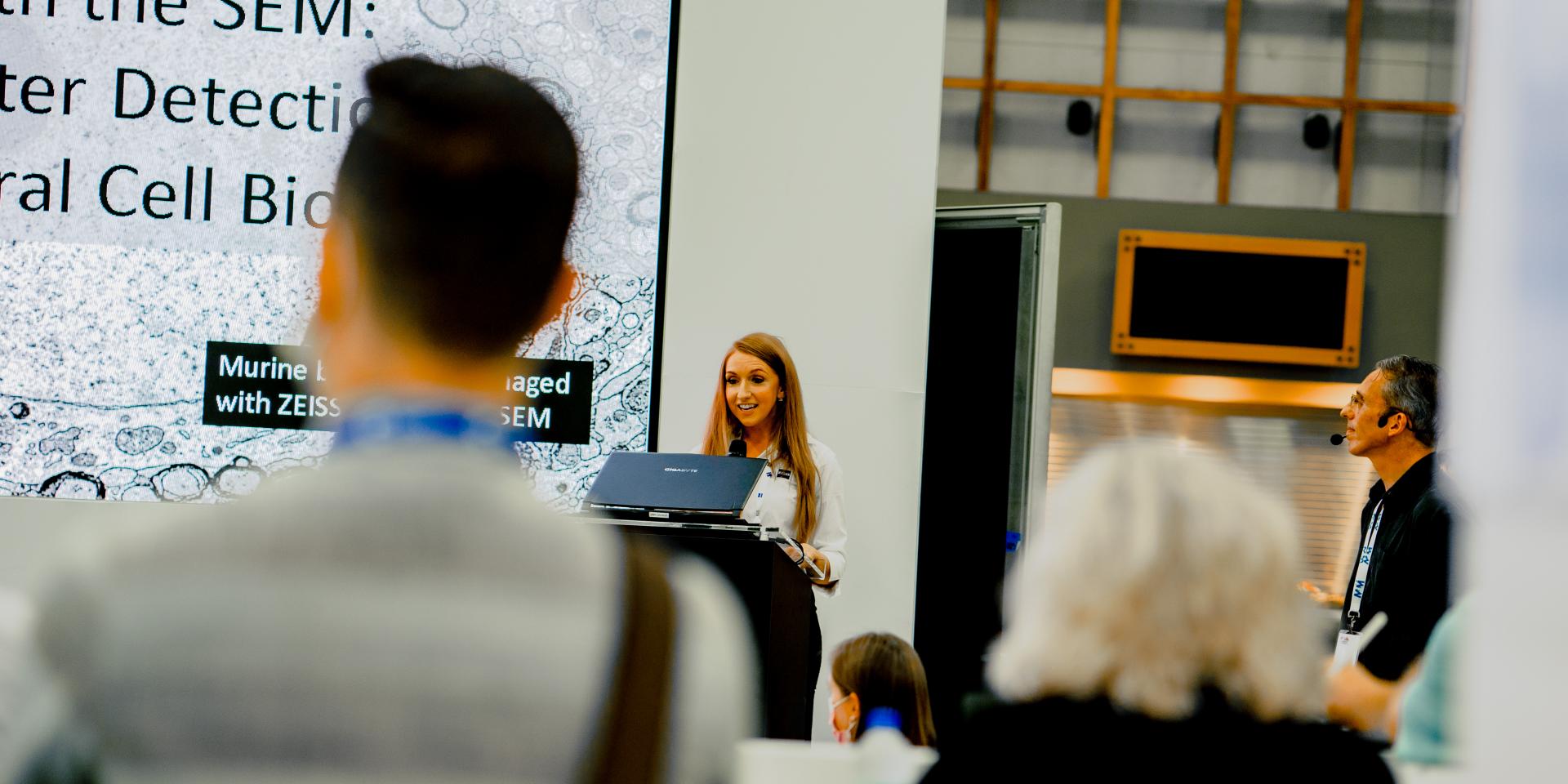

-
Abstract
The winds of the current geoeconomic climate are accelerating our need and ability to move innovations from the lab to the marketplace, often referred to as “The Valley of Death.” One significant area of demand is automated nanostructure characterization and process monitoring in R&D institutions. While purpose-built CDSEMs excel in mass production, they tend to be less flexible, more expensive, and more specialized in operation compared to versatile laboratory SEMs. This presentation will cover methods and results for pattern-driven automated metrology using the ZEISS Gemini560, featuring a 200mm capable sample chamber, a high-precision stage, and an integrated GenISys software kit, at the Penn State Materials Research Institute. We will focus on applications involving photonic devices, such as large-area gratings, photonic crystals, and meta lenses, along with the unique features of ZEISS FESEM technology that enable these applications.
We will Discuss:
- Challenges and solutions for acquiring and analyzing images in an automated workflow.
- Explore resilience strategies for addressing real-world limitations, including hardware issues and imaging artifacts.
- Examine examples from projects focused on process calibration and monitoring.
- Discover how to develop go/no-go criteria based on customer-driven specifications for devices.
- Validate FESEM measurements against device performance data to support findings.
- Utilize process monitoring tools, such as thresholds, shape fitting/comparison, and PV bands, to detect when a process is out of specification.
- The unique aspects of the ZEISS Gemini560 that make it well-suited for pattern-driven automated metrology.


Chad Eichfeld
Chad Eichfield is Operations Director at the Penn State Nanofabrication Materials Research Institute, where he specializes in advanced materials characterization and nanofabrication techniques. With a strong background in materials science and engineering, Chad focuses on the development and application of innovative methodologies for the fabrication and analysis of nanostructures. His work encompasses a range of cutting-edge technologies, including electron microscopy and automated metrology, aimed at enhancing the performance of next-generation electronic and photonic devices. Chad is dedicated to bridging the gap between laboratory research and real-world applications, contributing to the advancement of nanotechnology in various fields. He actively collaborates with interdisciplinary teams and is committed to mentoring the next generation of scientists and engineers in the realm of nanofabrication and materials research.

Steven Hernandez
Steven Hernandez serves as a Business Development Manager for Carl ZEISS Microscopy in North America. With over 15 years of expertise in electron microscopy and materials characterization, his experience spans roles such as lead microscopist at the University of Arizona’s EM Core Facility, high-volume photomask manufacturing at Intel, and electronic device failure analysis at Medtronic. In 2023, he joined ZEISS as a Product Applications and Sales Specialist. Currently, he focuses on collaborating with customers across various sectors to identify how ZEISS's advanced microscopy solutions can support their objectives.
-
Abstract
This talk will update the audience on current state-of-the-art technology in lab-based 3D X-ray Microscopy. 3D XRM is a powerful imaging technique when some or all of the following conditions apply:
- Samples under investigation contain features with 3D morphology/structure that matter to the processing, performance, or function
- Those feature(s) are hidden inside the object
- Critical features are in the size range of tens of nm to hundreds of microns
- There’s a benefit to keeping the sample intact (due to delicate structures and/or future sample investigations)
Such conditions often exist within various research fields including materials science, engineering, geology, and biology. In this talk, the latest advances in high resolution X-ray optics, image acquisition routines, novel reconstruction approaches, and powerful user interface tools will be presented. This will be followed by an introduction to how XRM is utilized to support research at and around the University of Utah, including a selection of case studies and how you can access the instrument to support your own work.

Will Harris, PhD
Dr. Will Harris is a Business Development Manager at ZEISS Microscopy. He holds a B.S. and PhD in Mechanical Engineering from the University of Connecticut, where he studied transport and degradation in electrochemical energy devices using synchrotron-based X-ray imaging and analysis techniques. He has 15 years of experience in high resolution 3D X-ray tomography, from synchrotron to lab-based instruments and covering a range of applications in materials science, engineering, and semiconductors. For the past 10+ years he has held several positions within ZEISS, all involved with supporting the X-ray imaging and materials science microscopy business, and currently focuses on engagement with the community in North America.
Brian Van Devener, PhD
Dr. Brian Van Devener is the manager of the Nanofab Electron Microscopy & Surface Analysis Lab (EMSAL) at the University of Utah. The EMSAL is the largest imaging and materials characterization lab in Utah, with $11.3M in instrumentation for EM and materials characterization, and serves over 200 on-campus and 80 off-campus researchers in industry and academia. Van Devener has over 20 years of experience in advanced characterization techniques, and is the technical and operational head of the facility. With a PhD in Physical Chemistry focusing on heterogeneous catalysis, he has authored/co-authored 36 peer reviewed publications.
-
Abstract
In situ microscopy experiments offer a fantastic means to directly discern linkages between materials’ processing, structure, and properties. However, such workflows have traditionally been very challenging to perform due to their time-consuming nature, the deep level of expertise required, and the complexity of coordinating between numerous sub-system components. This tutorial will present recent advances to help move past some of these roadblocks by:
- Using deep learning X-ray reconstruction (ZEISS DeepRecon Pro) to drastically accelerate data acquisition during time-lapse/4D in situ microtomography experiments, without compromising on data resolution or quality
- Making powerful in situ thermomechanical experiments in the SEM accessible to users of all experience levels (with ZEISS In Situ Lab SEM), by integrating hardware and automating software control of the microscope, loading stage, and analytical (EDS, EBSD) detectors.
This session will incorporate results generated on various additive manufactured metal samples, performed by the ZEISS Microscopy applications team in collaboration with scientists at NIST, as well as work performed in the Mechanical Engineering Department at Liberty University.
Nathan Johnson, PhD
Nathan Johnson earned his Ph.D. from the Colorado School of Mines and went on to complete a postdoctoral fellowship at Stanford University. Over the past decade, he has specialized in developing and applying laboratory and synchrotron-based X-ray characterization techniques. His research sits at the intersection of advanced manufacturing, advanced characterization, and laboratory automation, with a focus on accelerating materials discovery and innovation.

Prof. Mark Atwater, PhD
Prof. Atwater is an Associate Professor of Mechanical Engineering at Liberty University, and holds degrees in Manufacturing Engineering Technology, Mechanical Engineering, and Materials Science and Engineering. His research often combines these areas in fundamental microstructural analysis, bulk mechanical performance, and practical strategies to produce advanced materials in a scalable way. Ongoing research topics include microporous and nanoporous metals, surface hardening through patterned deformation, and non-equilibrium alloy development for functional applications.
-
Abstract
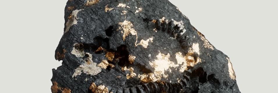
Ancient stone buildings and towering pyramids stand as testaments to humanity's obsession with technological advancement. Along our journey, as we transitioned from stone to simple metals, and from simple metals to advanced materials, we established a framework for material innovation—focusing on structure, properties, processing, and performance. Central to achieving the next material breakthrough is our ability to characterize these materials accurately and effectively.
Join us as we explore techniques, applications, and workflows utilizing the full spectrum of the ZEISS microscopy portfolio, which plays a crucial role in advancing materials science research across fields such as electronic and battery materials, metals and alloys, and materials for additive manufacturing.
Steven Hernandez
Steven Hernandez serves as a Business Development Manager for Carl ZEISS Microscopy in North America. With over 15 years of expertise in electron microscopy and materials characterization, his experience spans roles such as lead microscopist at the University of Arizona’s EM Core Facility, high-volume photomask manufacturing at Intel, and electronic device failure analysis at Medtronic. In 2023, he joined ZEISS as a Product Applications and Sales Specialist. Currently, he focuses on collaborating with customers across various sectors to identify how ZEISS's advanced microscopy solutions can support their objectives.


Live Microscopy Demos at M&M
Connect directly to our ZEISS Microscopy Customer Center. Find your demo of interest and reserve your spot

Connect with Our Microscopy Experts at M&M
Engage with our specialists virtually at the ZEISS Microscopy Customer Center located in Dublin, CA. Follow the link below to access our selection of workflows that showcase our most recent innovations.