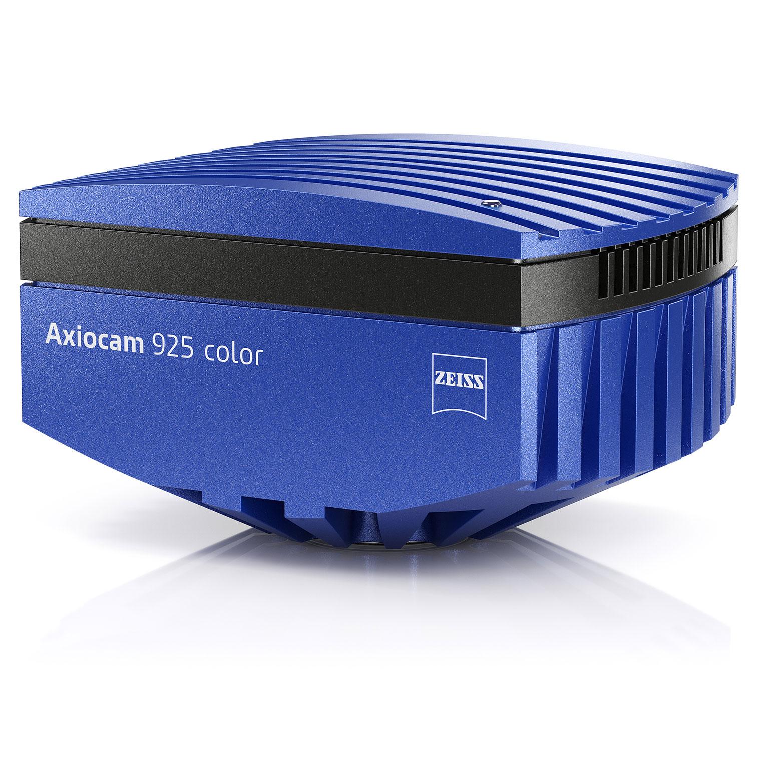

ZEISS Axiocam 925 color
A 25-megapixel microscope camera for color imaging across large fields of viewThe ZEISS Axiocam 925 color combines a 25-megapixel CMOS sensor with a 19.2 mm diagonal to capture large sample areas with excellent color fidelity. Its 2.74 µm pixels resolve fine details across microscope magnifications.
Capture full pixel count images at up to 58 fps for high-quality time-lapse imaging of dynamic objects. The 30 fps live image supports smooth sample navigation. Active sensor thermal stabilization helps ensure reproducible image quality, while HDR mode provides a dynamic range of up to 1:25,000.
Global shutter architecture supports distortion-free imaging of dynamic processes. The camera offers color and monochrome modes, high speed data readout, and seamless ZEN integration. Its large sensor can reduce the need for image tiling. The Axiocam 925 color is suitable for life science, pathology, and materials research.


Mouse vertebral tissue stained


Mouse vertebral tissue stained with Safranin O/Fast Green, revealing the intricate architecture of bone, cartilage, and bone marrow. The multicolor staining enables clear differentiation of tissue components and highlights structural details across the vertebral microenvironment. Courtesy of Core Facility Pathology & Tissue Analytics, Helmholtz Center Munich, Germany.
- Demanding color imaging, including fast, high-resolution live imaging and co-observation across a very large field of view
- Fast tile scanning and imaging across a broad range of intensities and exposure times
- Large-area imaging in pathology, cytology, and materials research
Mouse vertebral tissue stained with Safranin O/Fast Green, revealing the intricate architecture of bone, cartilage, and bone marrow. The multicolor staining enables clear differentiation of tissue components and highlights structural details across the vertebral microenvironment. Courtesy of Core Facility Pathology & Tissue Analytics, Helmholtz Center Munich, Germany.


Graph – Axiocam 925 color – Normalized spectral responsivity with IR cut filter
Graph – Axiocam 925 color – Normalized spectral responsivity with IR cut filter
Highlights of Axiocam 925 color
- 25-megapixel CMOS sensor with a 19.2 mm diagonal
- Up to 58 full pixel count images per second
- 2.74 µm pixels for resolving fine details across microscope magnifications
- Fast quad-port readout and global shutter for distortion-free imaging
- Excellent color fidelity in color mode, with both color and monochrome imaging modes available
- 30 frames per second in live image mode
- Active sensor thermal stabilization for reproducible image quality
- HDR mode with a dynamic range of up to 1:25,000
Application examples


Kidney cortex morphology in detail


H&E-stained kidney cortex tissue visualizes glomeruli and renal tubules, supporting evaluation of tissue morphology and organization.
Kidney cortex morphology in detail
H&E-stained kidney cortex tissue visualizes glomeruli and renal tubules, supporting evaluation of tissue morphology and organization.Sample courtesy of Core Facility Pathology & Tissue Analytics, Helmholtz Center Munich, Germany


Close-up view, more detail


A cutout of the original kidney image scaled up for a closer view.
Close-up view, more detail
A cutout of the original kidney image scaled up for a closer view.Sample courtesy of Core Facility Pathology & Tissue Analytics, Helmholtz Center Munich, Germany


