. The cell walls were stained with propidium iodide (white). Imaged with ZEISS LSM 780.")
. The cell walls were stained with propidium iodide (white). Imaged with ZEISS LSM 780.")
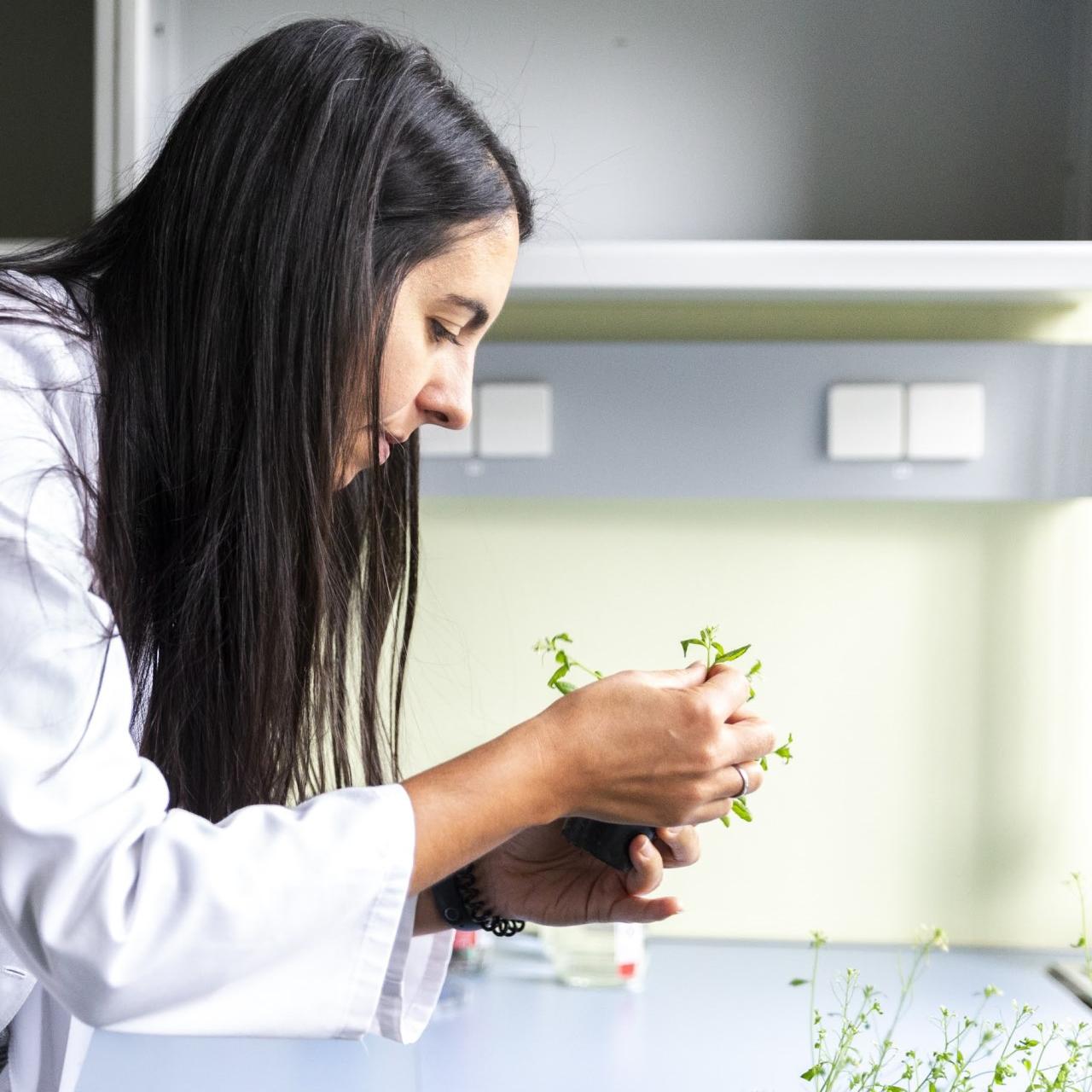
Arabidopsis Research
Discover a comprehensive toolkit for Arabidopsis thaliana research, supporting everything from basic plant development studies to advanced cellular and molecular analyses.
Arabidopsis at the Heart of Plant Innovation
Arabidopsis thaliana has become the cornerstone of modern plant biology. With its small genome, short life cycle, and ease of genetic manipulation, Arabidopsis enables researchers to uncover the molecular and cellular mechanisms that govern plant growth, development, and response to the environment. As global challenges like food security, climate resilience, and sustainable agriculture become increasingly urgent, Arabidopsis continues to play a critical role in translating fundamental discoveries into real-world solutions. To advance this work, plant scientists require precise, reliable imaging tools that can visualize dynamic biological processes across scales—from whole tissues to subcellular structures.
Imaging Arabidopsis and other plant systems presents unique challenges: thick, light-scattering tissues; strong autofluorescence; diverse sample sizes and geometries; and the need for both high-resolution and high-throughput analysis. Capturing dynamic processes like root development, subcellular trafficking, or pathogen interactions requires advanced imaging techniques that preserve sample integrity while delivering crisp, quantitative data. ZEISS microscopy solutions are purpose-built to meet these demands—offering high sensitivity, gentle imaging, deep tissue penetration, and intelligent automation. Whether you're imaging whole seedlings, scanning tissue sections, or tracking gene expression at the single-cell level, ZEISS helps you visualize plant biology with clarity and confidence.
Illuminate Arabidopsis Discoveries with ZEISS Microscopy
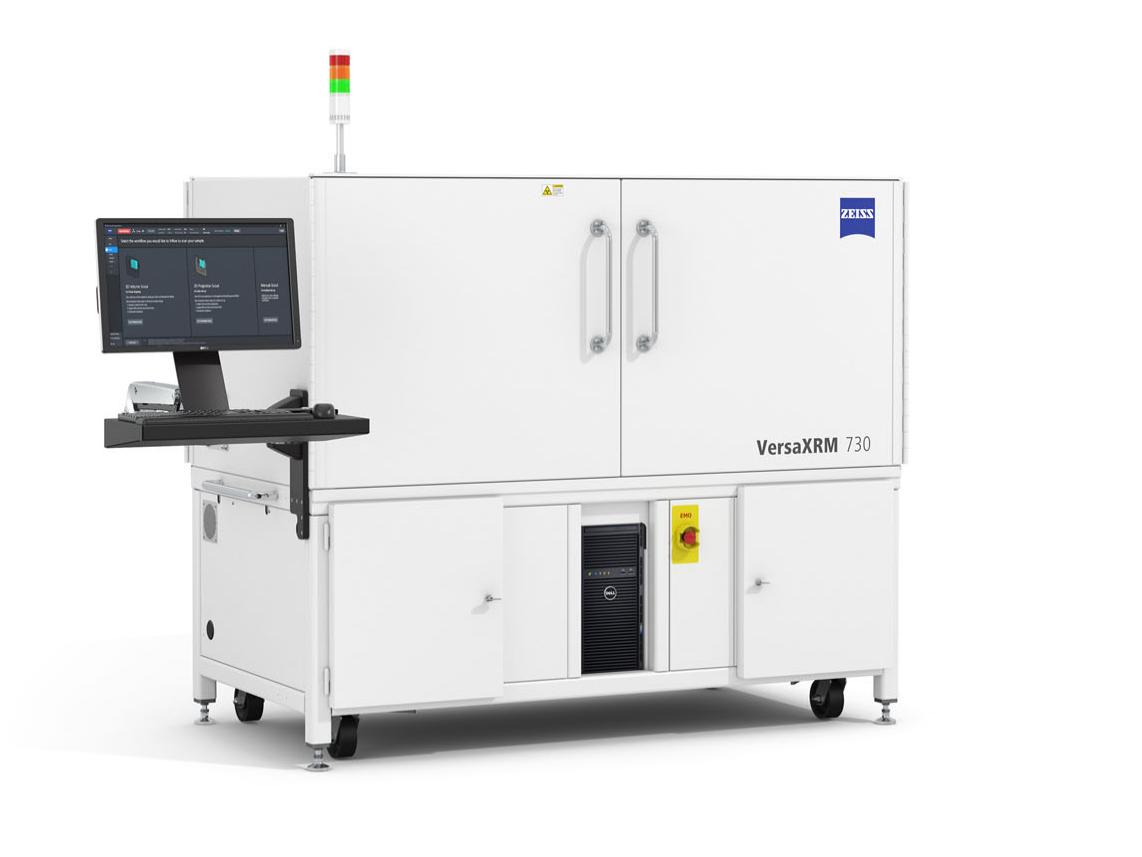
ZEISS microscope solutions support high-quality visualization of Arabidopsis to advance insights in plant biology, across diverse imaging workflows.Non-destructive 3D imaging at multiple resolutions, revealing floral architecture in rich structural detail
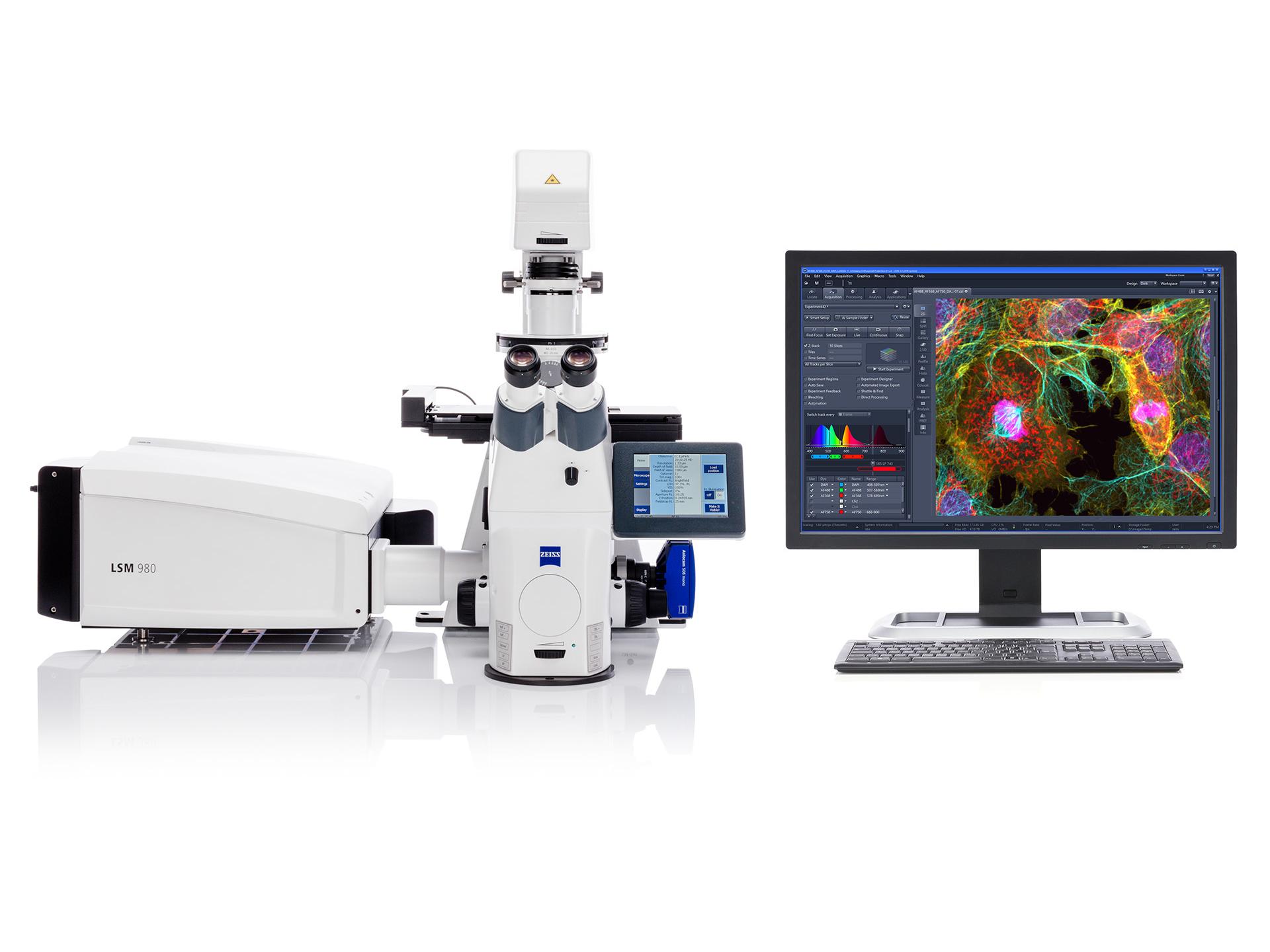
Precisely separate signals from ER, golgi, and chlorophyll, enabling clear visualization of subcellular structures
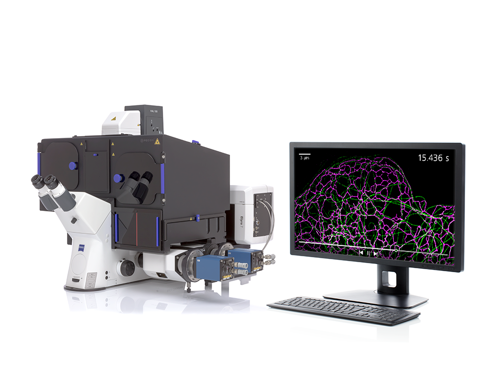
3D super-resolution imaging of microtubule networks in living leaf cells without photodamage

Recommended Products for Arabidopsis Research


ZEISS Lab Essentials


FAQs for Arabidopsis Research
-
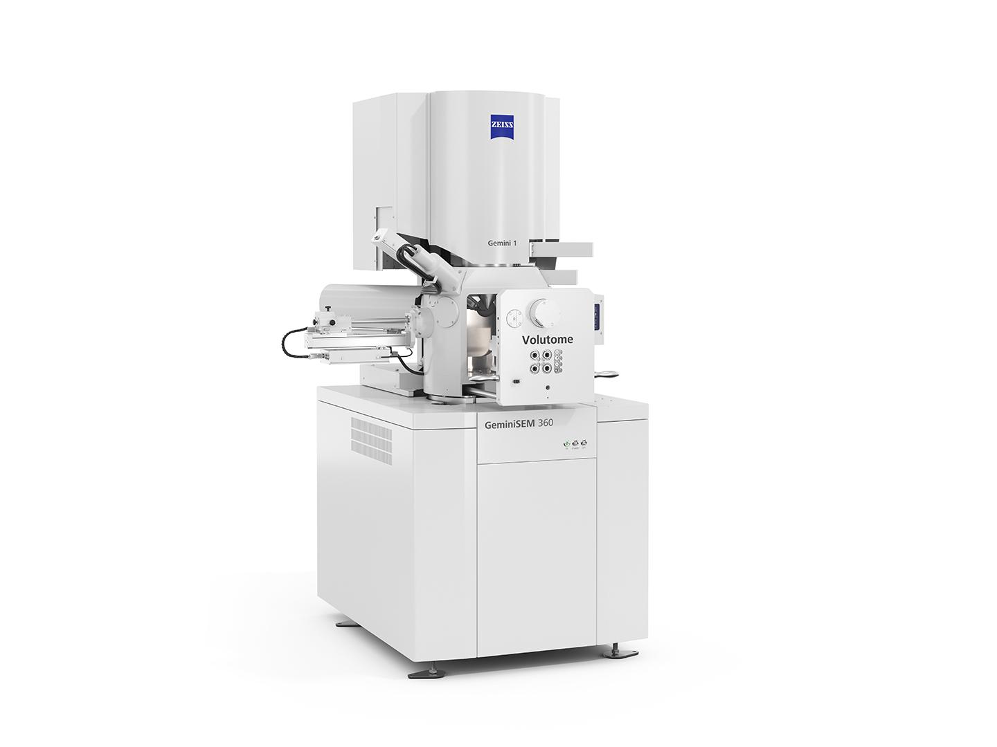
ZEISS provides a broad range of microscopy techniques suited to Arabidopsis research, including fluorescence microscopy, confocal microscopy, super-resolution microscopy, light sheet microscopy, and X-ray and electron microscopy. Each approach offers distinct advantages for visualizing specific structures, dynamics, and developmental stages.
For example, ZEISS LSM confocal systems with Airyscan provide high-resolution imaging of subcellular features and reporter lines in living tissues. Elyra 7 with SIM² enables super-resolution imaging of organelles and cytoskeletal elements. ZEISS Lightsheet 7 allows gentle, volumetric imaging of roots and embryos over time. For non-destructive 3D imaging of intact flowers or seeds, ZEISS VersaXRM delivers multiscale resolution. Whether you're investigating gene expression, root development, or seed architecture, ZEISS offers the right solution to support your Arabidopsis research from cell to whole plant.
-
For live imaging of Arabidopsis, ZEISS offers several systems optimized for speed, sensitivity, and minimal photodamage. ZEISS Lightsheet 7 is ideal for long-term, volumetric imaging of living roots, embryos, and tissues, providing fast acquisition with low phototoxicity. ZEISS LSM 990 with Airyscan enables high-resolution confocal imaging of dynamic cellular processes with enhanced sensitivity and resolution, even in thick samples. For fast 3D snapshot imaging of rapid events like protein re-localization, ZEISS Lightfield 4D captures entire volumes in a single snap. These platforms help researchers preserve sample viability while capturing detailed, time-resolved data.
-
ZEISS offers imaging solutions specifically designed to minimize photodamage in light-sensitive Arabidopsis samples. ZEISS Lightsheet 7 uses a thin sheet of light to illuminate only the imaging plane, drastically reducing light exposure and enabling gentle, long-term imaging of living tissues like roots or embryos. For confocal imaging, Airyscan technology on ZEISS LSM systems increases sensitivity, allowing you to use lower laser power while still achieving high resolution and signal quality. These tools help preserve sample health while capturing reliable, high-quality data.
-
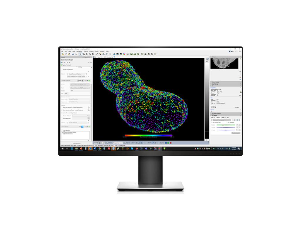
High-throughput phenotyping in Arabidopsis requires imaging workflows that are fast, reproducible, and scalable. ZEISS offers several solutions tailored to these needs. The ZEISS Celldiscoverer 7 provides automated imaging of seedlings, roots, or leaf disks with consistent focus, illumination, and positioning, ideal for side-by-side comparison of multiple genotypes. For fixed tissue sections or large sample sets, the Axioscan 7 enables rapid slide scanning with high image quality and batch processing capabilities. Combined with ZEISS ZEN software for image analysis and annotation, these systems support large-scale screens and time-efficient data collection for functional genomics, mutant characterization, and transgenic line evaluation.
-
Thick tissues in Arabidopsis, such as leaves, roots, and developing floral organs, scatter light and can make it difficult to maintain resolution and contrast, especially at depth. ZEISS offers multiple solutions designed to overcome this challenge:
- ZEISS LSM 990 or LSM Airyscan enhances resolution and sensitivity, allowing deeper imaging in complex tissues with less noise and photodamage.
- ZEISS Lightsheet 7 enables fast, gentle optical sectioning of living tissues in 3D, ideal for tracking developmental processes in roots, embryos, or whole seedlings.
- ZEISS Elyra 7 with Lattice SIM² provides super-resolution imaging in thick samples while preserving viability, making it ideal for studying cytoskeletal organization and protein localization in dense plant tissues.
- ZEISS Axio Observer with Apotome.3 offers structured illumination for optical sectioning in widefield systems, great for high-contrast imaging of thicker tissue sections when confocal is not required.
These platforms provide a range of options to help you visualize internal structures clearly, even in light-scattering or photosensitive Arabidopsis samples.