

ZEISS VersaXRM multi-scale 3D X-ray microscopy
Explore intact samples across scales, locate regions of interest, and move from broad structural understanding to targeted insight while preserving context.


Why choose VersaXRM
Explore broadly. Investigate precisely. Preserve context.ZEISS VersaXRM systems help connect whole-sample understanding with targeted internal analysis. Move efficiently from overview to detail, preserve the context of your sample, and generate 3D data you can interpret with confidence.

 Move from overview to detail

Connect whole-sample overview imaging with targeted high-resolution imaging for detailed analysis in a single workflow, so users can understand where features are and how they relate to the surrounding structure.
 Target regions of interest efficiently

Scout-and-zoom workflows, 3D navigation, and guided setup help locate buried defects, interfaces, and internal features with greater certainty.
 Preserve sample integrity

Investigate intact samples non-destructively before destructive preparation, supporting repeated analysis, correlative workflows, and better-informed downstream decisions.
 Adapt across applications and sample types

Support diverse imaging needs across materials and industrial research, electronics, batteries, life sciences, geology, and other complex fields.
 Extend visibility with advanced reconstruction

Leverage reconstruction technologies that improve image quality, increase throughput, enhance contrast, and reduce artifacts in challenging datasets.
 Turn 3D data into confident interpretation

Combine ZEISS optics, stable imaging platforms, guided workflows, and visualization tools to generate reliable datasets that support meaningful analysis.
The VersaXRM portfolio
Choose the right ZEISS X-ray microscope for your workflow


Supporting 3D X-ray microscopy across scales
VersaXRM systems support diverse applications by connecting whole-sample context with targeted, non-destructive 3D insight across scales.

Materials research in 3D X-ray microscopy


Three-dimensional rendering of recycled battery black mass with individual material phases segmented and color coded for analysis.
Materials research in 3D X-ray microscopy
Characterize microstructures, defects, and degradation in metals, composites, and battery materials non-destructively, connecting internal structure to performance through in situ, correlative, and large-volume 3D imaging.


Semiconductor & electronics failure analysis
3D X-ray microscopy rendering of a smartphone system-on-chip package, with a green inset highlighting the central chip region.
Semiconductor & electronics failure analysis
Inspect complex packages and assemblies in 3D, localize buried defects, and preserve critical evidence before destructive downstream workflows in failure analysis and package development.


Industrial research with non-destructive 3D imaging


Three-dimensional X-ray rendering of a metal additively manufactured manifold revealing complex internal channels and lattice structures.
Industrial research with non-destructive 3D imaging
Evaluate internal structures, defects, and failure mechanisms without destructive preparation, linking 3D sample information to product performance, process development, and validation workflows.


Life science 3D imaging with preserved context
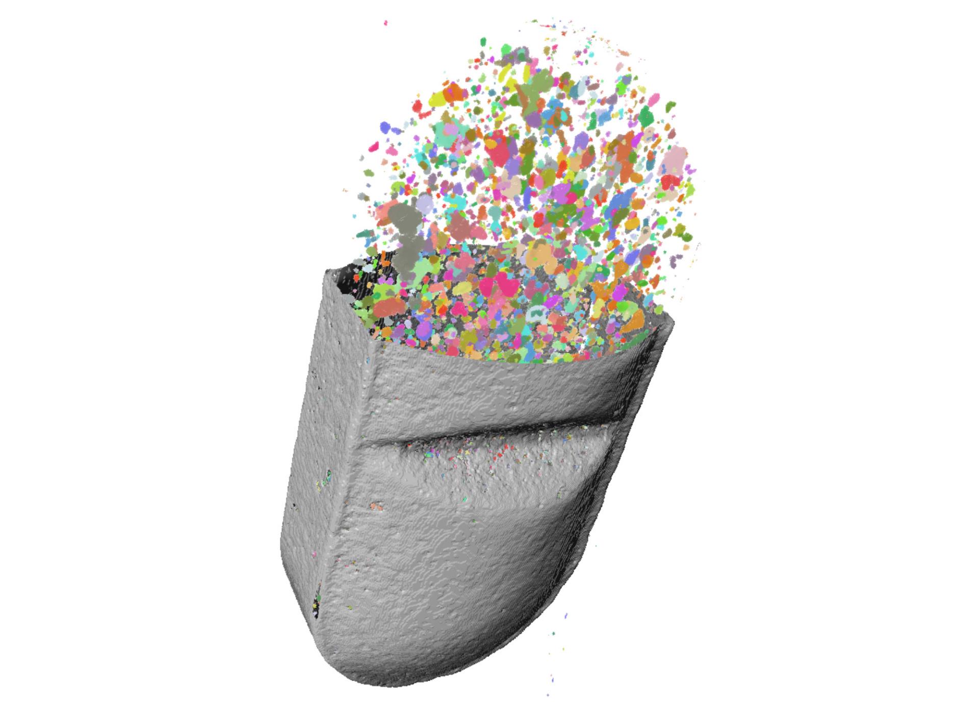

Three-dimensional rendering of an antihistamine tablet showing segmented internal particles and ingredient distribution.
Life science 3D imaging with preserved context
Explore intact biological structures in 3D, preserve spatial context across scales, and locate regions of interest for downstream correlative microscopy workflows.


Geological and mining 3D X-ray microscopy


High-resolution X-ray cross-section of a sandstone sample showing pores, mineral grains, and internal microstructure.
Geological and mining 3D X-ray microscopy
Analyze pore networks, mineral distributions, and internal structures in intact geological samples while preserving rare specimens and supporting in situ or fluid-flow studies.
Advanced Reconstruction Technologies


Extend what you can see, measure, and interpret
The ZEISS Advanced Reconstruction Toolbox (ART) improves image quality, throughput, and data interpretation across VersaXRM workflows. DeepRecon Pro uses AI-assisted reconstruction for faster, high-quality results, DeepScout enhances large-volume reconstruction, and MARS reduces artifacts in complex multi-material samples.
PhaseEvolve improves contrast in low- and medium-density materials, while OptiRecon helps balance image quality and throughput for different workflow needs.
Add-ons
ZEISS VersaXRM accessories help adapt imaging workflows to different sample types, research questions, and operational needs. From sample handling to in situ imaging and workflow automation, accessories can extend how VersaXRM systems support routine imaging, advanced experiments, and multi-user environments.


Autoloader


Autoloader compatibility enables unattended imaging of multiple mounted samples, improving throughput and supporting consistent, repeatable workflows in high-utilization laboratories.
Autoloader
Support multi-sample studies and higher system utilization with handling options designed to reduce manual intervention and help users position, stabilize, and image samples more effectively.


In situ imaging
Study samples under changing conditions while preserving the ability to return to the same region of interest. In situ workflows help teams investigate how structures evolve during heating, cooling, compression, corrosion, wetting, fluid flow, or other environmental conditions.


ZEISS Solutions Lab
Extend the capabilities of your VersaXRM system with customized workflows developed by the ZEISS Solutions Lab. From automated acquisition routines to application-specific analysis and software tools, our experts help streamline imaging processes, reduce manual intervention, and improve reproducibility for unique research and industrial workflows.
Reconstruction and analysis
Extend acquisition workflows with advanced reconstruction and analysis tools that support visualization, segmentation, interpretation, and multi-modal data workflows.

FDK reconstruction 1x


X-ray microscopy image of a ceramic matrix composite sample showing conventional FDK reconstruction.


FDK reconstruction 10x


X-ray microscopy image of a ceramic matrix composite sample showing conventional FDK reconstruction at 10x magnification.


DeepRecon Pro reconstruction 10x


X-ray microscopy image of a ceramic matrix composite sample showing DeepRecon Pro reconstruction, highlighting reduced artifacts and improved visibility of internal pores and microstructural features.
Comparison of reconstruction methods in a ceramic matrix composite (CMC) sample. Left: conventional FDK reconstruction at 1× magnification. Center: conventional FDK reconstruction at 10× magnification. Right: DeepRecon Pro reconstruction at 10× magnification, demonstrating reduced reconstruction artifacts and improved visualization of internal features.
© Sample courtesy of Dr. David Marshall, Colorado University Boulder, USA
Downloads






Services and support
Every ZEISS VersaXRM system is supported by ZEISS service, application, and training expertise. From installation and onboarding to workflow optimization and long-term maintenance, ZEISS helps labs build confidence, improve productivity, and keep systems performing reliably.
FAQ
-
X-ray microscopy is useful when internal sample structures need to be visualized in 3D while preserving the intact sample and its surrounding context. ZEISS X-ray microscopy (XRM) systems are especially valuable for understanding hidden defects, interfaces, pores, cracks, or buried regions of interest that may be missed or altered by destructive preparation, while providing high-resolution, non-destructive imaging across a wide range of sample types.
-
Resolution at a Distance™, or RaaD™, enables high-resolution imaging while maintaining working distance between the source and sample. This allows users to image fine details at high resolution even in larger or more complex samples, investigate regions of interest inside intact samples without trimming or refixturing, and supports in situ setups.
-
Yes. VersaXRM systems support workflows that begin with broad sample exploration and move toward targeted investigation of selected regions of interest. This helps users identify where meaningful features are located, then efficiently investigate those features at higher resolution while maintaining the surrounding sample context.
-
The right VersaXRM system depends on the application, sample types, throughput needs, and performance requirements. Choose VersaXRM 730 for ultimate performance across the most demanding samples and advanced workflows. VersaXRM 615 is a high-performance workhorse for routine and advanced non-destructive 3D imaging. VersaXRM 5 Insight is a high-value hybrid VersaXRM + microCT solution for efficient, insight-driven workflows.
-
VersaXRM systems support a wide range of samples across materials and industrial research, semiconductor and electronics, battery and energy materials, life sciences, geology, and mining. Example samples may include metals, composites, batteries, electronic packages, industrial components, biological tissues, geological specimens, and other complex materials or devices.
-
VersaXRM can be used to image intact samples non-destructively before downstream preparation or higher-resolution analysis. This helps users locate regions of interest, preserve spatial context, and make more informed decisions before moving to techniques such as light microscopy, electron microscopy, FIB-SEM, or other destructive workflows.
-
Advanced reconstruction technologies can improve throughput, image quality, contrast, artifact reduction, and visualization of challenging samples. Tools such as DeepRecon Pro, DeepScout, MARS, PhaseEvolve, and OptiRecon help users get more value from VersaXRM datasets and adapt reconstruction workflows to different sample and application needs.
-
Yes. VersaXRM workflows can support in situ and 4D imaging studies where users repeatedly image the same sample over time or under changing conditions. This is useful for studying material evolution, deformation, heating, cooling, corrosion, wetting, compression, fluid flow, and other dynamic processes while preserving sample context.