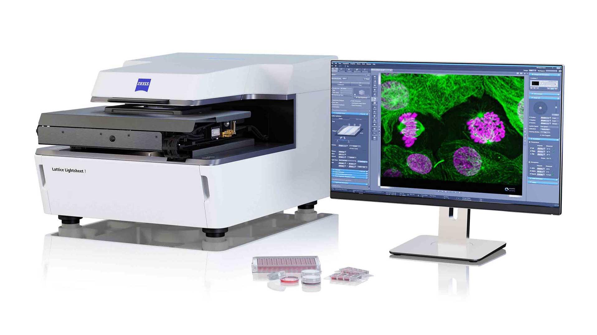
In order to reduce the data size of the original raw dataset (2 channels; 150 slices; 200 time points; 16-bit; ~ 40 GB), the images were cropped to include only the relevant cellular areas. These cropped images were then used to generate a stack subset containing only every second slice and the data were then transformed to 8-bit. The final data set had 2 channels, 22 slices and 200 time points, it was 8-bit and was ~ 1.2 GB. in size.
The image analysis in ZEISS arivis Pro consisted of three steps. Firstly, image processing to denoise the images was performed, with special emphasis on preserving spherical structures and removing striping artefacts. This was done using the image processing functions “Morphology Filter” and “Denoising” independently for both channels.
Secondly, vesicles in both channels were independently segmented using Watershed segmentation with minimum vesicle volume of 0.3 µm³. The Rab5a-positive vesicles were then filtered based on the “minimal” distance to the next Golgi7-positive vesicle and assigned to the group of Rab5a-positive/Golgi7-negative “endosomal” vesicles (if distance > 0). Rab5a-positive/Golgi-positive objects were assigned to “Golgi-associated” vesicles.
Finally, to derive the movement of vesicles over time, object tracking was performed with both vesicle types.
The sketch summarizes the image analysis procedure. In addition, the ZEISS arivis Pro pipeline to perform these operations is available for download at the bottom of this page as part of the case study data package ("Green-Magenta vesicle detection_Tracking_Rev4.xml”).


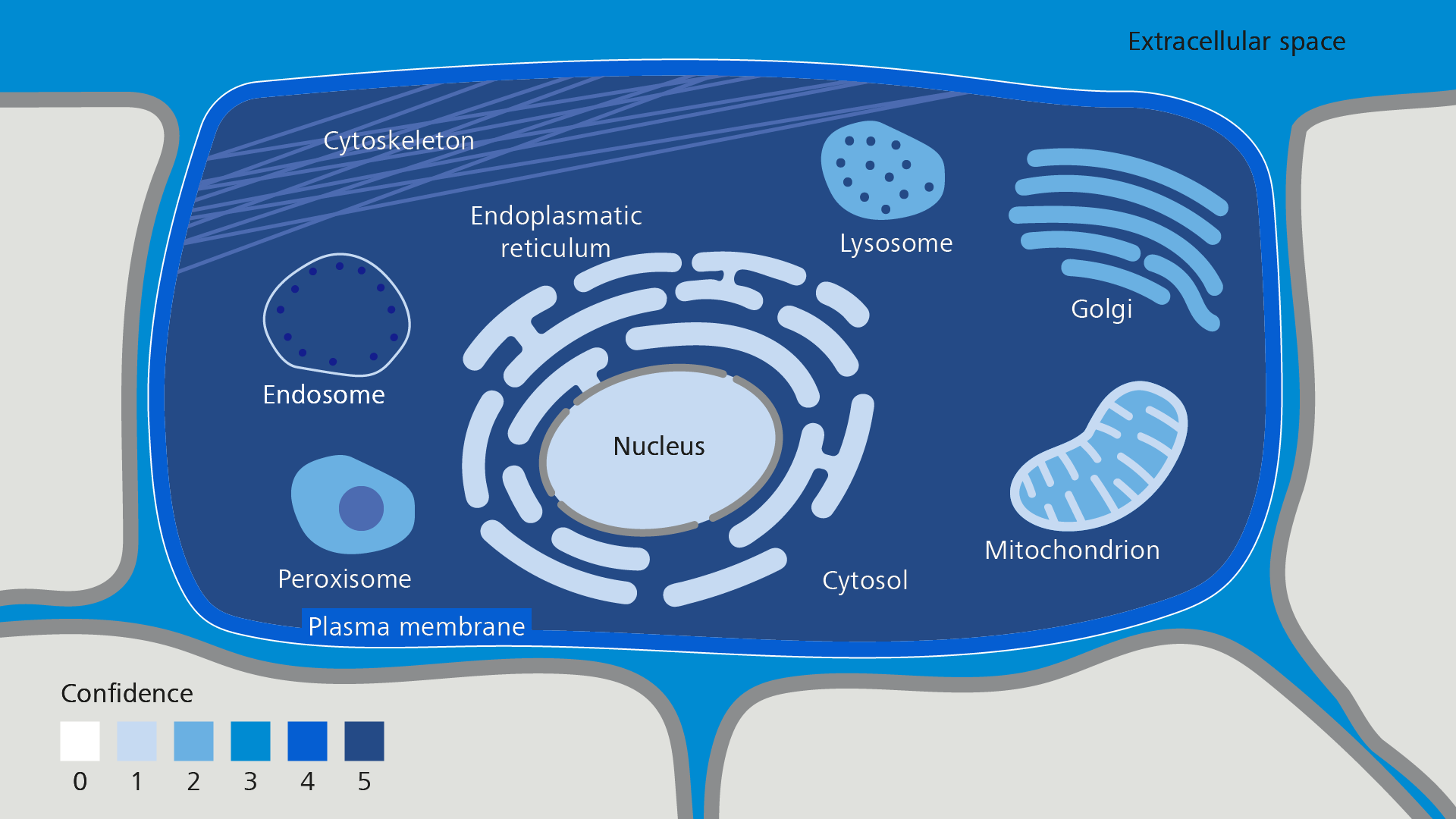


 overlap of Golgi7-labelled vesicles with a fraction of the Rab5a-labelled vesicles. Also note “striping artefacts” in the green channel.")
 overlap of Golgi7-labelled vesicles with a fraction of the Rab5a-labelled vesicles. Also note “striping artefacts” in the green channel.")
 overlap of Golgi7-labelled vesicles with a fraction of the Rab5a-labelled vesicles. Also note “striping artefacts” in the green channel.")
 overlap of Golgi7-labelled vesicles with a fraction of the Rab5a-labelled vesicles. Also note “striping artefacts” in the green channel.")


 on a scatter plot")
 on a scatter plot")
 on a scatter plot")
 on a scatter plot")


")
")
")
")


