Advanced Microscopy Techniques
New Dimensions in Neuroscience
Investigate the Brain in Unparalleled Resolution

Scroll animation items

Studying Synaptic Function
Overcome the diffraction limit by combining super-resolution imaging and advanced processing techniques to visualize synaptic components at the nanoscale.

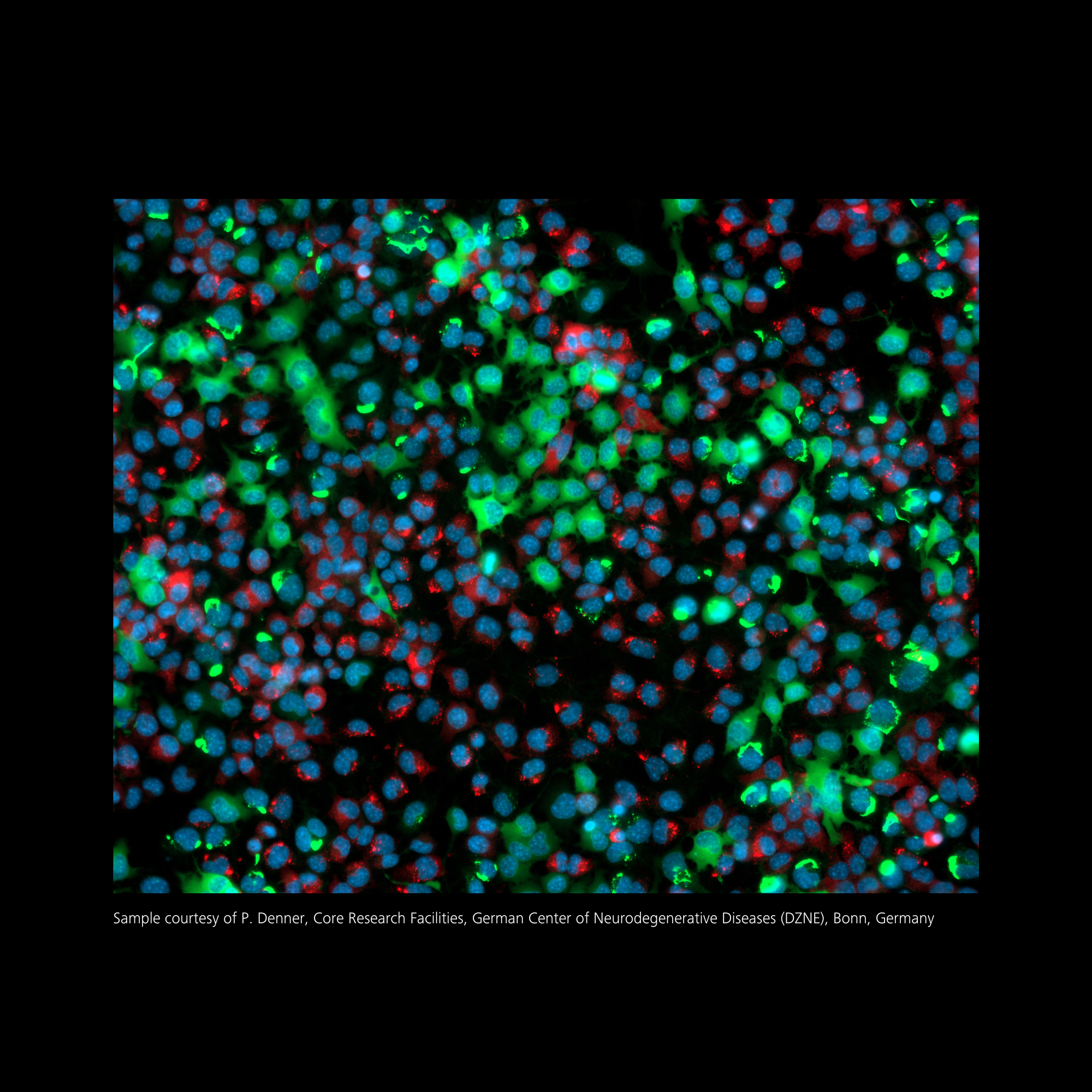
Cellular Dynamics of Neurodegenerative Disease
Visualize in real-time cellular processes underlying disease progression and identify the critical changes in behavior and morphology to gain a deeper understanding of neurodegeneration.


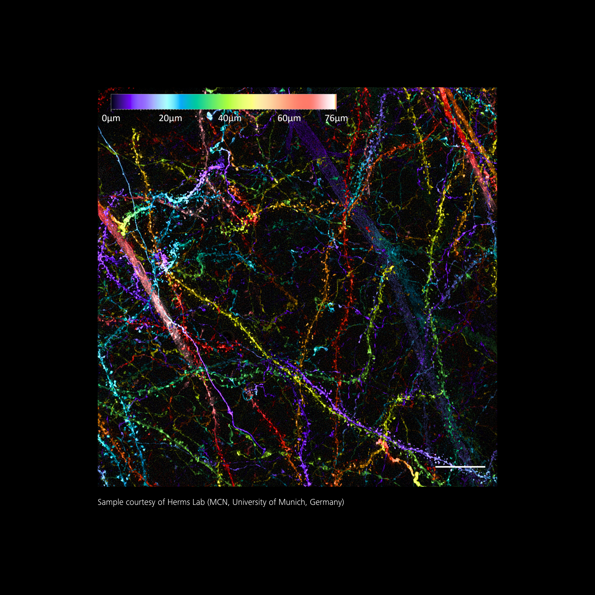
Studying the Role of Glial Cells
Enhanced optical sectioning capabilities can minimize our-of-focus light, allowing for clearer visualization of glial cell processes and their interactions with neurons and vasculature.
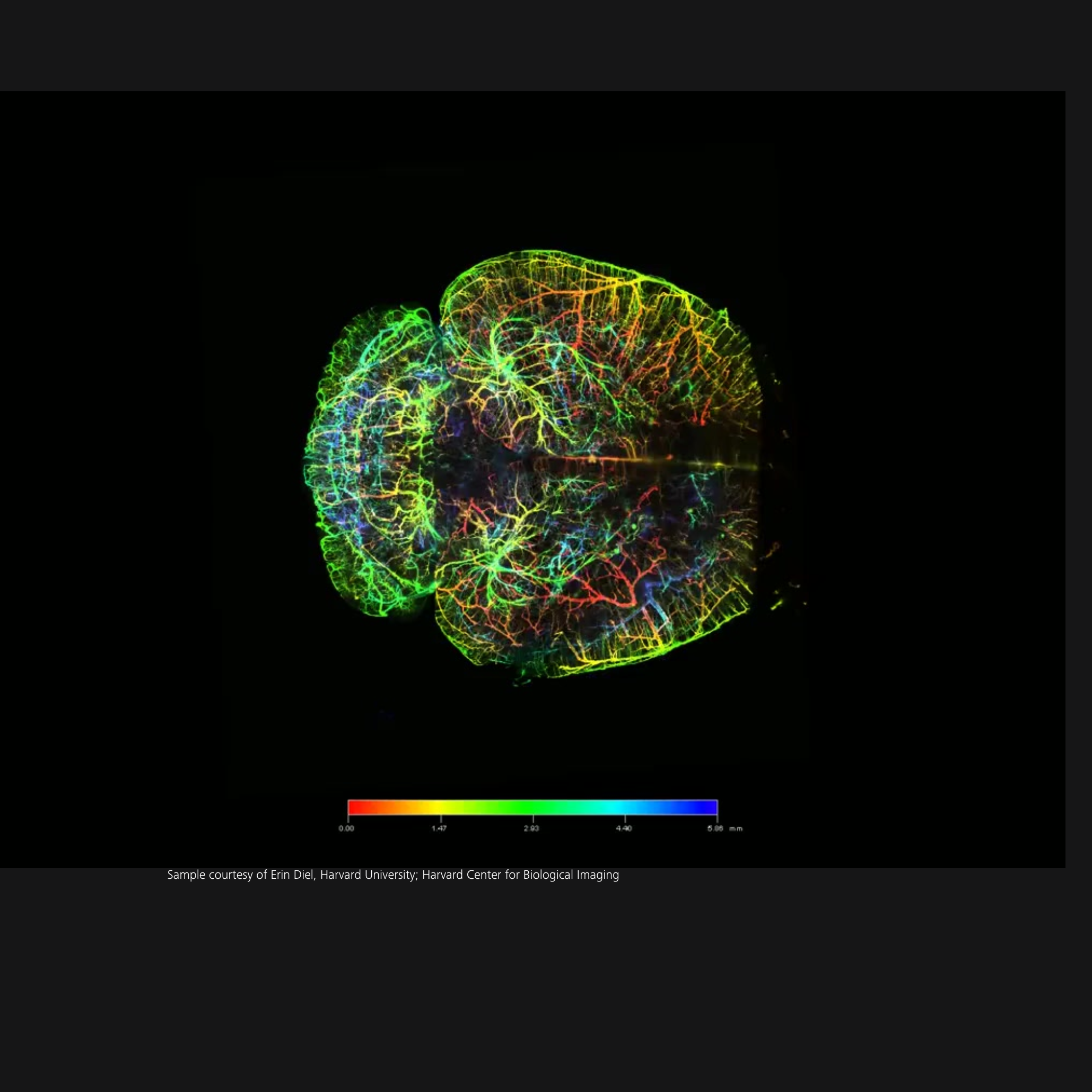

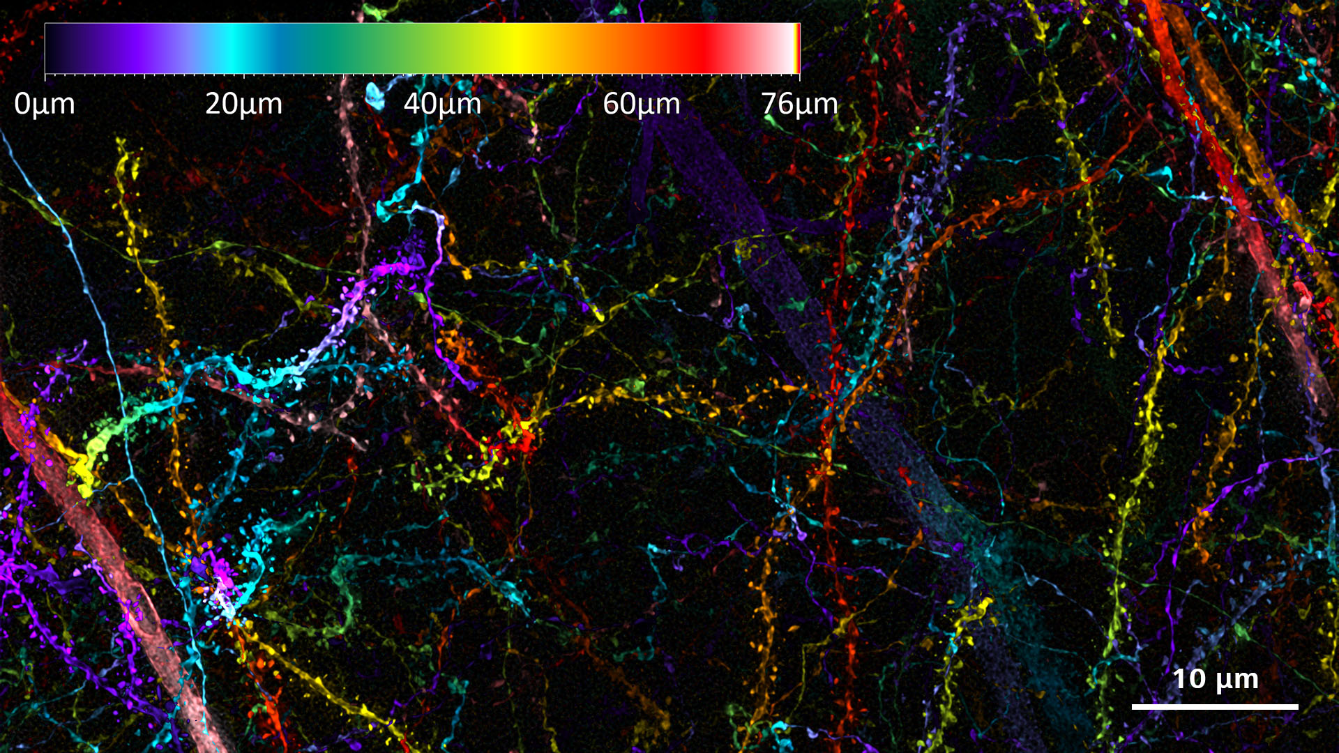
Understanding Neural Circuitry
Light sheet imaging captures data from multiple angles, allowing for thee-dimensional imaging of large samples and whole rodent brains so that researchers can map entire neural circuits and brain vasculature.

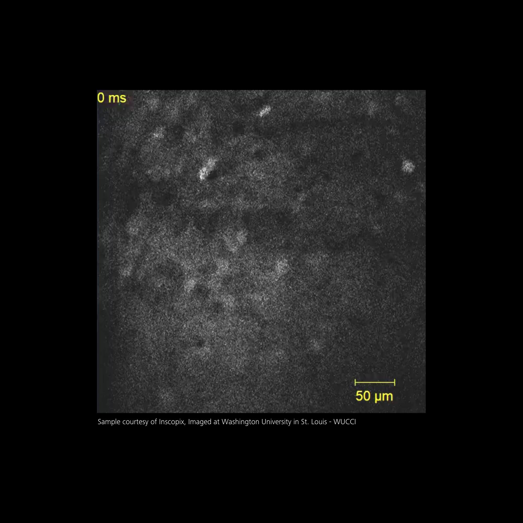
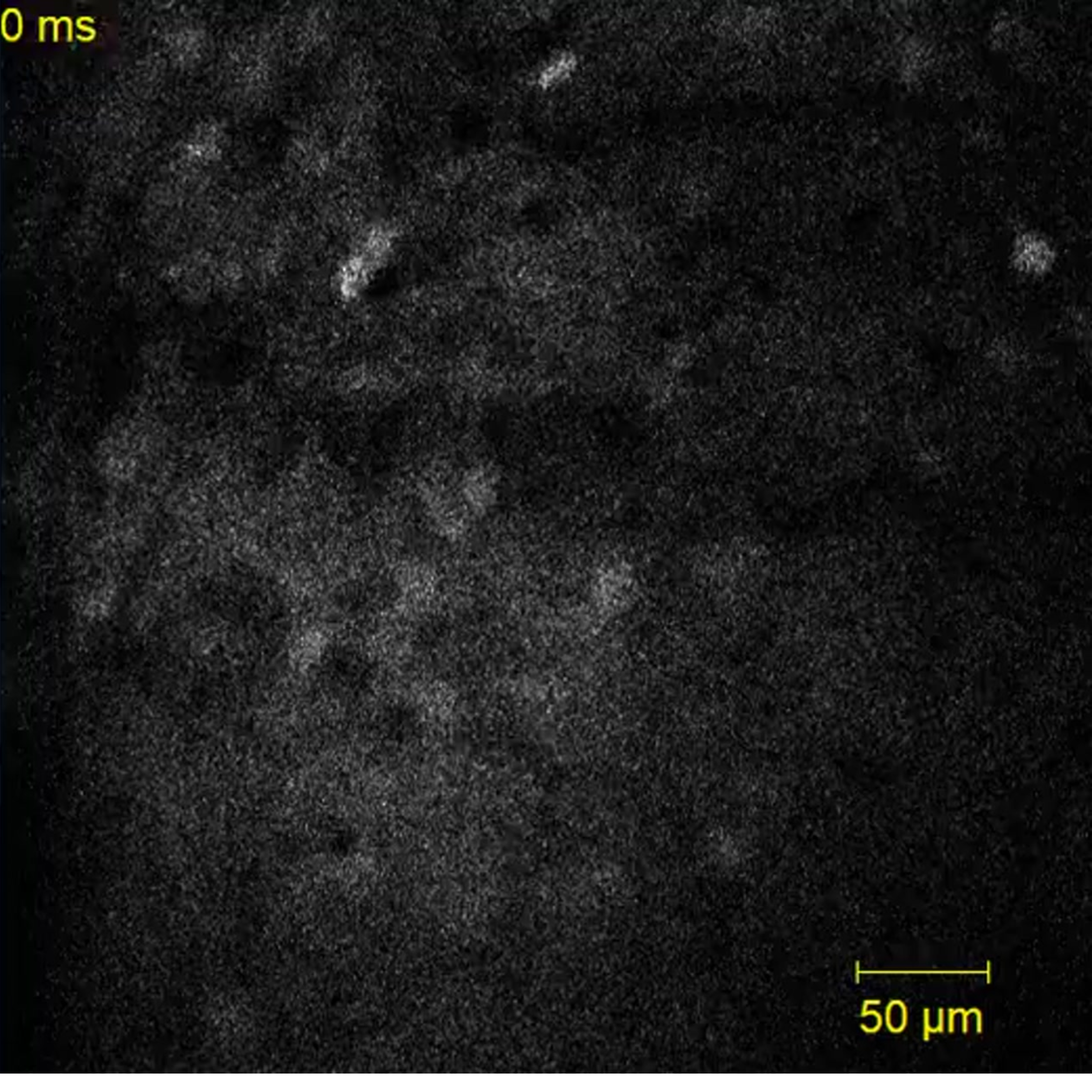
Visualize Deep Brain Activity
Image deep in the living brain, obtaining high-resolution images at speed allowing researchers to observe complex dynamics in real-time, mapping circuits to external stimuli.
Scroll animation items

Are you leveraging the latest innovations?
Discover the techniques that are advancing neuroscience
Visualize Deep Brain Activity
Image deep in the living brain, obtaining high-resolution images at speed allowing researchers to observe complex dynamics in real-time, mapping circuits to external stimuli.

Studying Synaptic Function
Overcome the diffraction limit by combining super-resolution imaging and advanced processing techniques to visualize synaptic components at the nanoscale.
Understanding Neural Circuitry
Light sheet imaging captures data from multiple angles, allowing for thee-dimensional imaging of large samples and whole rodent brains so that researchers can map entire neural circuits.


Cellular Dynamics of Neurodegenerative Disease
Visualize in real-time cellular processes underlying disease progression and identify the critical changes in behavior and morphology to gain a deeper understanding of neurodegeneration.
Studying the Role of Glial Cells
Enhanced optical sectioning capabilities can minimize our-of-focus light, allowing for clearer visualization of glial cell processes and their interactions with neurons and vasculature.
Scroll animation items
Are you leveraging the latest innovations?
Discover the techniques that are advancing neuroscience
Studying Synaptic Function
Overcome the diffraction limit by combining super-resolution imaging and advanced processing techniques to visualize synaptic components at the nanoscale.
Cellular Dynamics of Neurodegenerative Disease
Visualize in real-time cellular processes underlying disease progression and identify the critical changes in behavior and morphology to gain a deeper understanding of neurodegeneration.
Studying the Role of Glial Cells
Enhanced optical sectioning capabilities can minimize our-of-focus light, allowing for clearer visualization of glial cell processes and their interactions with neurons and vasculature.
Understanding Neural Circuitry
Light sheet imaging captures data from multiple angles, allowing for thee-dimensional imaging of large samples and whole rodent brains so that researchers can map entire neural circuits and brain vasculature.
Visualize Deep Brain Activity
Image deep in the living brain, obtaining high-resolution images at speed allowing researchers to observe complex dynamics in real-time, mapping circuits to external stimuli.
Why You Do What You Do
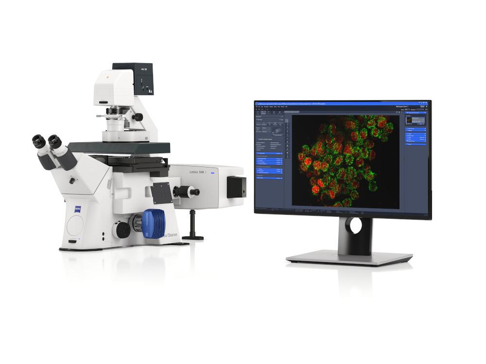
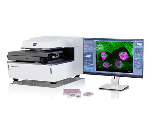
Recommended Products for Neuroscience
Buy Online, Get a Quote or Trial


FAQs for ZEISS in Neuroscience
-
Yes, ZEISS provides various case studies and application notes that showcase the use of their microscopy systems in neuroscience. Examples include:
- Imaging Synaptic Structures: Using confocal microscopy to study synaptic connections and plasticity.
- Neurodegenerative Disease Research: Employing two-photon microscopy to observe changes in neuronal behavior in live animal models.
- Brain Mapping: Utilizing advanced imaging techniques to visualize brain connectivity and function.
-
ZEISS microscopy solutions enhance neuroscience research by providing:
- High Resolution: Advanced optics and imaging technologies allow for detailed visualization of neuronal structures and processes.
- Live Imaging: Many systems are designed for live-cell imaging, enabling the observation of dynamic processes in real-time.
- Multimodal Imaging: The ability to combine different imaging modalities (e.g., fluorescence and electron microscopy) allows for comprehensive analysis of samples.
These features facilitate a deeper understanding of neural mechanisms and contribute to advancements in neuroscience.
-
ZEISS offers a variety of microscopy techniques tailored for neuroscience research, including:
- Confocal Microscopy: Ideal for imaging thick samples and obtaining high-resolution images of cellular structures.
- Two-Photon Microscopy: Particularly useful for imaging living tissues at greater depths, minimizing photodamage.
- Electron Microscopy: Provides ultra-high resolution for detailed visualization of neuronal structures.
These techniques enable researchers to explore complex neural networks and cellular interactions in detail.