User Story
Brain Tumor Microenvironment Spatial Changes Uncovered by High-Throughput Hyperplexed Immunofluorescence Imaging
A New Hyper-Multiplex Workflow using ZEISS Axioscan Enables High-Throughput Imaging of Fragile Tissues with 40-50 Markers over Large Regions and High Magnifications
The tumor microenvironment is comprised of cellular and non-cellular components that can engage with cancerous cells and impact tumor behavior. Professor Johanna A. Joyce leads a lab at the University of Lausanne, Switzerland, with the goal of understanding the complex cellular interactions and spatial biology of the tumor microenvironment, including in primary and metastatic brain tumors. Her team is working to find new ways to exploit the immune and stromal cell populations to develop new treatments to improve the lives of patients.
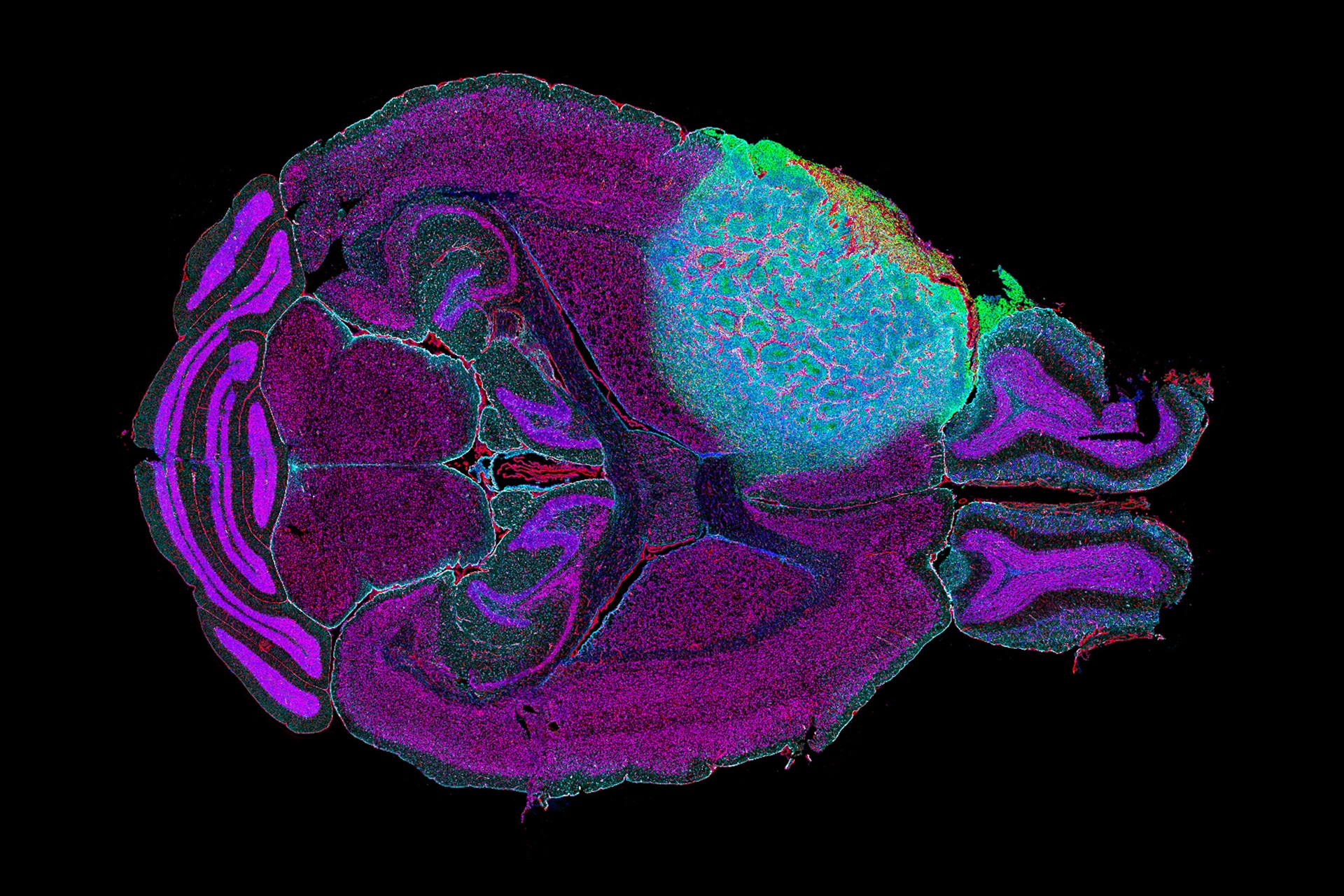
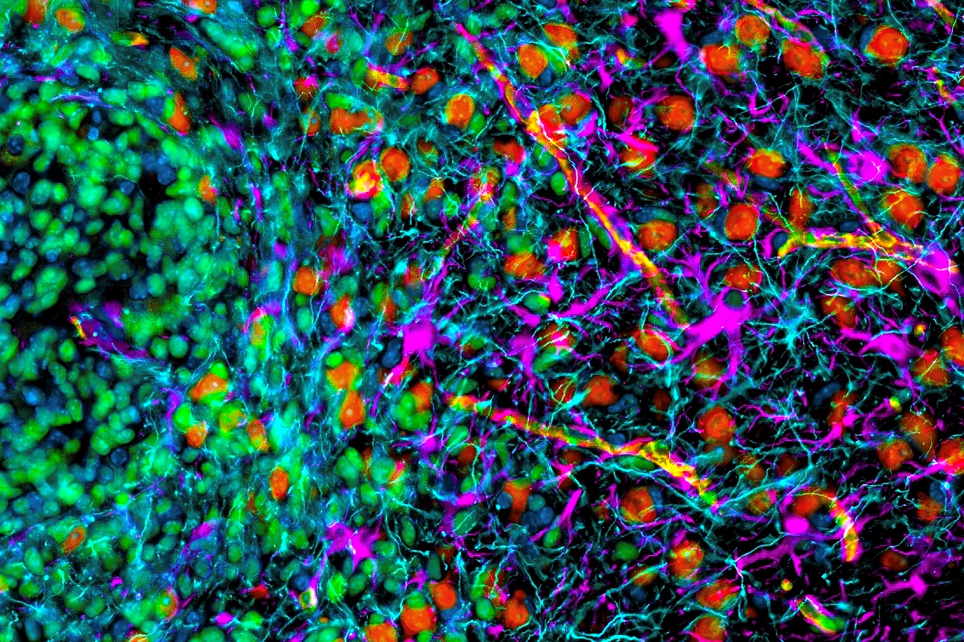
Dr. Spencer S. Watson, a Research Fellow in Professor Joyce’s lab, published an article investigating how the brain tumor microenvironment responds to radiotherapy, not just in terms of changes in different cell populations, but also regarding how the cells spatially organize themselves. In order to do this, the team worked to create a new multiplex workflow, called Hyperplexed Immunofluorescence Imaging (HIFI), to study the spatial biology of tumors before and after treatment. Their workflow, which utilizes the ZEISS Axioscan digital slide scanner, allowed them to achieve 40-50 multiplexed markers with extremely fragile tissue over large regions and high magnifications, and revealed interesting differences in spatial reorganizations post-radiotherapy treatment.
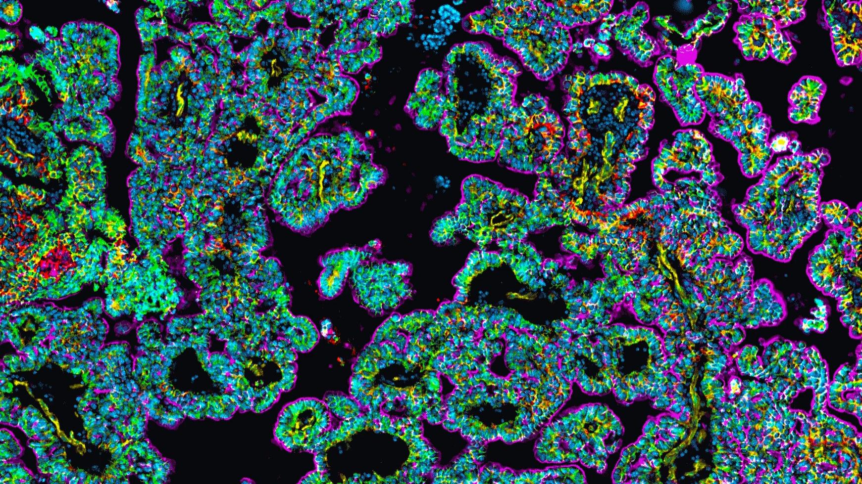


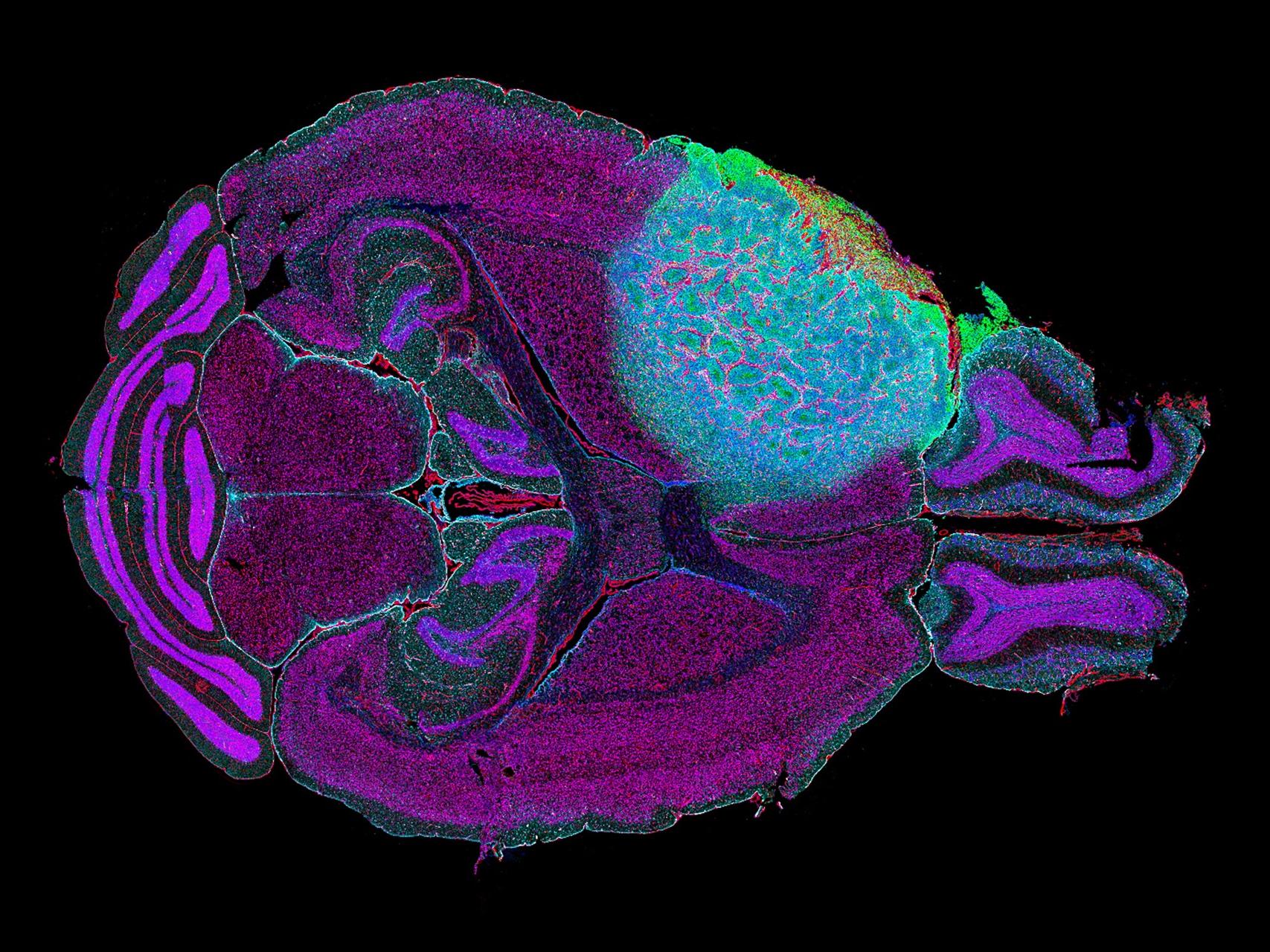

 image exploring the spatial biology of a mouse brain glioblastoma focused on the dynamic interface between the tumor and the surrounding brain.")
 image exploring the spatial biology of a mouse brain glioblastoma focused on the dynamic interface between the tumor and the surrounding brain.")


 image exploring the spatial biology of a mouse brain glioblastoma focused on the dynamic interface between the tumor and the surrounding brain.")
 image exploring the spatial biology of a mouse brain glioblastoma focused on the dynamic interface between the tumor and the surrounding brain.")
 image exploring the spatial biology of a mouse brain glioblastoma focused on the dynamic interface between the tumor and the surrounding brain.")
 image exploring the spatial biology of a mouse brain glioblastoma focused on the dynamic interface between the tumor and the surrounding brain.")


