

Developing the Future of Energy Conversion & Storage
By understanding structure in electrochemical materials


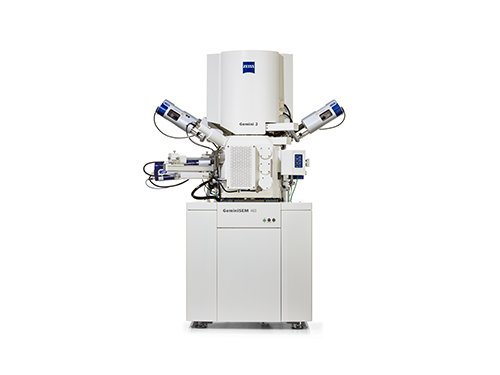
Solid Oxide Fuel Cells and Electrolyzers
Solid oxide cells, for energy generation or electrolysis, contain electrodes with complex, porous, and often multi-phase 3D microstructures. The discrete morphology of these structures directly impacts the efficiency of transport processes, electrochemical activity, and device longevity.
3D FIB tomography performed by ZEISS Crossbeam to reconstruct, utilizing electron imaging and simultaneous EDS elemental mapping, a portion of an SOEC cell. Sample courtesy of: M. Cantoni, EPFL Lausanne, Switzerland.


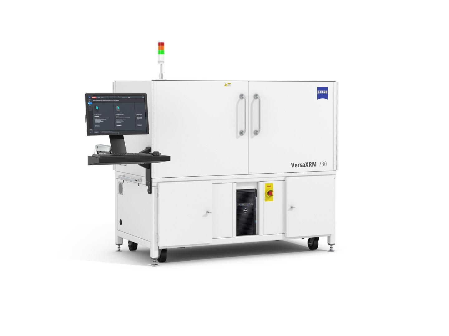
Polymer Electrolyte Fuel Cells
Low temperature fuel cells, typically consisting of a polymer (Nafion) membrane sandwiched between porous catalyst layers and carbon fiber-based gas diffusion electrodes, represent a challenging system for designing and managing coupled reactive flow phenomena. Between gas transport pathways, water generation and flooding, and catalyst dispersion/agglomeration, microscopy can help better understand the key micro and nano-structural features driving performance in these cells.
3D X-ray microscopy data obtained by ZEISS VersaXRM of a portion of a PEM fuel cell membrane electrode assembly.
Recommended Products for Electrochemical Materials Research


ZEISS Materials Research Lab Essentials


What types of microscopy techniques are available for research of electrochemical materials?
ZEISS offers a variety of microscopy techniques tailored for energy materials design and engineering including:-
ZEISS offers a variety of microscopy techniques tailored for studying electrochemical materials and devices:
- Optical Microscopy: Addresses all typical optical inspection and analysis tasks using upright, inverted, zoom, digital, and confocal systems. Useful for sample preparation, quick inspection, or large area imaging of cross sections.
- Electron Microscopy: Provides ultra-high resolution for nanoscale visualization of material surfaces. Provides various types of signals (SE, BSE) and is often equipped with an EDS detector for elemental analysis.
- FIB-SEM Microscopy: Extends SEM analysis to the sub-surface with the addition of a gallium focused ion beam for targeted milling. Enables site-specific cross sections, serial sectioning 3D tomography with imaging and analytics, and preparation of samples for other methods like nanoscale XRM. By combining FIB-SEM with a femtosecond (fs) laser, researchers cover a huge range of material ablation demands.
- 3D X-ray Microscopy: Delivers nondestructive 3D imaging by high resolution X-ray tomography, both at the micro and nano-scales. See inside energy devices in their native state or during cycling. Create 3D maps of electrode morphology for detailed parameter measurement or electrochemical modeling.
These systems come together to form a versatile portfolio, empowering you to investigate the diverse range of features that exist within modern electrochemical devices. -
The ZEISS microscopy portfolio is designed to address three key types of characterization challenges often faced by researchers studying electrochemical materials and devices:
- Multiscale Imaging: Many electrochemical systems display hierarchical structures, with critical features spanning from the macro- to nano-scales. The extensive optical, electron, and X-ray portfolio of microscopes from ZEISS is designed not only to cover this range of needs, but help researchers connect their samples and image data to navigate through the different lengthscales and instruments in a coordinated and intelligent manner.
- 3D Characterization: The complex structures found in energy materials (electrodes, multi-layer systems, porous materials) usually exist in three dimensions, therefore it can be critical to work with imaging techniques that reflect and capture that complexity. ZEISS offers microscale and nanoscale 3D X-ray microscopy, and FIB-SEM tomography techniques to ensure materials’ structures are visible in all three spatial dimensions.
- In Situ and In Operando Experiments: To best understand how a material will perform in a given device, scientists are often interested in observing how microstructures react under real operational conditions (cycling, aging, etc) or under imposed stimulus/load (thermal, electrical bias, etc). The ZEISS microscopy portfolio offers a variety of in situ and in operando setups to easily facilitate such workflows.
-
The ZEISS microscopy portfolio addresses imaging needs for electrochemical devices including:
- Li Ion Batteries: ZEISS microscopes address the full range of characterization needs for Li ion batteries, from characterization of raw powders, to electrode morphology, to intact and functional cells. Microscopy data can help reveal insights for both traditional chemistries (based on Ni, Mn, Co, and graphite) as well as emerging variants using Zn, Na, S, silicon, and more.
- Solid Oxide Fuel Cells: High temperature solid oxide fuel (and electrolyzer) cells have complex nano- and micro-scale electrode morphologies. Nanoscale XRM and FIB-SEM tomography help capture realistic 3D image volumes of these discrete structures.
- Polymer Membrane (PEM) Fuel Cells: PEM fuel cells contain complex and highly porous electrode structures consisting of particle and fiber geometries. Nondestructive imaging, like that provided by microscale ZEISS Versa X-ray microscope, is critical to understanding the relationships between flow and electrochemical reactive processes in 3D.
-
ZEISS X-ray microscopes are perfect instruments for noninvasive, nondestructive imaging of the interior of intact cells. Using the tomography technique, these XRM systems produce high resolution 3D images of the distribution of particles, pores, electrode layers, membranes, and more all without the need to ever disassemble the device. This makes it possible to examine functional devices numerous times, such as before and after charge cycles or extended operation, or even to observe devices in operando to visualize the structure under real operation conditions.