
Scanning Electron Microscopy and X-ray Imaging in Life Science
Microscopy provides an abundance of information about life science specimens. Whether it be live visualization of developmental processes, the capture of ultrastructure at nm resolution or anything in between, the resulting data provides valuable insights into both structure and function of biological specimens.
This webinar series is focused on scanning electron microscopy (SEM) and X-ray imaging. Working at different length-scales and providing complementary information, these two imaging approaches are used both separately and in combination to tackle numerous biological questions.
Each module focuses on a different topic and together these form a solid basis to introduce you to the technologies and application possibilities:
- Learn how scanning electron microscopy and X-ray imaging work
- See how you can use these technologies to provide exciting experimental insights in your research
- Understand the key considerations associated with optimizing your results
Fundamentals of Scanning Electron Microscopy (SEM) in Life Sciences
Presented by:
Aubrey Funke, Product Marketing Manager Life Science Electron Microscopy, ZEISS North America
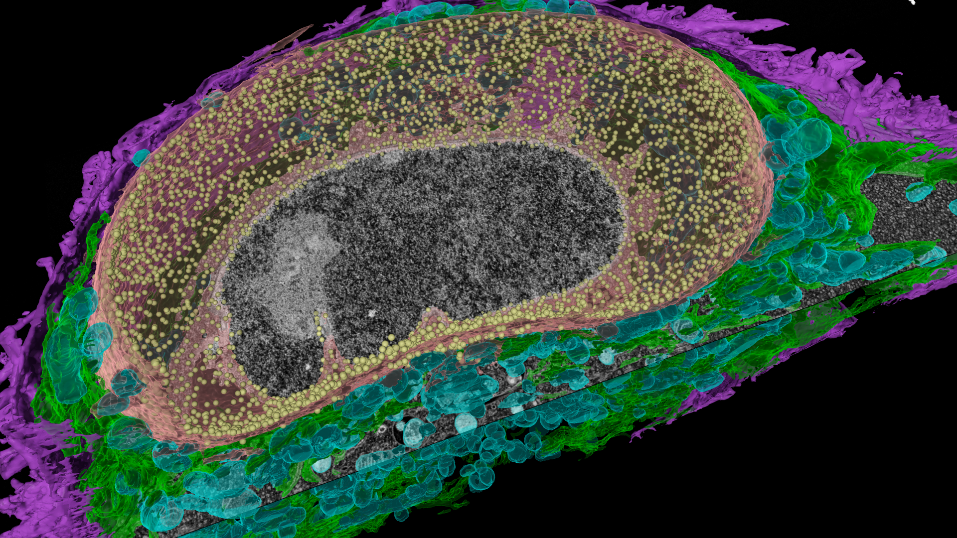
Scanning Electron Microscopy (SEM) has been long established as a powerful and valuable technique for life science research for samples as varied as insects, tissues, organoids, viruses, cells and plants. Using a beam of electrons to scan the surface of a plethora of sample types, SEM provides a wealth of information about biological structures, high resolution imaging of ultrastructure across large fields of view, compositional information, and the ability to correlate other types of microscopy to link structure and function. Scanning electron microscopes are classically known for imaging structural details on the surface of biological samples but have evolved into providing detailed volume and subcellular information.
Key Learnings:
- Learn how SEM works and the considerations you need to make when selecting the right imaging parameters
- See some of the applications that can be addressed using SEM approaches
- Hear about some of the challenges that can limit the quality of the images you generate using SEM and how you can avoid them
Fundamentals of X-ray imaging in Life Sciences
Presented by:
Dr. Rosy Manser, Solution Manager Life Science X-ray Microscopy, ZEISS
Dr. Liz Duke, Team Leader for Biological X-ray Imaging, EMBL Hamburg, Germany
Unlike SEM, imaging with X-rays is non-destructive. X-rays penetrate through specimens and as such are able to capture structural details without physically cutting the sample. Relative to SEM, X-ray imaging is not as well-established in life science research, but the power of the possibilities now offered by instruments based in the lab and at the synchrotron is rapidly changing this balance. The increasing availability of suitable staining and mounting protocols is also aiding the increase in use of X-ray imaging for life science research questions.
Key Learnings:
- Learn how X-ray tomography works and the numerous instruments that use similar approaches
- Experience the wide range of applications that can benefit from X-ray imaging, both in the lab at the synchrotron
- Hear about some of the considerations around image quality that come with X-ray imaging and how you can avoid them
Preparing your Life Science sample for Scanning Electron Microscopy (SEM)
Presented by: Dr. Kirk Czymmek, Director of the Advanced Bioimaging Laboratory, Donald Danforth Plant Science Center, USA
The very high resolution offered by the SEM comes together with some stringent sample preparation requirements and readying samples for electron microscopy has been known as something of a dark art! Generating the right level of fixation, a smooth embedding and perfect staining takes expertise and practice and many written protocols are not self-explanatory if you don't have previous experience.
Key Learnings:
- Learn about the most common sample preparation approaches for SEM in Life Science
- Hear about some of the common challenges that can limit the quality of the resulting prepared specimen
- Make yourself familiar with the fundamentals of Volume EM sample preparation
Expanding your Scanning Electron Microscopy (SEM) to 3-Dimensions and beyond
Presented by:
Dr. Philipp Bastians, Sales Manager Life Science Electron & X-ray Microscopy, ZEISS North America
The value of SEM in providing high resolution structural information in a wide range of Life Science samples is well appreciated. Developments in technology and sample management capabilities also enable the scaling up of these insights into far larger 2D areas as well as 3D volumes. This expansion of SEM acquisitions provides exciting opportunities for new insights and experiments.
Key Learnings:
- Learn how you can use different approaches for increasing the area or volume you capture using SEM
- See some examples of how each of these approaches have expanded the scope of biological questions that can be addressed
- Hear about the different opportunities that each approach provides
Associated Training Guides
Download our training guides below


Contact us
Excited to learn more? Contact us to speak directly with a ZEISS representative about the specific needs of your facility.