Visualize cellular structures and processes at unprecedented detail.
Multimodal Imaging Techniques
Observe dynamic biological processes in real-time and analyze complex interactions within plant cells.
Advanced Software Solutions
With features like 3D reconstruction and automated image processing, researchers can efficiently analyze large datasets, accelerating discoveries in plant science.
From Cells to Ecosystems
Harnessing cutting-edge techniques in plant science empowers researchers to delve into the complex mechanisms of plant growth and development. This innovative approach enhances our comprehension of plant physiology and ecology, paving the way for breakthroughs in sustainable agriculture and environmental conservation. By unraveling the genetic and biochemical pathways that govern plant responses to stressors, we are advancing efforts to cultivate resilient crops and promote biodiversity in our ecosystems.
Scroll animation items
Want to visualize the unseen world of plant biology?
Discover techniques to transform your research
High Resolution and Contrast
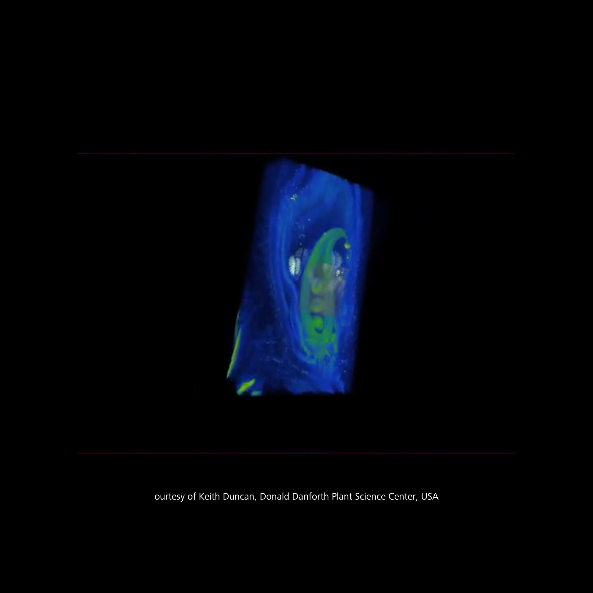
ZEISS Lattice Lightsheet 7 provides high spatial resolution, which can be an essential for studying fine structures like pollen morphology.
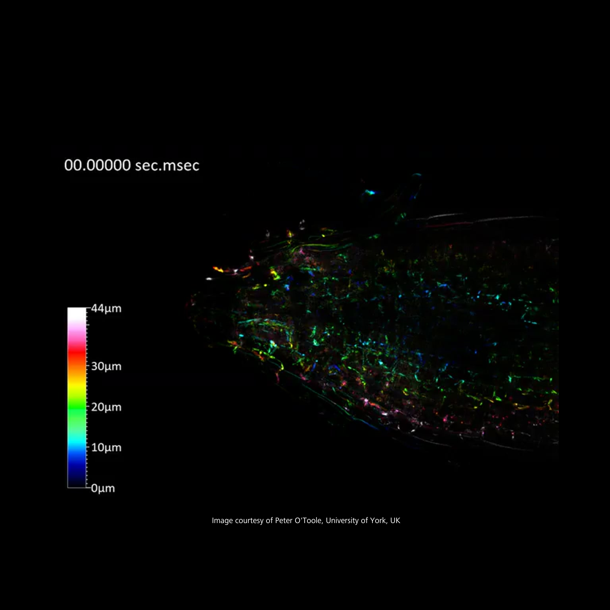
The 3D imaging capabilities of the VersaXRM allow for an effective appreciation of the spatial relationships among these critical reproductive structures. By visualizing the positioning of the ovary, ovules, and anthers in three-dimensional space, researchers can gain valuable insights into the reproductive biology of soybeans, enhancing our understanding of their development and function.
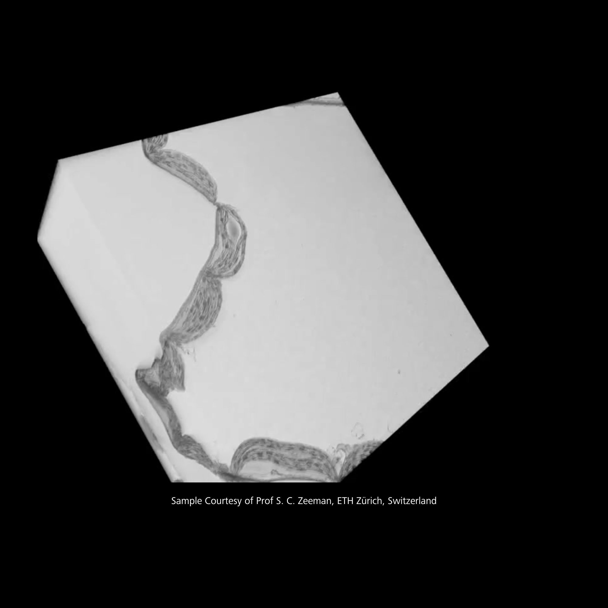
ZEISS Volutome enables efficient data acquisition, allowing researchers to capture high-resolution images quickly. This speed is particularly beneficial when working with sensitive biological samples that may change over time or require immediate analysis..
Yes, ZEISS provides various case studies and application notes that showcase the use of their microscopy systems in neuroscience. Examples include:
Imaging Synaptic Structures: Using confocal microscopy to study synaptic connections and plasticity.
Neurodegenerative Disease Research: Employing two-photon microscopy to observe changes in neuronal behavior in live animal models.
Brain Mapping: Utilizing advanced imaging techniques to visualize brain connectivity and function.
ZEISS microscopy solutions enhance neuroscience research by providing:
High Resolution: Advanced optics and imaging technologies allow for detailed visualization of neuronal structures and processes.
Live Imaging: Many systems are designed for live-cell imaging, enabling the observation of dynamic processes in real-time.
Multimodal Imaging: The ability to combine different imaging modalities (e.g., fluorescence and electron microscopy) allows for comprehensive analysis of samples.
These features facilitate a deeper understanding of neural mechanisms and contribute to advancements in neuroscience.
ZEISS offers a variety of microscopy techniques tailored for neuroscience research, including:
Confocal Microscopy: Ideal for imaging thick samples and obtaining high-resolution images of cellular structures.
Two-Photon Microscopy: Particularly useful for imaging living tissues at greater depths, minimizing photodamage.
Electron Microscopy: Provides ultra-high resolution for detailed visualization of neuronal structures.
These techniques enable researchers to explore complex neural networks and cellular interactions in detail.