5 μm FFPE section of mouse kidney simultaneously stained with 6 IF markers plus DAPI. Imaged on Axioscan 7 spatial biology for spatial omics applications
ZEISS Spatial Biology Solutions

Spatial Biology Imaging

From Complexity to Clarity: Workflow Automation at Scale

Unlock clarity in complexity and accelerate your spatial biology research. ZEISS solutions for spatial biology seamlessly integrate multiplexed tissue imaging and analysis into your workflows, enabling scalable, reproducible, and high-throughput spatial profiling. By visualizing cellular interactions in their native tissue context, our technologies empower researchers to gain deeper insights into disease mechanisms and transform the way pathology and translational science are approached.

  • Seamless sample to report experience
  • Automated Whole Slide Imaging for consistent results
  • Advanced analysis software for insightful data interpretation
Automated multiplex immunofluorescence imaging of up to eight biomarkers across 100 slides per run—ideal for high-throughput clinical research and drug development.

Unlock the Power of Spatial Biology

Are you working in translational research, immuno-oncology, or a service lab seeking a high-throughput tissue imaging workflow that ensures accuracy and reproducibility across large cohorts?
The ZEISS tissue multiplexing workflow for biopharma, academic medical centers, and CROs scales spatial biology to routine histopathology settings. By integrating automated slide scanning, multiplex staining, and streamlined analysis, laboratories can generate rich biomarker data with high consistency and reproducibility across studies.


Standardized and automated methods ensure consistency across multi-site clinical studies and longitudinal projects. The workflow also streamlines analysis and reporting through integrated software, enabling researchers to evaluate multiple biomarkers simultaneously and translate data into actionable biological and clinical insights.

Schedule Your Demo

See ZEISS Technology in Action

Request a demo tailored to your needs—whether virtual or in-person at the ZEISS Microscopy Customer Center.

Choose the experience that works best for you!

End-to-End Spatial Biology Workflow Starter

A Seamless Sample to Report Experience
Diagram explaining the end-to-end spatial biology workflow starter bundle ​
Staining Ultivue 4-Plex T-Act Kit​
Image of Ultivue 4-Plex T-Act Kit​

Pre-optimized panel with verified reagent performance and superior staining protocol for best speed and ease-of-use​.

Imaging Axioscan 7 spatial biology

Optimized configuration with best performance for routine spatial biology proteomics workflows​

Imaging SlideStream annual license​
Screenshot of SlideStream, intuitive workflow manager software for scan and analysis automation

Streamlined, fully automated image acquisition experience, with seamless integration for Ultivue scan protocols and automated image transfer to mindpeak platform.​

Analysis Mindpeak starter license​

- Includes access and Spatial Biology research platform, Image Viewer, Image Storage and Data Analysis Platform​

- AI-Powered Image Analysis Algorithms​

- 6 months , 0.5Tb , 25 images can be analyzed​

Configure your Axioscan 7 for Spatial Biology

Workflow automation for spatial biology at scale

The new Axioscan 7 Spatial Biology Configuration is designed to maximize efficiency, accuracy, and reproducibility for multiplex immunofluorescence (mIF) and high-throughput spatial biology workflows.

Compatibility with Leading Reagents

The system supports both DNA-barcoded multiplex immunofluorescence (mIF) and Tyramide Signal Amplification (TSA)-based chemistries. Researchers can also deploy a wide variety of fluorophores – spanning industry standards like OPAL™ dyes as well as next-generation TSA dyes from Spatomics – to achieve robust, high-resolution imaging in 8-plex assays and beyond. Compatible reagent solution leaders include:
  • InSituPlex

    Ultivue by Vizgen

  • SignalStar® mIHC

    Cell Signaling Technology

  • StreptaClick® TSA panels

    Kromnigon

Reagents - Tailored for Tissue Multiplex Staining

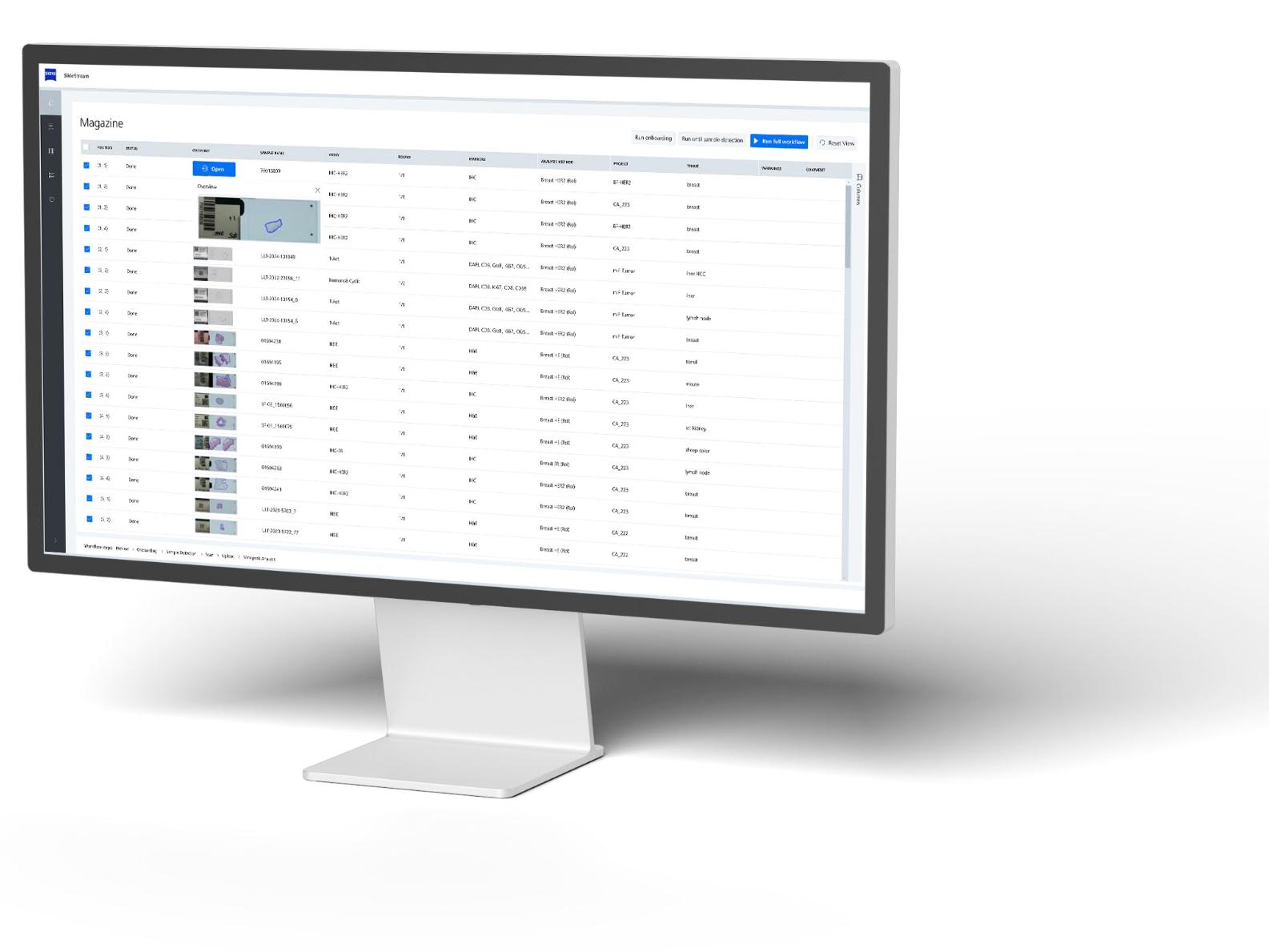
The starter bundle explicitly includes an OmniVUE™ reagents kit for “T-Act” biomarker analysis from Ultivue, including co-optimized StarVUE™ AI-analysis. At the heart of Ultivue are two technology platforms:​

InSituPlex®
Massively parallel single-molecule amplification

  • Highly configurable multiplexed biomarker detection​
  • Pre-optimized, clinical-grade assay performance​
  • Fast and gentle high-throughput workflow

STARVUE™
Deep learning enabling faster image-to-insight​

  • Biomarker morphology and signal intensity-driven cell classification​
  • Highly scalable and adaptable image processing​
  • Enables precise accurate results faster at a lower cost

The integration of Ultivue’s assay solutions into the new ZEISS End-to-End workflow offering, put you on the fast track to generate accurate, reliable and reproducible data.

SlideStream Inuitive Workflow Manager Software

Simplify the process of scanning, analyzing, and managing high-volume microscopy slides
  • ZEISS' SlideStream Assay Management Software, transforms multiplex imaging into a compatible, scalable, and reproducible spatial biology workflow.
  • ZEISS' spatial biology bundle has transformed the way we generate spatial phenotypic data for our clinical research.

    Prof. Christoph Kuppe, MD, PhD University Clinic RWTH Aachen
  • ZEISS is addressing a key bottleneck in whole-tissue scanning through their workflow and we are benefitting from the efficiency gain.

    Christopher Mills Senior Scientist, Concept Life Sciences (CRO in United Kingdom)
  • project overview
  • project image overview ims
  • Image viewer with quality accepted
  • marker display and results qc
  • phenotypes
  • phenotypes zoom in
  • Screenshot of a Mindpeak landing page with a ZEISS projects overview
    Screenshot of a Mindpeak landing page with a ZEISS projects overview
  • Screenshot of a Mindpeak project image overview named mIF Tumor Screen
    Screenshot of a Mindpeak project image overview named mIF Tumor Screen
  • Screenshot of an image viewer in Mindpeak, showing image quality accepted
    Screenshot of an image viewer in Mindpeak, showing image quality accepted
  • Screenshot of an image viewer in Mindpeak, showing the marker display and the results
    Screenshot of an image viewer in Mindpeak, showing the marker display and the results
  • Screenshot of an image viewer in Mindpeak, showing phenotypes
    Screenshot of an image viewer in Mindpeak, showing phenotypes
  • Screenshot of an image viewer in Mindpeak, with phenotypes zoomed in
    Screenshot of an image viewer in Mindpeak, with phenotypes zoomed in

Mindpeak Cloud-Based Analysis Solutions for Highest Precision in Biomedical Research1

Elevate your multiplexing IF and IHC workflows with ZEISS Axioscan 7 for Spatial Biology combined with the power of Mindpeak, leaders in the field of digital pathology. Our new Spatial Biology analytics platform enables effortless transition to cloud-based image analysis—giving researchers remote access to their images, project organization tools, and automated AI-driven workflows. Powered by Mindpeak’s foundation AI models for advanced image analysis, the system delivers robust results and standardized reporting, without the need for expertise and efforts to adjust or develop algorithms, while still offering the flexibilities to analyze a growing set of common biomarkers used in oncology and immuno-oncology including customizable phenotype definitions for precision in biomedical assays.​

  • Image QC and AI triggering Workflow

    Image QC and AI triggering Workflow

  • Custom Phenotype Tool

    Custom Phenotype Tool

  • Image QC and AI triggering Workflow
  • Custom Phenotype Tool

Our Partners

  • Partner logo: mindpeak
    mindpeak
  • Partner logo: Ultivue
    Ultivue by vizgen
  • Partner logo: Kromnigon
    Kromnigon
  • Partner logo: Cell Signaling Technology
    Cell Signaling Technology
  • Partner logo: Concept Life Sciences
    Concept Life Sciences

FAQs: Axioscan 7 Spatial Biology

  • The Axioscan 7 spatial biology can separate 8 fluorescence channels, currently supported by Kromnigon SpectraClick reagents. A typical panel could consist of 7 target markers plus DAPI. Supported fluorophores include DAPI, CFP, GFP, DsRed, Cy3, Cy5, Cy7 and fluorophores with similar spectral properties. Single round staining and single shot imaging can be done for 8-channels. For going to higher marker numbers (e.g. 14, 28, etc.) cyclic staining and imaging can be performed.

  • The physical slide capacity of the system is 100 slides (25 trays, 4 slides each). The system can image over 100 slides per day thanks to the high level of automation and high scan speed. 10 mm2 tissue area can be scanned in 3 to ~10 minutes pure scan time for 5- or 8-Plex respectively.

  • Yes. A single run can include up to 100 slides, each potentially containing multiple tissue sections or cores. Pre-configured scan profiles for mIF and chromogenic assay stained tissue can be selected per sample, allowing users to load and run diverse experiments in one batch, in a single automated workflow.

FAQs: SlideStream Workflow Manager

  • No prior microscopy experience is required. The software and scanner are designed for ease of use, with an intuitive interface and automated workflows that enable any operator to run the system efficiently, with minimal or no training.

  • The Axioscan 7 connects to the LIMS by scanning QR codes or barcodes printed on the slide labels. The scanner reads the slides unique ID from the code on the sample, retrieves the required additional information from the LIMS, which will be used to automatically populate the SlideStream interface with the relevant details, streamlining the workflow and reducing the need to manually re-enter pre-existing data.

FAQs: Image Management System and data analysis platform

  • For optimal performance, we recommend using the latest version of Google Chrome, a stable internet connection with at least 10 Mbps bandwidth, and a computer running a current, up-to-date operating system. These requirements ensure smooth and responsive operation of the platform.

  • Yes, local deployment is possible. For customers handling large image volumes or requiring local data storage, Mindpeak offers hybrid deployment options. In this setup, data is stored on the customer’s server while processing is performed on Mindpeak’s secure cloud infrastructure, helping reduce storage costs while maintaining greater control over sensitive data.

  • Storage limitations are defined by the IT infrastructure of customers as defined in their purchased packages. Additional storage capacities can always be acquired ad hoc in alignment with customer needs and budget requirements.

  • We partner exclusively with ISO 27001-certified cloud providers, such as AWS and Hetzner, to ensure enterprise-grade data protection. All data is encrypted both at rest and in transit, and regular security audits are performed to safeguard your sensitive research. These measures ensure your data remains secure, private, and fully under your control.