ZEISS Axio Imager scan
Automated acquisition and advanced data analyses for high-throughput screening applicationsEquip yourself with automated imaging acquisition tools and AI-powered software analyses to break through previous experimental barriers that come with manual imaging and analysis workflows. ZEISS Axio Imager scan was designed to increase the efficiency of your large-scale data collection workflows. Selected hardware and software components create a streamlined process from acquisition to evaluation of relevant data for your research needs.
Bundle components
-
Microscope
- Axio Imager 2 (upright widefield microscope stand)
- Scanning stage 130 × 100
- Motorized condenser NA 0.9
- Apotome 3
Light source / camera
- Viluma 7
- Filter sets 38, 43, 50, 70, 96
- Axiocam 305 color
- Axiocam 820 mono
Objectives
- EC Plan-Neofluar 5× / 0.16
- Plan-Apochromat 10× / 0.45 , 20× / 0.8
- LD LCI PApo 40× / 0.95 Corr
Workstation
- Z8 Workstation with 128 GB RAM and nVidia Quadro RTX A4000 16 GB DP
- Axio Imager 2 (upright widefield microscope stand)
-
Motorized Acquisition
Acquire images and control motorized components in multi-dimensional experiments.
Smart Acquisition
Create flexible and intelligent image acquisitions workflows with the Experiment Designer, Guided Acquisition and Experiment Feedback tools.
2D Toolkit
Create flexible automatic measurement programs for the analysis of your 2D images.
AI Toolkit
Make use of powerful AI tools. With this package, you can train AI models for various applications.


Looking for an inverted microscope configuration?
This widefield bundle for screening applications can also be configured based on the inverted microscope ZEISS Axio Observer 7.
 to identify macrophages and hematoxylin counterstain")
 to identify macrophages and hematoxylin counterstain")
, CD3 (TRITC), PD-L1 (Cy5), CD68 (Cy7), and DNA (DAPI).")
, CD3 (TRITC), PD-L1 (Cy5), CD68 (Cy7), and DNA (DAPI).")
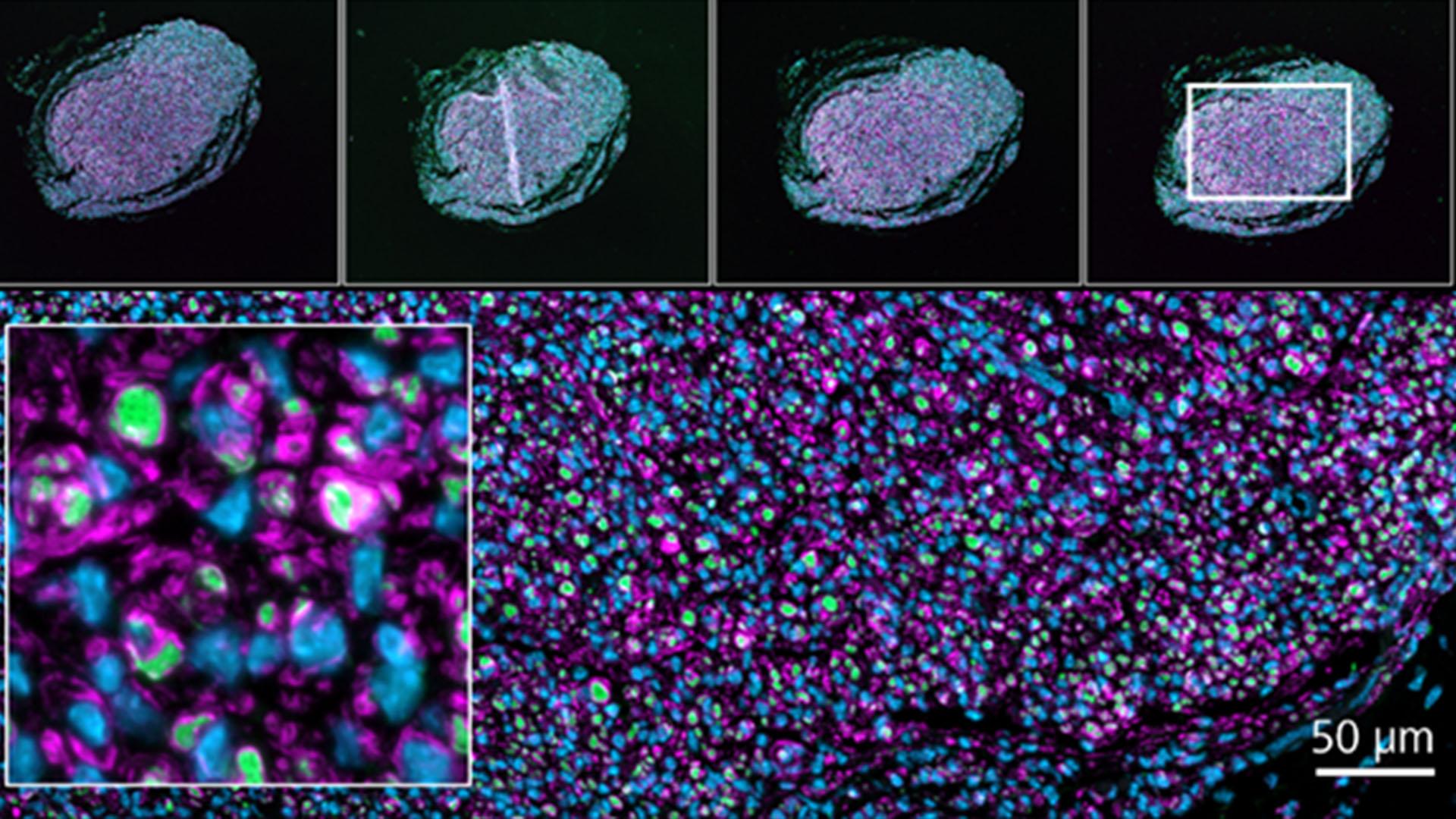
 and Schwann cells (magenta), nuclei with DAPI (blue).")
 and Schwann cells (magenta), nuclei with DAPI (blue).")