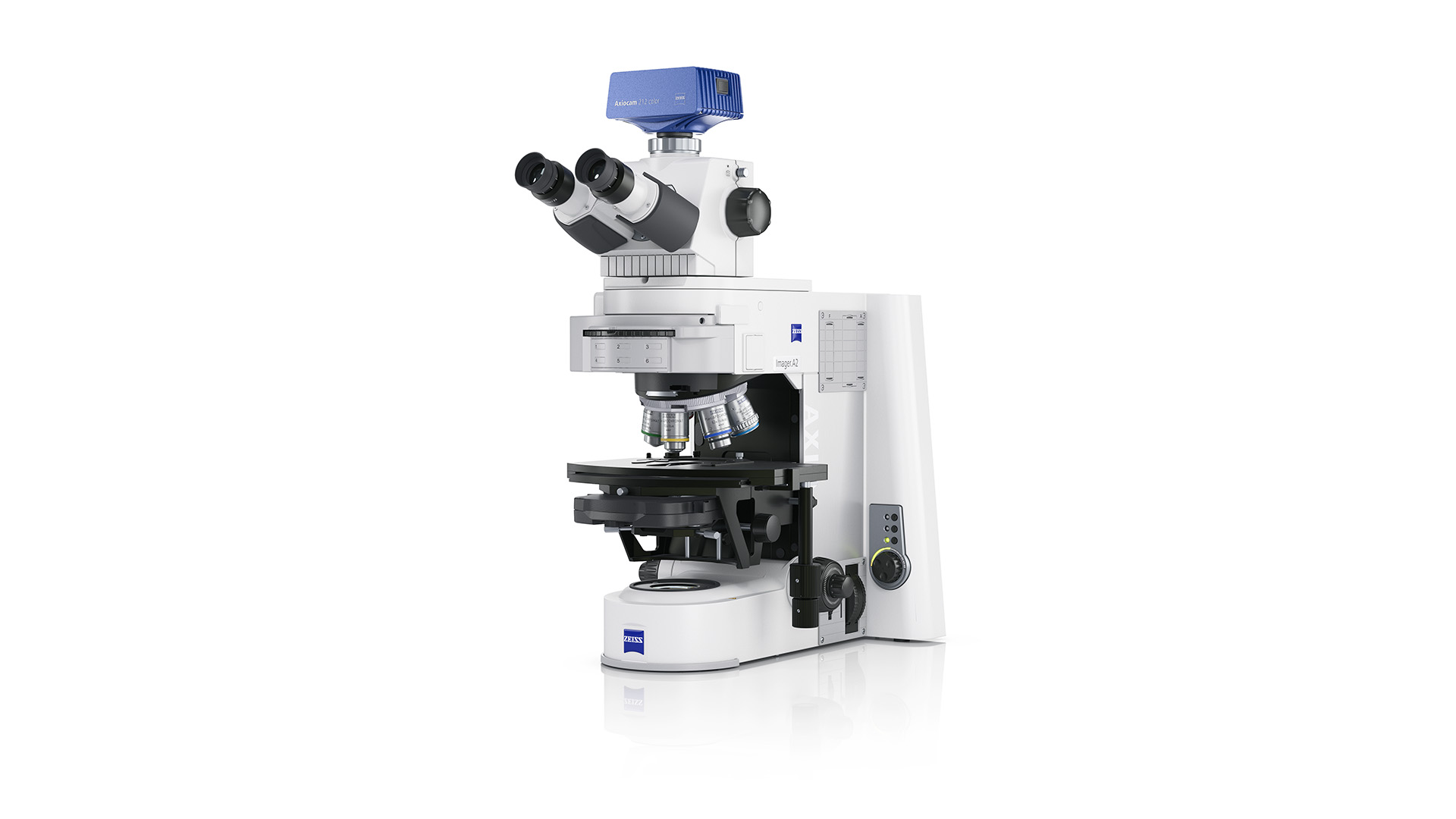
ZEISS Axio Imager 2
Upright microscope platform for productive high-resolution imagingExplore cellular structures and processes of your biological specimens at unprecedented levels of detail. Whether you are conducting cellular biology studies, histological examinations, or cancer research, you can be ensured with the versatility and performance needed to enhance your research outcomes.


U2OS cells stained with Phalloidin
High-performance optics with 25 mm field of view allow you to simply see more in every image. Detect more cells per shot or scan large areas faster for highest productivity. U2OS cells stained with Phalloidin (green, actin) and Hoechst (blue, DNA), acquired using a Plan-Apochromat 20× / 0.8 objective.
Optical excellence
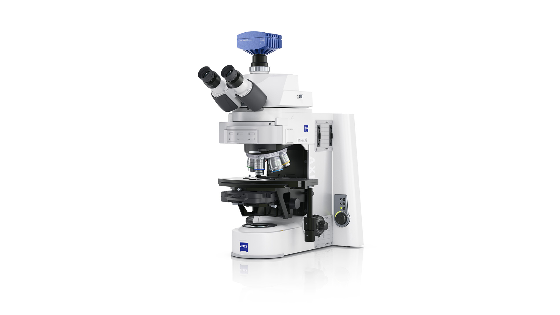
Highest contrast and resolution across a large field of viewA stable imaging system with high-quality optics is essential to achieve the clarity and resolution required to understand biological processes. With ZEISS Axio Imager 2, you can rest assured that you have the best optics to support your research. The z-drive features a 10 nm step size, ensuring precise focusing of your sample and maintaining its position. The stable imaging cell was designed for a vibration-free proximity between the filter cubes, camera, and objective, ensuring highest image quality and reproducibility. The 25 mm field of view allows you to image larger areas of your sample faster.
Caption: High-performance optics with 25 mm field of view allow you to simply see more in every image. Detect more cells per shot or scan large areas faster for highest productivity. U2OS cells stained with Phalloidin (green, actin) and Hoechst (blue, DNA), acquired using a Plan-Apochromat 20× / 0.8 objective.


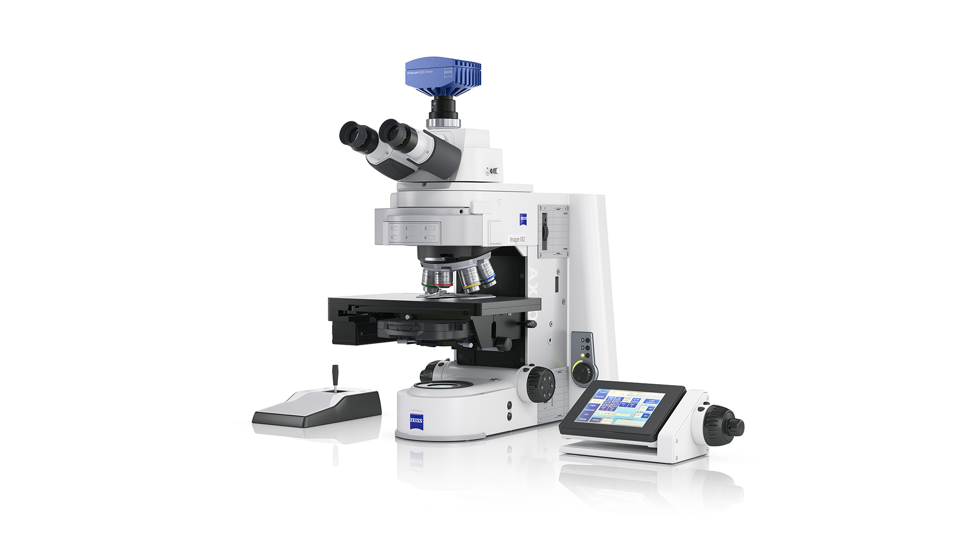
Ergonomic phototubes
Ergonomic excellence
Human-centered design for comfortable observationTailor the setup to your personal preferences with ergonomic phototubes for comfortable viewing. Customize the microscope for quick access to frequently used functions or user-specific settings. ZEISS Axio Imager 2 is also designed to work without a connected PC. With a smart Axiocam camera, you can display images on an external monitor or TV and save your images directly to a memory stick.


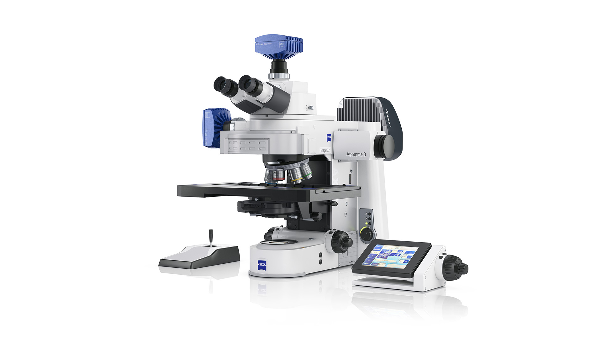
ZEISS Axio Imager 2 with LSM 990 Airyscan and incubator
Modular excellence
Custom setups designed for efficient imagingZEISS Axio Imager 2 can be adapted to your application requirements with a high degree of freedom. It is compatible with fast and sensitive cameras of the ZEISS Axiocam portfolio as well as with cameras from other manufacturers. Choose from bright and efficient multi-LED light sources from the ZEISS Viluma family and pair them with up to 10 filter sets for maximum spectral flexibility. Expand your configuration with Apotome 3 or an LSM confocal—supplemented by an XL incubator for imaging at elevated temperatures if required.


, DNA (blue) and actin (magenta)")
, DNA (blue) and actin (magenta)")
, panCK (green), CD3 (orange), PD-L1 (red), CD68 (purple)")
, panCK (green), CD3 (orange), PD-L1 (red), CD68 (purple)")