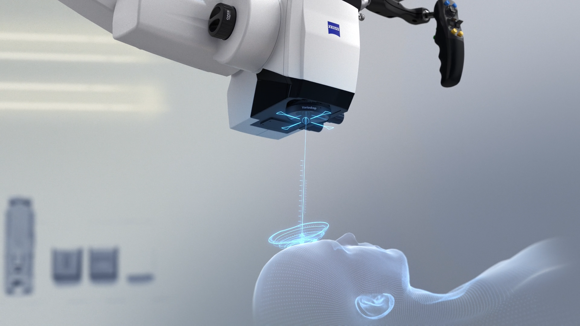
First evaluation report: ZEISS KINEVO 900 with ZEISS QEVO
Comprehensive collection of cases evaluating transcallosal, retrosigmoid, pterional and transsphenoidal approaches in a lab settingBackground
The prototype evaluation of the newly developed Robotic Visualization System® – KINEVO® 900 from ZEISS produced by Carl Zeiss Meditec AG (Oberkochen) took place at the Anatomical Institute of the University of Regensburg. The surgical-technical evaluation was carried out by Prof. Dr. med. Karl-Michael Schebesch and Dr. med. Julius Höhne, both doctors at the Department of Neurosurgery (University Medical Center of Regensburg). For a short period of time, the head of the department, Prof. Dr. med. Alexander Brawanski, was also present.
Method
The application, the movement and the adjustment to the surgical field of the KINEVO 900 is intuitive. If one looks at KINEVO 900 system, the similarity of handling compared to OPMI PENTERO 900 is striking. In particular, this applies to the microscope suspension, the handgrips and the microscope head and is important for any neurosurgeon adopted to the handling of conventional surgical microscopes.
Conclusion
At the Anatomical Institute logistics and spatial relations were optimal for the surgical technical evaluation. All surgical instruments were available (e.g. electric Trepan, osseous saw, instruments, expendable items) to create a realistic simulation of the surgical approaches. The operations were performed on an anatomical section table; illumination was provided by the light of the Robotic Visualization System – ZEISS KINEVO 900. Delivery, setup and connection of the components were carried out exclusively by employees of Carl Zeiss Meditec AG. The examined cadavers were all deep-frozen human heads. On both days 10 cadaver heads were evaluated and then refrozen. Each head was taken out of the freezer 24 hours prior to examination, allowing adequate thawing time at room temperature. On the preparation, some icy patches were encountered, however the bony structures and cerebral membranes were intact. Cranial nerves were in a very good state and comparable with in vivo findings. Even though bloodless, preparation of vascular structures was authentic. The parenchyma of the brain of all specimens appeared clearly atrophied and partially liquefied. However, there was a wide variation in the state of preservation. In all approaches that are mainly intraparenchymal (above all the transcallosal approach) only well-preserved specimens were chosen. Once the ventricular system was entered, all anatomical structures could clearly be visualized.