On-demand webinar
How to obtain excellent confocal laser endomicroscopy (CLE) images and interpret them
3 May 2021
· 52 min watch


Author
Evgenii Belykh MD, PhD
Neurosurgery Resident, Rutgers University, New Brunswick, New Jersey, USA
SUMMARY
Peer-to-peer experience sharing including ex vivo study results and comprehensive summary
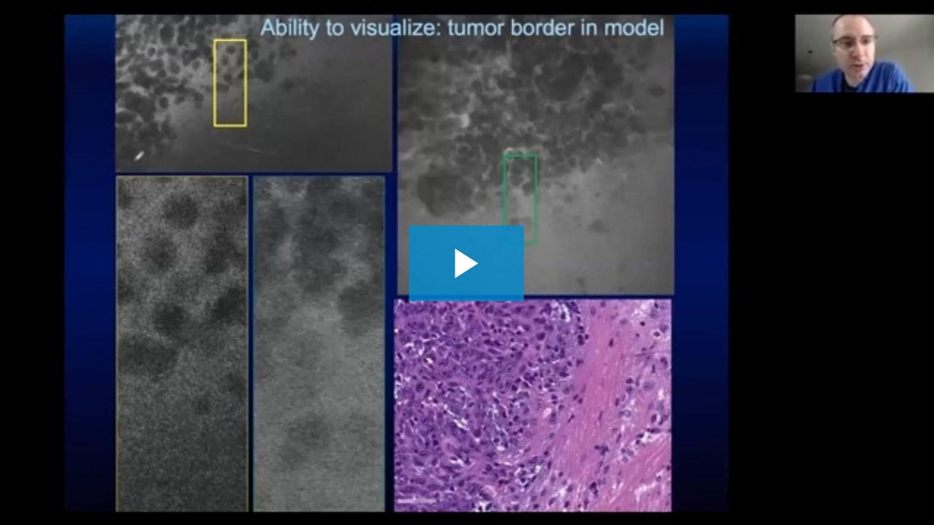
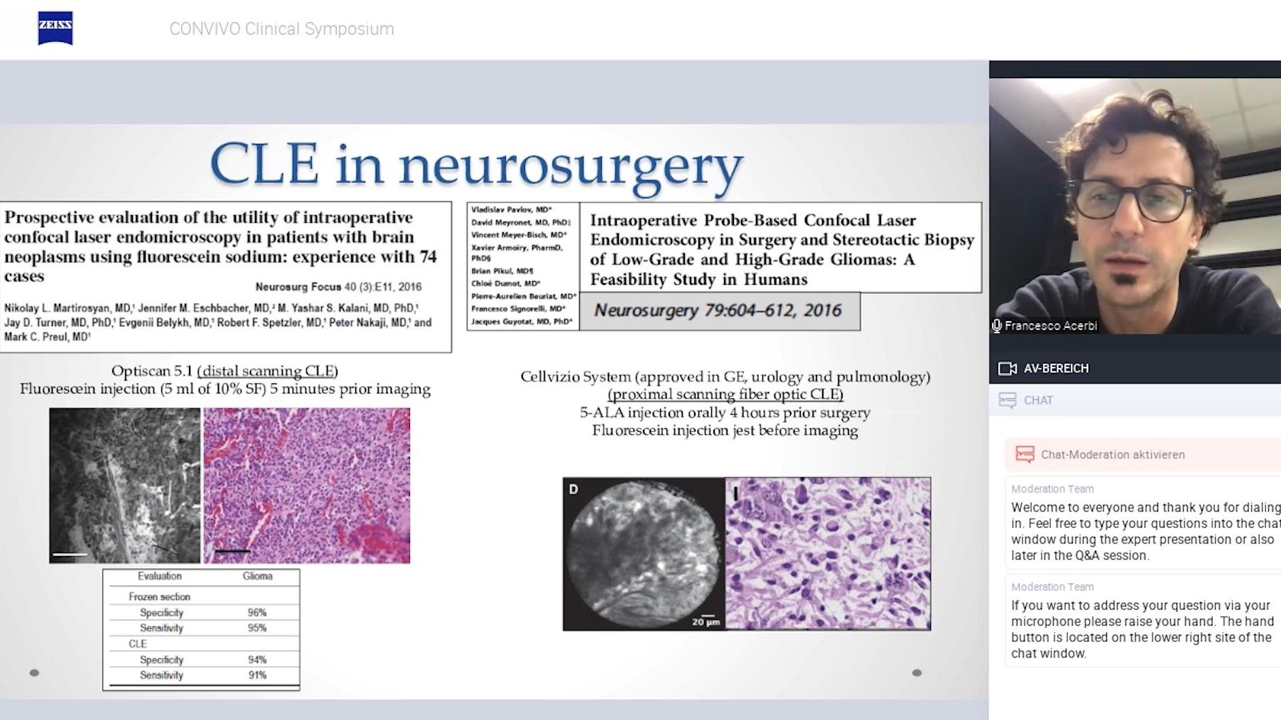
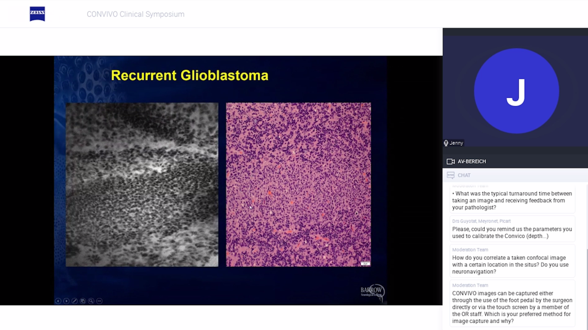
Dr. Evgenii Belykh’s talk is dedicated to the question of how excellent probe-based confocal laser endomicroscopy (CLE) images can be obtained and interpreted.
Following an introduction on fluorescence-guided neurosurgical oncology, Dr. Belykh explains (starting at minute 07:08) why it could be beneficial for the interpretation of confocal imaging to add cellular movement using in vivo case examples. Furthermore (starting at minute 23:55), ex vivo study results are presented, followed by a comprehensive summary based on years of clinical experience with CLE in neurosurgery at the Barrow Neurological Institute in Phoenix, Arizona.