
Micro-CT Scanners
Revolutionizing Non-Destructive 3D Imaging in Research and IndustryMicro-Computed Tomography (micro-CT) scanners are powerful tools that allow you to see inside objects without causing damage. These machines use x-ray technology to produce high-resolution 3D images of samples.
This technology unlocks new opportunities for research and industry. It assists scientists in studying the internal structure of materials and living tissues. Doctors can even examine lab-grown or biopsied tissues to improve medical treatments.
Engineers use it to inspect a wide variety of parts. Thousands of manufacturers trust ZEISS industrial CT products to ensure quality. They deliver high-resolution 3D imaging that reveals details you never knew existed.
Whether you're in research, manufacturing, or healthcare, micro-CT scanning can improve your workflow.
Key Takeaways
• Micro-CT scanners create detailed 3D images of all types of samples using x-rays
• You can use this technology to examine internal structures without destructive techniques
• ZEISS industrial CT products offer high-resolution imaging for various applications

Principles of Micro-CT Technology
Micro-CT technology uses a series of x-rays and computer processing to create detailed 3D images of objects. The results allow you to see inside parts without needing to cut them open.
X-Ray Source and Detection
Micro-CT scanners use a specialized x-ray tube to produce an x-ray beam. This beam passes through the object being scanned.
Some x-rays go through easily, while others are blocked. The thicker and denser the material is, the fewer x-rays will pass through.
A detector on the opposite side of the object detects the passing x-rays and measures their intensity, resulting in a 2D image of the object's interior.
The scanner takes many of these pictures as it either rotates around the object or rotates the object.
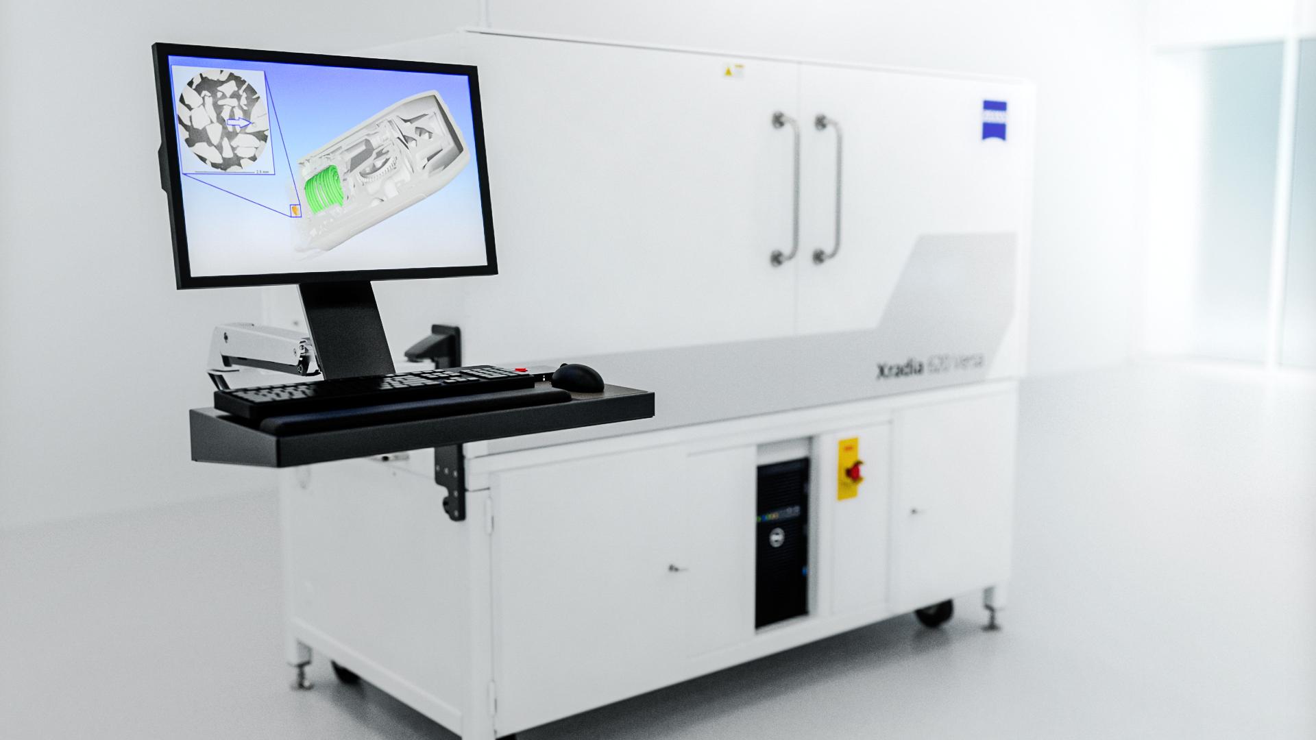
ZEISS micro-CT systems use powerful x-ray sources to capture high-resolution images of small details inside objects.
As the scanner spins, it captures hundreds or even thousands of 2D x-ray images. Each image shows the object from a slightly different angle.
A computer then combines all these 2D images to create a 3D model. This process is called computed tomography, or CT.
The more pictures taken, the better the final 3D image will be.
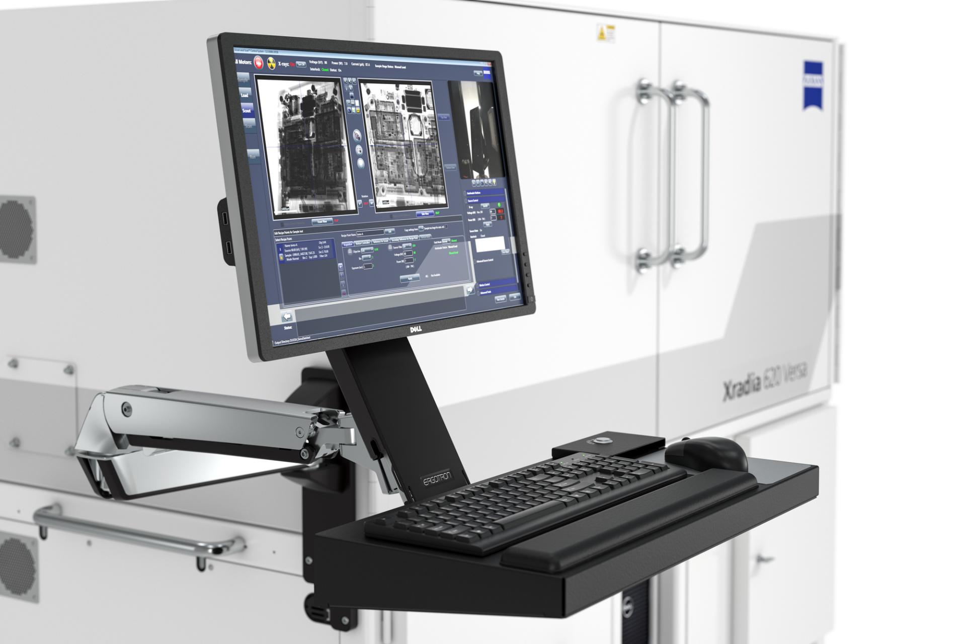
ZEISS INSPECT X-Ray software helps you maximize the value of your CT data.
Image Reconstruction
The last step is to convert all those 2D pictures into a 3D model that you can view on a computer. This process is called image reconstruction.
Special software processes the raw data from the scanner and converts it into a 3D image.
You can then rotate, slice, and measure this 3D model on your screen.
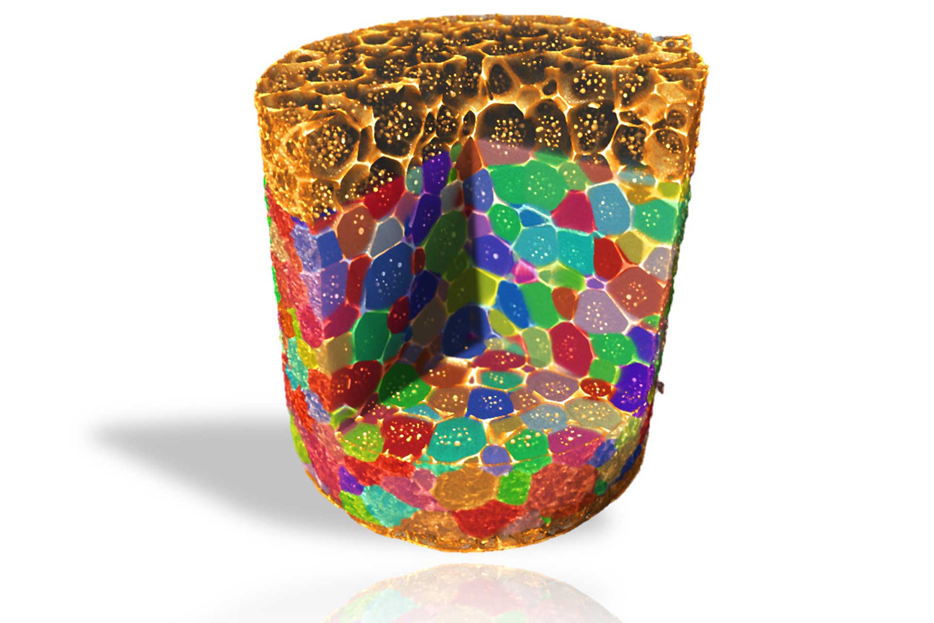
The software can also color various parts of the image based on their density. This helps you visualize the different materials inside an object.
Micro-CT Scanner Components
Micro-CT scanners are made up of several essential parts that work together to generate detailed 3D images. These components enable high-resolution scanning with precision and accuracy.
X-ray Tubes
X-ray tubes are central to micro-CT scanners. They produce the x-rays necessary for imaging.
Most tubes use a tungsten target to generate x-rays when struck by electrons. The tube's voltage and current can be adjusted to improve image quality for various materials.
A higher voltage x-ray beam can penetrate denser objects. Voltages of micro CT tubes can range from less than 100 kilovolts (kV) for biological materials and plastics to over 300 kV for many metals and alloys.
There is often a trade-off between x-ray power and resolution. The more power a tube provides, the larger the focal spot size becomes. It's similar to a flashlight—a bigger flashlight can emit more light and cover a wider area. Therefore, the focal spot size of the tube influences image sharpness. Smaller spots yield clearer images but may restrict power output.
Geometric Magnification
The details that can be detected depend heavily on your image's magnification. You can adjust magnification by moving the sample closer to the x-ray source. Higher magnification allows you to see finer details but decreases the field of view.
Some scanners utilize x-ray optics to focus the beam, enabling very high magnification of tiny samples. ZEISS leads the way in applying optics to enhance resolution in CT imaging. X-ray microscopy (XRM) is a unique form of CT imaging that achieves resolutions as fine as 50 nanometers.
Detectors
Detectors capture x-rays passing through the sample. Most micro-CT scanners utilize flat-panel detectors. These can vary in type, size, and speed, but all share the goal of converting x-rays into visible light. The light is then captured and digitized.
The number and size of pixels on a detector influence the field of view, image resolution, and scan times. Smaller pixels can produce higher-resolution images but tend to be slower and less efficient. X-ray imaging often involves balancing scan time with image quality.

Applications of Micro-CT Scanning
Micro-CT scanning has transformed research and analysis in many fields. This advanced technology enables detailed 3D imaging of small objects without causing any damage.
Biological and Medical Research
Micro-CT scans allow detailed study of biological samples. You can analyze plant structures, tissues, and organs without damaging them. This method supports long-term research since samples remain intact. One important use of micro-CT is examining tumors and their growth patterns.
ZEISS micro-CT scanners provide high-resolution imaging of soft tissue and bone samples. They help track disease progression and treatment effects over time. These scans have supported the development of new drug therapies and surgical techniques.
Researchers use micro-CT to analyze bone density and structure, aiding in understanding conditions like osteoporosis. It also allows for precise examination of dental implants and root canal treatments.
Material Science and Engineering
In material science, micro-CT scanning is essential for nondestructive testing. It allows inspection of the internal structure of materials without causing damage. This technique is valuable for quality control in manufacturing.
ZEISS industrial CT systems help you examine porosity in metals and plastics. You can identify tiny cracks, voids, and defects that may impact product performance. This technology is essential in the aerospace and automotive industries.
Micro-CT scans reveal the microstructure of foams, ceramics, and polymers. You can study how materials behave under stress and improve their properties. This solution enables the creation of stronger, lighter materials for various applications.
Archaeology and Paleontology
Micro-CT scanning has revolutionized the way you study ancient artifacts and fossils. It allows you to examine delicate specimens without touching or damaging them, preserving valuable historical and scientific information.
Using ZEISS micro-CT technology, you can examine inside mummified remains or sealed pottery. You'll reveal hidden details and structures that were previously unknown. This method aids in dating artifacts and helps to understand ancient cultures.
Paleontologists use micro-CT to study fossilized bones and teeth. You can reconstruct extinct species and analyze their anatomy in 3D. This process gives new insights into evolution and prehistoric life.
Automation and Machine Learning
New micro-CT systems can operate independently. You just place your samples inside, and the machine handles everything else. This saves time and lets you scan more items.
Intelligent software increases inspection speed by analyzing images automatically, detecting cracks, measuring pores, or counting cells.
Machine learning is improving micro-CT. It can reduce noise in images or fill in missing data. Some software can even detect issues in scans automatically.
These advanced tools help you maximize your scans. They continually expand the usefulness of micro-CT across more fields every day.
Frequently Asked Questions
Micro-CT scanning is a powerful imaging tool with many applications. Let's explore some common questions about this technology.
What factors influence the cost of a micro-CT scanner?
The cost of a micro-CT scanner depends on several things. Resolution, scanning speed, and sample size all affect the price. Higher-end models, such as ZEISS Xradia systems, can be more expensive due to their advanced features and capabilities.
How does micro-CT imaging differ from conventional CT imaging?
Micro-CT offers much more detailed images than regular CT scans, revealing tiny structures as small as a few micrometers. This capability makes it ideal for examining small samples or detecting tiny defects in materials.
What is the resolution capability of micro-CT scanners?
Top micro-CT scanners can achieve resolutions below 1 micrometer. ZEISS Xradia Versa systems can deliver sub-micron detail across various sample sizes and materials.
Which micro-CT scanning services are available for research and clinical uses?
Many labs and companies offer micro-CT scanning services. These are useful if you don't need to buy your own scanner. Some places allow you to send in samples, while others perform the scanning on-site.
What are some limitations or drawbacks of using micro-CT scanning?
Micro-CT has some downsides. It can be slow when used on large samples.