
From 3D Light to 3D Electron Microscopy
Hosted at VIB in Ghent - Belgium- 00 years
- 00 months
- 00 days
- 00 hours
- 00 minutes
- 00 seconds
This event on Correlative Light & Electron Microscopy (CLEM) is jointly organized by ZEISS, The Francis Crick Institute, EMBL and VIB.
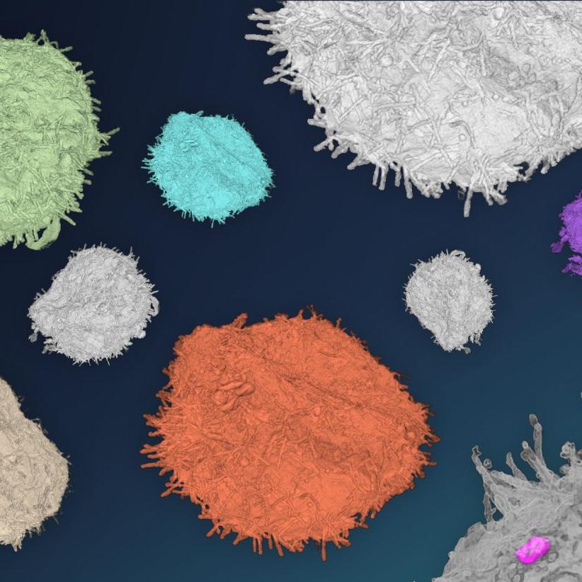
Life exists in 3D. In the last two decades, imaging techniques have been pushed forward and X-ray, light and electron microscopy can now be used to image larger volumes of biological samples with higher resolution. Therefore, the interest in these technologies is increasing and the Nature magazine named "volume EM" as one of the seven technologies to be watched in 2023.
When using correlative, or multimodal microscopy, a large variety of different imaging techniques can be combined. New and improved workflows combining two, or more imaging modalities are enabling more detailed insights into biological processes than ever before.
The datasets generated by these new workflows are increasingly large and complex. New developments in data management not only focus on image processing and visualization of these large datasets, but also on i.e. file compatibility, big data handling and storage.
This event will combine scientific sessions with a broad range of workshops where correlative workflows, tools, tips & tricks will be shared between participants and trainers/speakers.


Agenda
|
Day 1
Tuesday, 19 March, 2024
|
|
|---|---|
|
15:30 - 16:30 |
Registration & Coffee |
|
16:30 - 17:00 |
Welcome |
|
17:00 - 18:30 |
Keynote Session |
|
17:00 - 17:45 |
Keynote Speaker: Kirk Czymmek - Donald Danforth Plant Science Center (USA) |
|
17:45 - 18:30 |
Keynote Speaker: Eija Jokitalo - University of Helsinki (Finland) |
|
18:30 - 22:00 |
Dinner @ Oude Vismijn |
|
Day 2
Wednesday, 20 March, 2024
|
|
|---|---|
|
08:20 - 08:50 |
Registration & Coffee |
|
08:50 - 09:00 |
Welcome |
|
09:00 - 12:45 |
Session 1: CLEM Workflows Nalan Liv, UMC Utrecht (Netherlands). |
|
11:15 - 12:45 |
Workshop 1-10
|
|
12:45 - 14:00 |
Lunch & Poster session |
|
14:00 - 15:30 |
Workshop 1-10 |
|
16:00 - 17:15 |
Session 2: Imaging Technologies Elizabeth Duke, EMBL Hamburg (Germany) |
|
16:00 - 19:00 |
Workshop 1-10 |
|
|
Free Evening |
|
Day 3
Thursday, 21 March, 2024
|
|
|---|---|
|
08:30 - 09:00 |
Welcome Coffee |
|
09:00 - 11:15 |
Session 3: Diverse Biological Samples Antentor O. Hinton, Vanderbilt School of Medicine (USA). |
|
11:15 - 12:45 |
Workshop 1-10 |
|
12:45 - 14:00 |
Lunch & Poster session 2 |
|
14:00 - 15:30 |
Workshop 1-10 |
|
16:00 - 17:30 |
Session 4: Data Management / Handling Kedar Narayan, Frederick National Laboratory for Cancer Research (USA). |
|
17:30 - 19:00 |
Workshop 1-10 |
|
19:00 - 20:00 |
Get together @ Technologiepark |
|
Day 4
Friday, 22 March, 2024
|
|
|---|---|
|
08:30 - 09:00 |
Registration & Coffee |
|
09:00 - 12:00 |
Session 5: Molecular Tools Ben Giepmans, University of Groningen (Netherlands). Closing words. |
Workshops 1 -10
Workshop 1: X-ray targeted volume EM From Versa and Ultramicrotome (Crosshair) to Volutome
Christel Genoud - UNIL, CH & Paolo Ronchi, EMBL
This workshop is designed to showcase the application of X-ray imaging in conjunction with serial block-face scanning electron microscopy (SBF-SEM) for targeted acquisition of a large 3D dataset in a piece of tissue.
Key techniques:
- X-ray tomography.
- Serial Block-face imaging (SBF-SEM).
- Ultramicrotomy.
- The “Crosshair Workflow” according to Meechan et al., 2022 (https://doi.org/10.7554/eLife.80899).
Workshop 2: fsLaser milling of biological tissues for nanoscale imaging
Carles Bosch & Christopher Peddie (The Francis Crick Institute, UK)
In this workshop we explore the use of fsLaser milling to generate sample arrays from a parent ~4.5 mm3 volume of mouse brain by analysing pre-milling X-ray CT scans, sharing insights on the milling process, manipulating pillars, and by imaging an extracted pillar with FIB-SEM.
Main techniques:
- X-ray tomography.
- FIB-SEM with a Femtosecond Laser.
Workshop 3: Correlative Array Tomography
Jemima Burden (UCL, UK) & Ian White (UCL, UK)
This workshop will cover many aspects of the correlative array tomography workflow, with several of the key techniques being demonstrated. Ultrathin sections of a sample will be collected onto coverslips using an ultramicrotome before EM imaging with a scanning electron microscope using Atlas Array Tomography.
Key Techniques:
- Array Tomography.
- Scanning Electron Microscopy.
- Ultramicrotomy.
Workshop 4: Power hour with molecular tools and spatial analysis experts
Anneke Kremer (VIB, BE), Evelien Van Hamme (VIB, BE), Ben Giepmans (UMC, NL)
For this power hour, we’ll delve into the diverse spectrum of markers that can be used for CLEM workflows (‘the CLEM toolbox’) – such as whether fluorescent, peroxidase-based or gold-labeled; and structure, protein and/or RNA markers.
Key Learnings:
- Gain insights into CLEM markers.
- Explore uUsage of endogenous information of the biosample for analysis like RAMAN techniques or elemental analysis.
Workshop 5: Alignment, visualization and sharing of correlative data
Julian Hennies (EMBL, DE) & Benjamin Pavie (VIB, BE), & Tatiana Woller (VIB, BE)
This workshop will delve into the basics of how to navigate large 3D datasets using the MoBIE browser, including how to set up a MoBIE project with data of correlative light and electron microscopy experiments.
Key topics:
- MoBIE.
- BioImage Archive.
- EMPIAR.
Workshop 6: Power hour on AI tools for segmentation
Kedar Narayan (NIH, US) & Ilya Belevich (University of Helsinki, FI)
This workshop will address some basic concepts in deep learning and will be guided through several open-source plugins and tools, such as MIB, SAM and empanada, that help tackle the segmentation problems of volume EM datasets. At the end of the session, participants will learn how to deploy some of these tools.
Key learnings:
- MIB, SAM and empanada for tackling volume EM data segmentation.
- Deployment of these tools.
Workshop 7: Power hour on different light microscopes for correlative microscopy
Eef Parthoens (VIB, BE) & Nikky Corthout (VIB, BE), Amanda Goncalves (VIB, BE) & Jeremy Verbeke (VIB, BE)
This workshop on Correlative Light and Electron Microscopy (CLEM) will primarily focus on the light microscopy component of CLEM. It will provide attendees with practical knowledge to enhance their CLEM methodologies, specifically in the realm of light microscopy.
Key techniques:
- Laser branding
- Gridded coverslips
- Fiducial markers
Workshop 8: Starting CLEM in a core facility setting
Saskia Lippens (VIB, BE) & Paul Verkade (University of Bristol, UK)
This workshop endeavors to explore several aspects of implementing and developing complex protocols, procedures, and technologies associated with CLEM.
Key learnings:
- Balance for adaptability to diverse biological inquiries with the facility's overarching principles of robustness and efficiency.
- Convergence of two technology fields of light microscopy and electron microscopy in order to perform CLEM and the critical roles different experts and scientists play in executing such projects within a core setting.
- Comprehensive understanding of how best to navigate the integration of diverse imaging technologies for life science research.
Workshop 9: High-throughput imaging with ZEISS MultiSEM
Helmstaedter Lab (MPI BR, Frankfurt), Anna-Lena Eberle, ZEISS
This workshop will explore the high-throughput serial section acquisition workflow with the ZEISS MultiSEM. Ultra-thin serial sections are produced with the ATUMtome before they will be imaged sequentially with the multibeam SEM. The MultiSEM utilizes up to 91 electron beams in parallel to increase the image acquisition.
Key technique:
- MultiSEM.
- ATUMtome.
Workshop 10: In Resin Fluorescence Preservation
Katlijn Vints (KU Leuven, BE) & Dumisile Lumkwana (The Francis Crick Institute, UK) & Malgorzata Sliwinska (VIB-KU, BE)
In this workshop, we will demonstrate a standard, successful working protocol for maintaining fluorophores in a wide variety of resin embedded samples using a High-Pressure Freezer, as well as a new protocol that does not require a high-pressure freezer to produce IRF blocks.
Key learnings:
- High-Pressure Freezer.
- Visual Proteomics CLEM-KIT.

Organizing committee
Erin Tranfield, VIB Ghent (Belgium).
Anneke Kremer, VIB Ghent (Belgium).
Alexandra Elli, Zeiss Research Microscopy Solutions (Germany).
Lucy Collinson, The Francis Crick Institute (United Kingdom).
Yannick Schwab, EMBL Heidelberg (Germany).
Paolo Ronchi, EMBL Heidelberg (Germany).
Location
VIB Ghent
Technologiepark-Zwijnaarde 71
9052 Ghent