Choanoflagellate rosette colony. Image courtesy of Kayley Hake. King Lab. University of California, Berkeley.

X-ray 현미경 (XRM)

고분해능 3D X-ray 현미경 및 CT
  • 재료의 특성과 거동을 비파괴 방식으로 분석

  • 미세구조의 세부 정보를 3차원(3D)으로 확인

  • 모델을 개발·검증하거나 구조적 세부 정보를 시각화

  • 비교적 큰 시료에서도 높은 콘트라스트와 서브마이크론 분해능 구현

ZEISS VersaXRM

멀티스케일 3D X-ray 현미경

시료를 손상 없이 다양한 스케일에서 탐색하고 관심 영역을 찾아 전체 구조에 대한 이해부터 타겟 인사이트까지 하나의 워크플로우를 유지하며 분석할 수 있습니다.

다양한 스케일을 아우르는 VersaXRM 시스템

전체 시료의 맥락과 타겟 비파괴 3D 인사이트를 다양한 스케일에서 연결하여 폭넓은 어플리케이션을 지원합니다
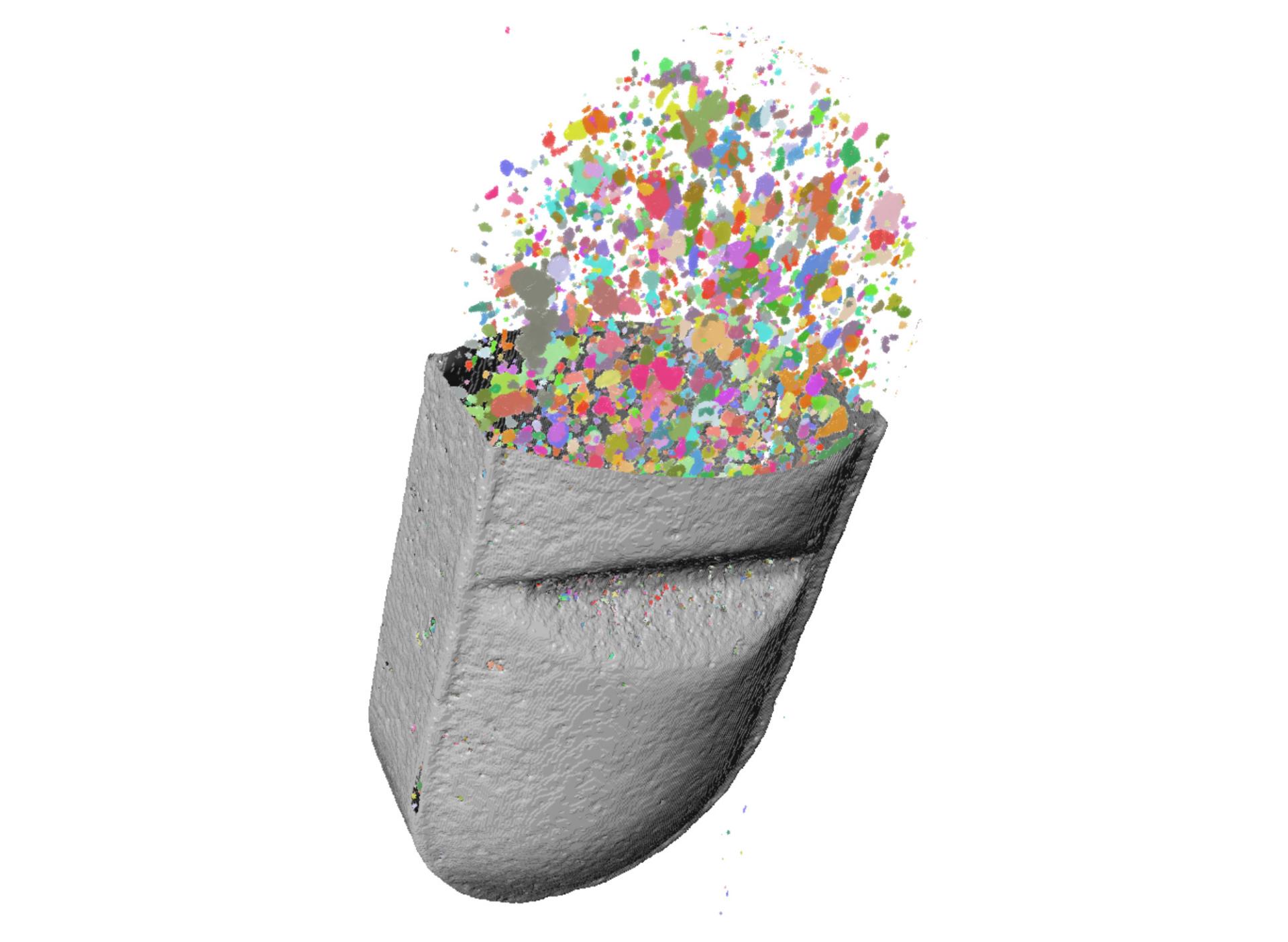
  • Three-dimensional rendering of recycled battery black mass with individual material phases segmented and color coded for analysis.

    Three-dimensional rendering of recycled battery black mass with individual material phases segmented and color coded for analysis.

    3D X-ray 현미경을 활용한 재료 연구

    금속, 복합재료 및 배터리 소재의 미세구조, 결함 및 열화를 비파괴 방식으로 특성화할 수 있습니다. in situ, 상관 분석 및 대용량 3D 이미징을 통해 내부 구조와 성능 간의 연관성을 확인할 수 있습니다.

  • 3D X-ray microscopy rendering of a smartphone system-on-chip package, with a green inset highlighting the central chip region.

    3D X-ray microscopy rendering of a smartphone system-on-chip package, with a green inset highlighting the central chip region.

    반도체 및 전자 분야의 고장 분석

    복잡한 패키지와 조립 구조를 3D로 검사하고 내부에 매립된 결함의 위치를 파악하며, 고장 분석 및 패키지 개발 과정에서 파괴적인 후속 분석을 수행하기 전에 중요한 증거를 보존할 수 있습니다.

  • Sample courtesy of Penn United Technologies Inc

    Three-dimensional X-ray rendering of a metal additively manufactured manifold revealing complex internal channels and lattice structures.

    Sample courtesy of Penn United Technologies Inc

    Three-dimensional X-ray rendering of a metal additively manufactured manifold revealing complex internal channels and lattice structures.

    비파괴 3D 이미징을 활용한 산업 연구

    파괴적인 시료 준비 없이 내부 구조, 결함 및 고장 메커니즘을 평가하고 3D 시료 정보를 제품 성능, 공정 개발 및 검증 워크플로우와 연계할 수 있습니다.

  • Three-dimensional rendering of an antihistamine tablet showing segmented internal particles and ingredient distribution.

    Three-dimensional rendering of an antihistamine tablet showing segmented internal particles and ingredient distribution.

    공간적 맥락을 유지하는 생명과학 3D 이미징

    생물학적 구조를 손상 없이 3D로 탐색하고 서로 다른 스케일에서도 공간적 맥락을 유지하며, 후속 현미경 연계 분석 워크플로우를 위한 관심 영역을 찾을 수 있습니다.

  • High-resolution X-ray cross-section of a sandstone sample showing pores, mineral grains, and internal microstructure.

    High-resolution X-ray cross-section of a sandstone sample showing pores, mineral grains, and internal microstructure.

    지질 및 광업 분야의 3D X-ray 현미경

    희귀 시료를 보존하면서 온전한 지질 시료 내부의 기공 네트워크, 광물 분포 및 내부 구조를 분석할 수 있으며 in situ 또는 유체 흐름 연구도 지원합니다.

ZEISS Xradia Ultra

연구실에서 탐구하는 나노스케일 X-ray 이미징

싱크로트론 X-ray 나노단층촬영은 나노스케일에서 비파괴 3D 이미징을 가능하게 하지만, 매우 제한적인 빔타임을 확보하기 위해 별도의 절차가 필요합니다. 하지만 더 이상 싱크로트론 사용 시간을 기다릴 필요가 없다면 어떨까요? 연구실에서 자체적으로 싱크로트론 수준의 기능을 활용할 수 있다고 상상해 보세요.

ZEISS Xradia Ultra 제품군은 나노스케일 분해능과 싱크로트론에 준하는 품질을 제공하는 3D 비파괴 X-ray 현미경(XRM)을 연구실에서 직접 활용할 수 있도록 합니다. 두 가지 모델 중에서 선택할 수 있습니다. ZEISS Xradia 810 Ultra와 ZEISS Xradia 800 Ultra는 모두 자주 사용하는 어플리케이션에서 최적의 이미지 품질을 얻을 수 있도록 설계되었습니다.

  • 시료의 원래 환경을 유지한 비파괴 이미징
  • 최대 50 nm의 공간 분해능과 16 nm voxel size를 구현하는 나노스케일 3D X-ray 이미징
  • 3D 및 4D in situ 실험
  • 나노구조의 정량화 및 모델링 입력 데이터로 활용
  • 경질 및 연질 재료 탐구
  • 에너지 소재

    리튬 이온 배터리

    Lithium ion battery cathode pore network and simulated diffusion through carbon binder domain. Imaged with Xradia 810 Ultra (Sample width 71 µm).

  • 엔지니어링 소재

    아연 입자

    In situ compressive indentation failure in a SiC:BN composite fiber. Imaged with Xradia 810 Ultra & Ultra Load Stage (Sample width 65 µm).

  • 폴리머 및 연질 소재

    엘라스토머

    Elastomer at different stages of compression during an in situ load stage experiment. (left: uncompressed, center: compressed, right: decompressed). Imaged with Xradia 810 Ultra.

  • 전자 분야

    구리 마이크로범프

    Copper microbump and interconnect visualization and defect inspection. Imaged with Xradia 800 Ultra (Sample width 52 µm).