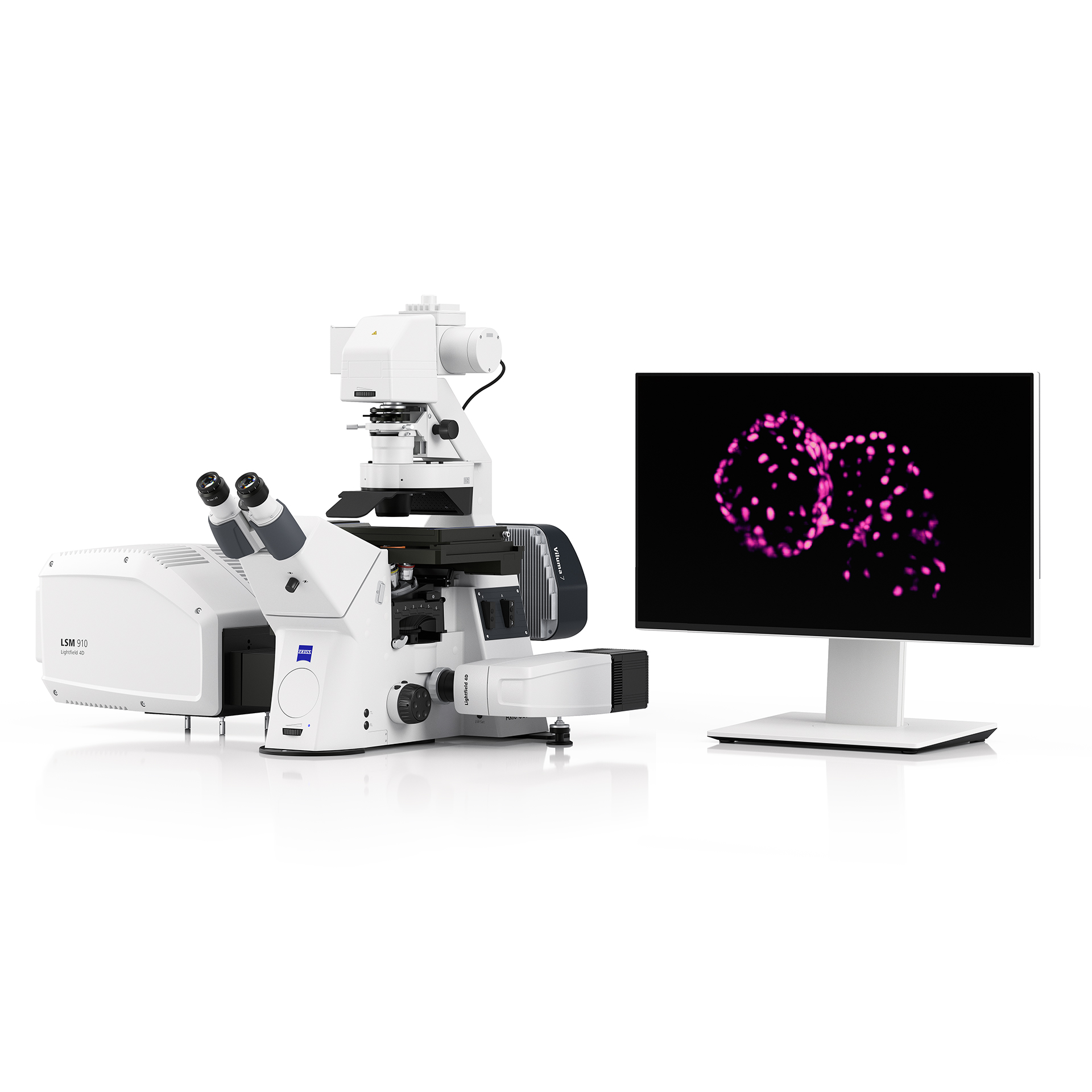
Product image of ZEISS LSM Lightfield 4D
upgrade promotion

Your LSM, Now in 4D

여러분의 LSM, 이제 4D로 확장하세요.

신규 장비 도입 없이 현재 보유한 LSM 900/980의 활용 범위를 초고속 볼륨 이미징으로 확장할 수 있습니다. Lightfield 4D로 빠르게 변화하는 생명 현상을 4D로 관찰하세요!

  • 기존 LSM 900/980 사용자를 위한 Lightfield 4D 업그레이드 패키지
  • Confocal의 활용 범위를 초고속 4D 이미징으로 확장
  • 빠르게 변화하는 생명 현상을 최대 80 volumes/s로 관찰
  • 한정 기간 제공되는 프로모션 혜택

ZEISS Lightfield 4D

살아있는 유기체의 고속 이미징을 위한 차세대 Confocal 현미경의 혁신

ZEISS Lightfield 4D는 살아있는 샘플의 빠른 3D 변화를 한 번의 촬영으로 포착하는 고속 볼륨 이미징 기술입니다. 시간에 따라 끊임없이 변화하는 생명 현상을 4D로 관찰하며, 기존 이미징 방식으로는 놓치기 쉬웠던 순간까지 생생하게 확인할 수 있습니다.

기존 LSM 900/980에 Lightfield 4D를 더하면 새로운 장비 도입 없이 현재 보유한 Confocal 시스템의 가능성을 초고속 4D 이미징으로 확장할 수 있습니다. 익숙한 LSM 플랫폼을 더 빠르고 부드러운 라이브 이미징 워크플로우로 업그레이드해 보세요!
ZEISS Lightfield 4D 주요 기능 한눈에 보기

  • ZEISS Lightfield 4D - Capture Spatial Signals & Fast Dynamics

    공간 신호 & 빠른 Dynamics 동시 포착

    시공간 정보를 온전히 유지하면서, 초당 최대 80 volumes의 속도로 유기체 전체의 가장 빠른 움직임을 포착하세요. . Lightfield 4D의 '한 번의 촬영, 하나의 Volume' 방식은 중요한 생체 활동을 놓치거나 왜곡 없이 정확히 기록합니다.

  • ZEISS Lightfield 4D - Gently Observe Organisms Over Time

    손상 없이, 장시간 Organisms 관찰

    단 한 번의 조명으로 전체 Z-stack을 획득하여 빛 노출과 광독성 영향을 최소화합니다. 이를 통해 살아있는 샘플도 높은 시간 해상도로 장시간 안정적으로 이미징할 수 있습니다.

  • ZEISS Lightfield 4D - Speed Up Acquisition & Throughput

    획득 속도 & 처리량 동시 향상

    Lightfield 4D의 탁월한 속도로 다채널 volume을 캡처하면 실험 생산성이 획기적으로 향상됩니다. 한 세션에서 이전보다 더 많은 샘플을 이미지화하고 분석하거나, 다양한 샘플 코호트를 효과적으로 비교해보세요.

  • ZEISS Lightfield 4D - Combine Light-Field with Confocal Imaging

    광학과 Confocal 이미징의 결합

    초고해상도 및 스펙트럼 이미징, 대용량 샘플의 optical sectioning, 분자 dynamics 측정 등과 같은 LSM의 다양한 기능에 Lightfield 4D의 저광독성·즉시 볼륨 이미징을 결합하여, 실험의 수준을 한 차원 끌어올리세요.

기존 LSM 900/980 사용자라면 누구나!

여러분의 Confocal을 4D로 확장하세요
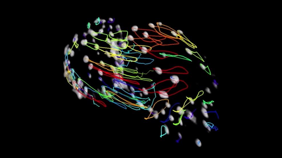
  • The movie shows 3 full heartbeats in 1.2 seconds, during which cardiomyocytes are temporally and spatially resolved

    Zebrafish 심혈관 발달 연구자

  •  Zebrafish blood flow

    Zebrafish 혈류 및 혈관 발달 연구자

  • Flow insect blood cells in the hemolymph of Drosophila melanogaster white pre-pupae

    Drosophila 면역세포 흐름 연구자

  • Sensitive 3D imaging of Drosophila muscle activation

    Drosophila 근육 활성 및 신경근 기능 연구자

  • Transgenic, 3-day old Arabidopsis thaliana hypocotyl

    식물 광반응 및 단백질 동역학 연구자

  • Drosophila 조직 형성 및 대사 발달 연구자

  • Zebrafish 감각기관 형태형성 연구자

  • Drosophila 배아 장기 발달 연구자

  • 3D spheroid 이미징 및 정량 분석 연구자

  • Cancer organoid 기반 약물 반응 연구자


  • 발달 중인 zebrafish 심장의 형태와 심장벽 움직임 관찰

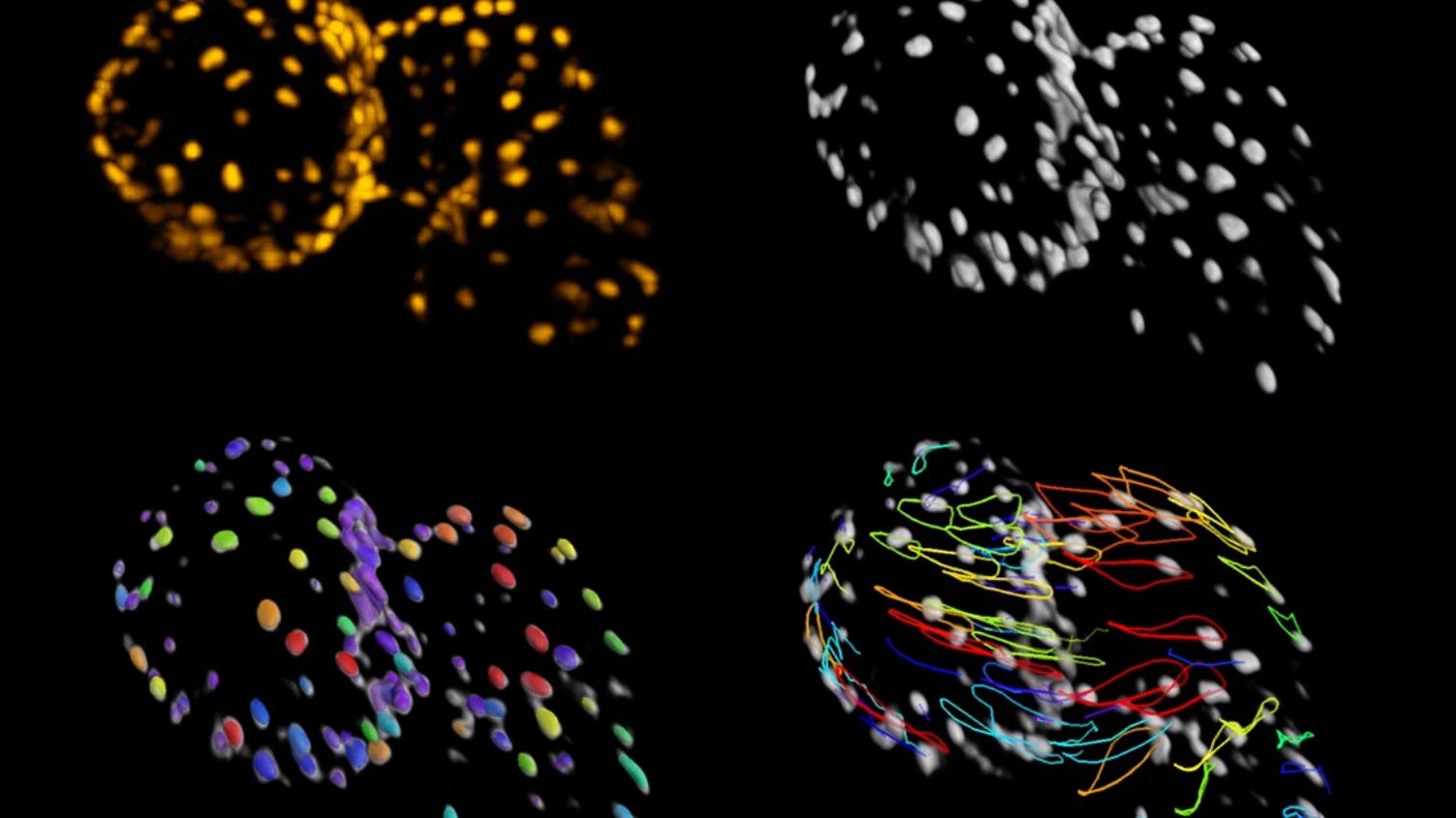
    Analyzing embryonic heart morphology and movement in 3D is challenging as the heart is continually beating. The data was recorded from a 3 days post fertilization zebrafish larvae embedded in agarose. ZEISS Lightfield 4D allowed to image the heartbeat with 80 volumes per second. The movie shows 3 full heartbeats in 1.2 seconds, during which cardiomyocytes are temporally and spatially resolved. This allows for cell segmentation and tracking using ZEISS arivis Pro. It is clearly visible that the cardiomyocytes follow exactly the same trajectory in every heartbeat.

    Courtesy of Stone Elworthy and Emily Noël, School of Biosciences, University of Sheffield, UK. Data acquired at Wolfson Light Microscopy Facility in the School of Biosciences at the University of Sheffield.

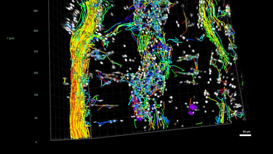
  • 발달 중인 zebrafish 배아의 꼬리 혈관을 따라 흐르는 적혈구 실시간 관찰

    Imaging the behavior of the circulatory system in real time in three dimensions is far not possible, as the heart pumps blood through the circulatory system at a speed of 3 beats per second, giving cells too fast of speeds to capture their movement in more than 2 dimensions at a time. Lightfield 4D with speeds up to 80 volumes per second offer the first opportunity to track blood cells throughout this network in 3D and visualize just exactly how this network is organized with respect to relative speed and directionality of blood in different vessels throughout the volume.

    Courtesy of Toby Andrews and Rashmi Priya, The Francis Crick Institute, London, UK

  • Drosophila 혈 림프 내 곤충 혈구세포 hemocyte의 흐름 관찰

    Investigating flow of hemocytes, the insect blood cells, through the hemolymph in vivo was almost impossible for researchers due to the fast three-dimensional movement. Lightfield 4D offers the unique opportunity to image a large volume fast enough to follow this process under physiological in vivo conditions. Thanks to the unparalleled imaging speed of 80 volumes per second, cells are reliably resolved both spatially and temporally. The acquired data allow subsequent segmentation and automated tracking using ZEISS arivis Pro.

    Courtesy of Iwan Robert Evans, University of Sheffield, UK. Data acquired at Wolfson Light Microscopy Facility in the School of Biosciences at the University of Sheffield.

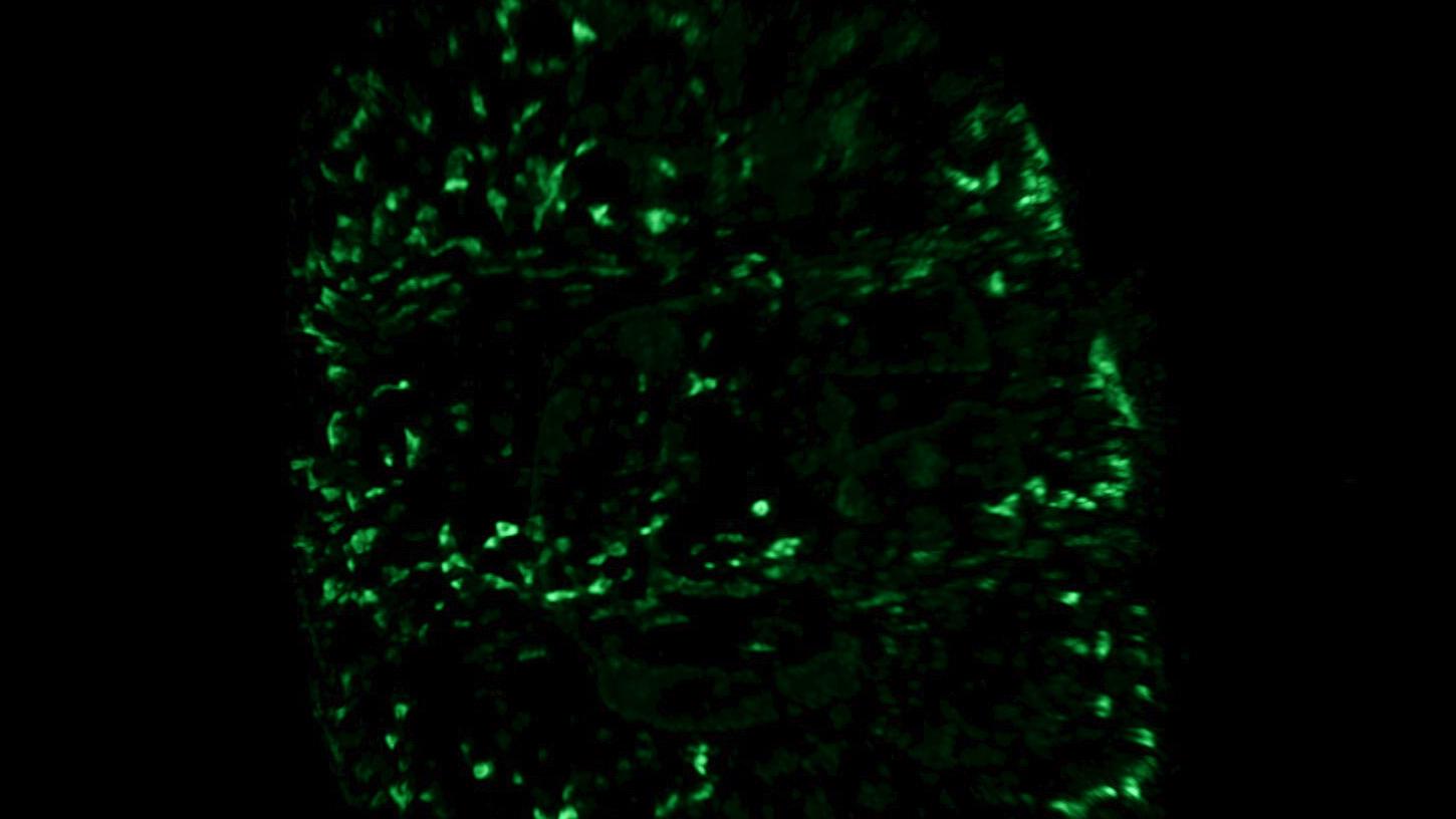
  • 민감한 Drosophila 근육 활성 3D 이미징

    Drosophila melanogaster 1st instar larvae, semi-constrained, expressing Calcium sensitive fluorescent proteins in all body wall muscles. Analysis with ZEISS arivis Pro to visualize muscle activity.

    Courtesy of Sean Sweeney, Department of Biology, University of York, UK

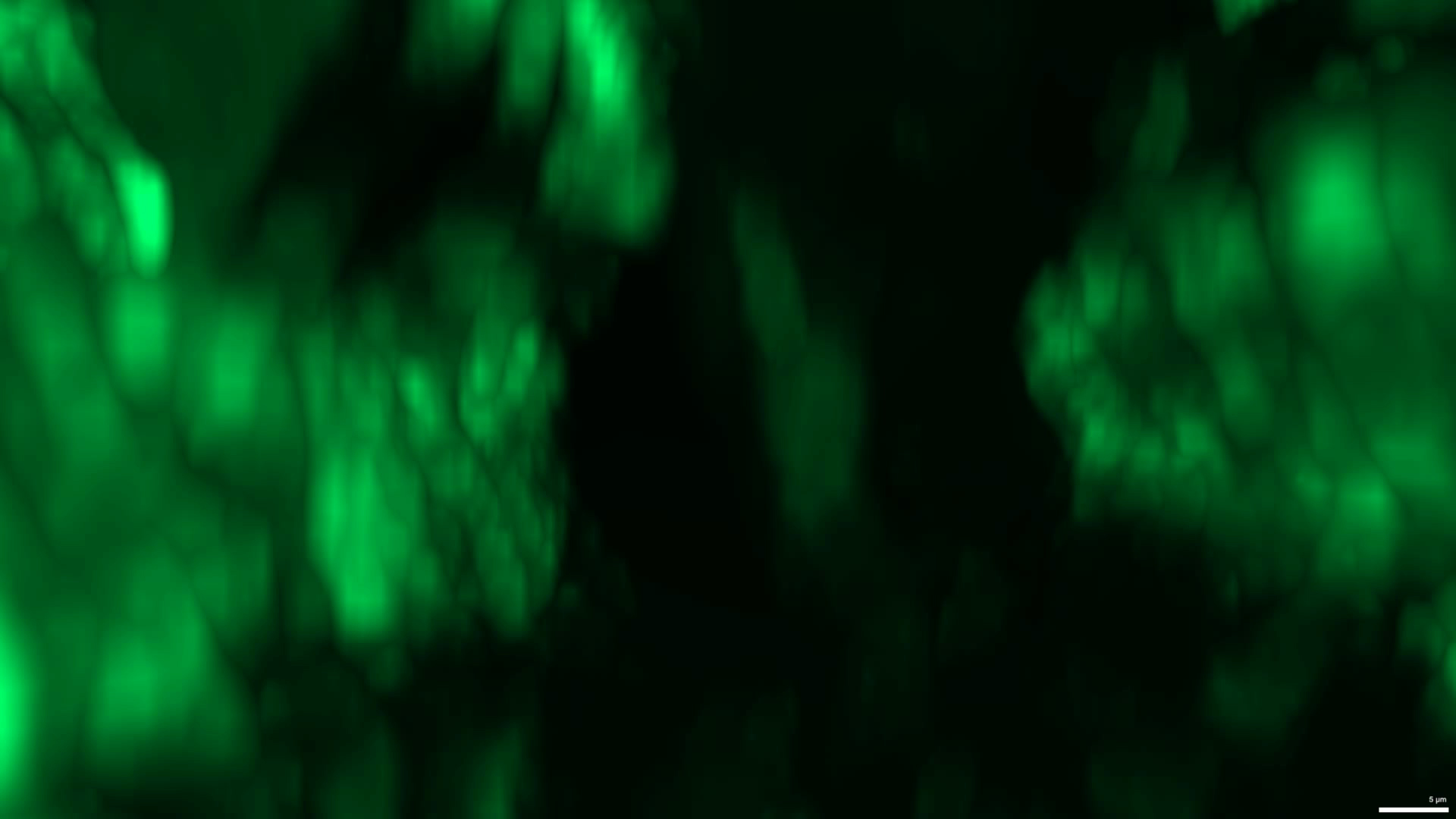
  • Arabidopsis thaliana에서 빛에 의해 유도되는 단백질 재분포 포착

    Investigating how plants respond to light appropriately on the molecular level is important for understanding how they carry out many of their light-dependent functions. In this situation, a re-localization of a mobile Arabidopsis thaliana protein upon blue-light stimulation is observed in the hypocotyl (stem) of a 3-day old seedling. The molecular dynamics are too fast for traditional confocal imaging. The high speed of Lightfield 4D enables an entire 3D volume to be imaged in a single snap, perfectly preserving the spatial localizations of protein aggregates. This provides reliable positioning information, which enables accurate reconstructions.

    Courtesy of Hannah Walters, Cellular Analysis Facility, MVLS-Shared Research Facilities, University of Glasgow. Data acquired at the Cellular Analysis Facility, University of Glasgow, UK

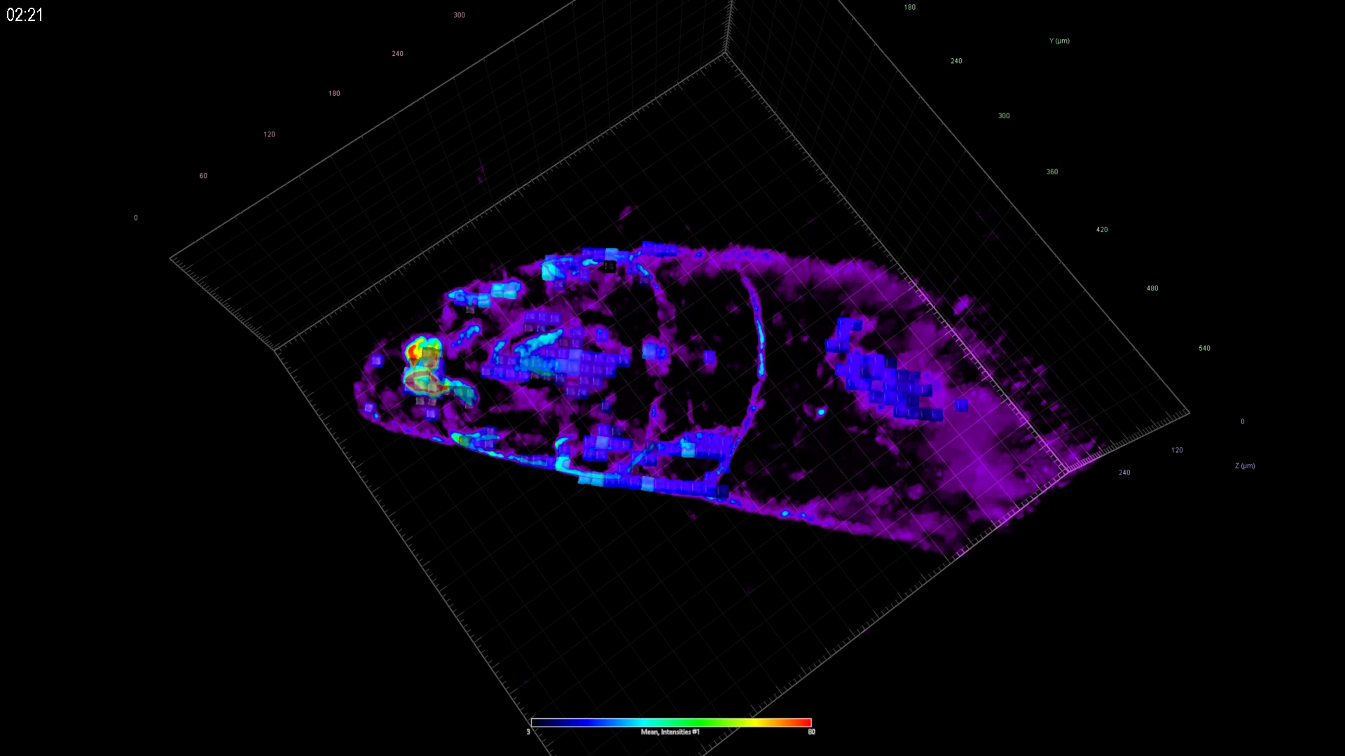
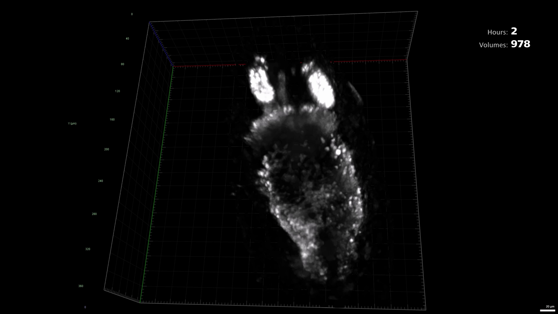
  • 발달 중인 Drosophila 번데기에서 지방체 조직 형성 관찰

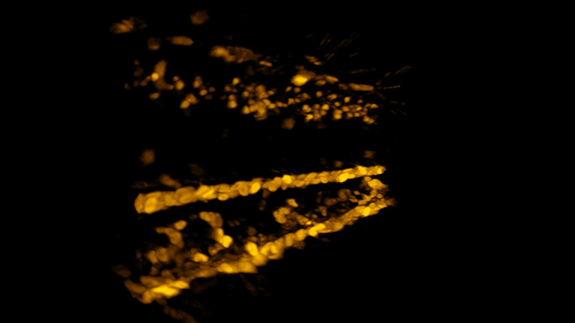
    Visualizing development of tissues and organs in intact animals enables better understanding of factors involved in their regulation and dysfunction. One example is the developing fat body forming during the pupal stage of Drosophila. Lightfield 4D allows to keep pace with the cell movement, to provide robust data for 4D tracking. Illumination is gentle enough to image overnight without sacrificing organism viability or fluorophore strength.

    15-hour overnight imaging including 12 positions and 10 animals, 500 ms exposure times per volume with 2-minute intervals.

    Courtesy of Ignacio Manuel Fernández Guerrero, Cellular Analysis Facility, MVLS-Shared Research Facilities, University of Glasgow. Data acquired at the Cellular Analysis Facility, University of Glasgow

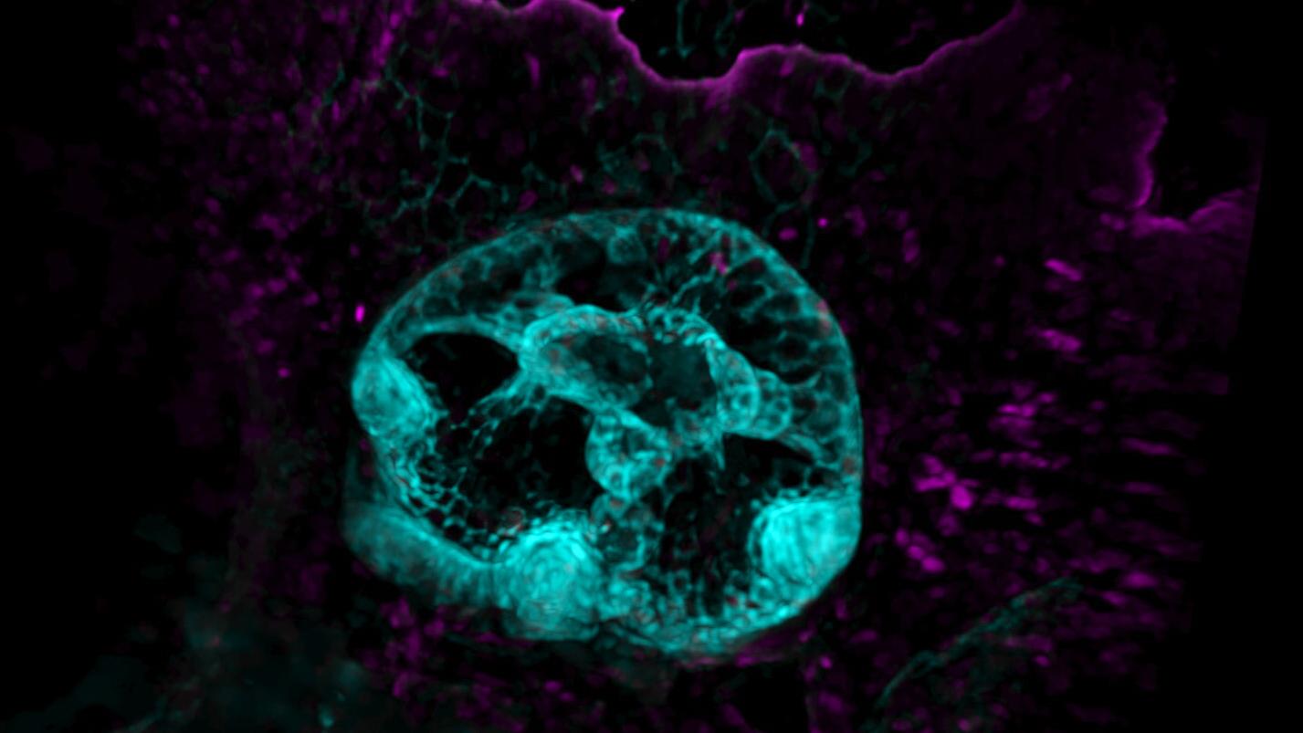
  • 발생 형태형성이 진행 중인 zebrafish 귀 관찰

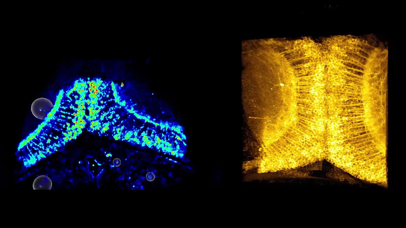
    Morphogenesis of developing organs requires a complex coordination of varying regulators and genomic elements. Lightfield 4D enables acquisition of light-sensitive processes with sufficient resolution to track epithelial cell morphological patterning. Its one-snap-one-volume imaging not only ensures that no developmental processes are missed or lost amidst the z-stacks but even allows multiple animals to be imaged in batch mode to capture all events and increase experimental throughput.

    Zebrafish embryo, timelapse movie of developing otic vesicle, 2 – 3 days post fertilization. Every 2 minutes, volumes of 4 different zebrafish embryo ears were imaged over the course of 16 hours.

    Courtesy of Tanya Whitfield, Sarah Baxendale, School of Biosciences, University of Sheffield, UK. Data acquired at the Wolfson Light Microscopy Facility, University of Sheffield.

  • Drosophila 배아의 장 발달 과정 추적

    The Drosophila embryonic midgut is particularly challenging to live image due to its depth combined with the different refractive indices of the eggshell and surrounding tissues. The current practice is to either use multiphoton microscopy and risk phototoxicity, or to image mutants in which the refractive index of the embryo is shifted and risk biological side effects during characterization.

    Lightfield 4D allows to image the formation of the midgut throughout much of embryogenesis without any risk of phototoxicity, and with unparalleled clarity. The dataset was recorded for close to 7 hours with 1 volume every 10 seconds.

    Courtesy of Andrew T Plygawko, School of Biosciences, University of Sheffield, UK. Data acquired at the Wolfson Light Microscopy Facility, University of Sheffield.

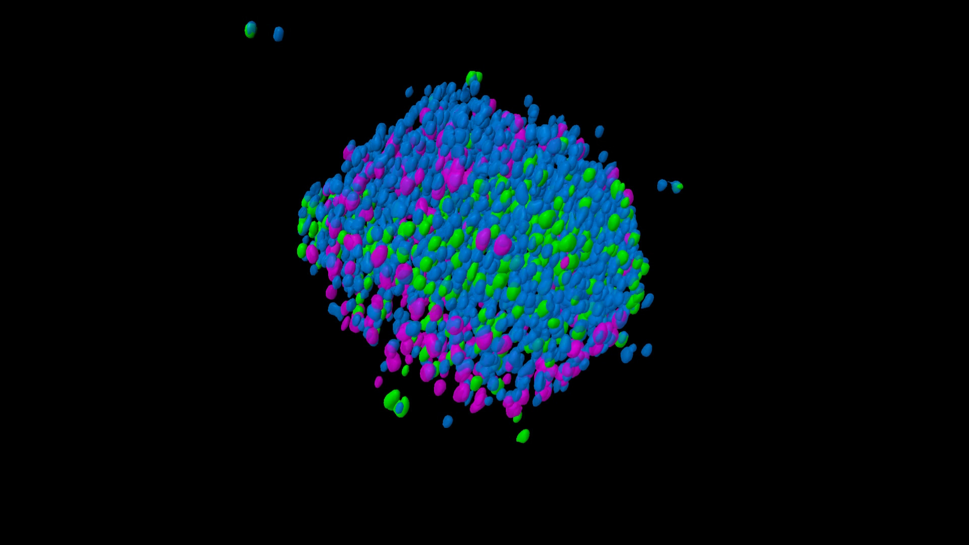
  • Cleared spheroid의 효율적인 볼륨 이미징 및 후속 세포 카운팅

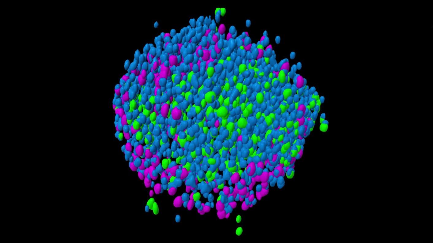
    Traditional acquisition methods like confocal point scanning or the use of spinning disk systems take considerable time for the acquisition of z-stacks. The speed of Lightfield 4D imaging enables advanced screening applications where higher throughput is required and facilitates faster screening of many spheroids under similar and different conditions, like in compound screens and drug treatments.

    Cleared spheroid of a co-culture of HCT-116-GFP (colon cancer) / NIH-3T3-RFP (fibroblasts) cells stained with Hoechst for nuclei. Imaged in an InSphero Akura plate. Dataset was segmented using arivis Pro.

    Sample courtesy of InSphero AG. Schlieren, Switzerland

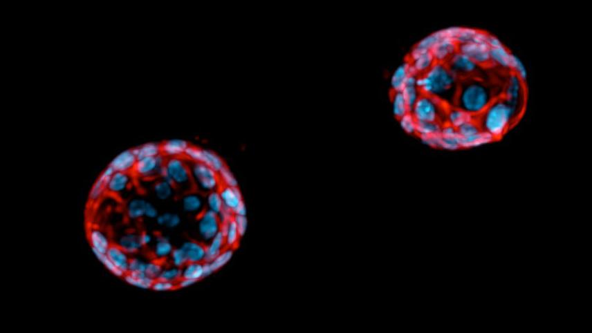
  • Perturbation 평가를 위한 cancer organoid 고속 이미징

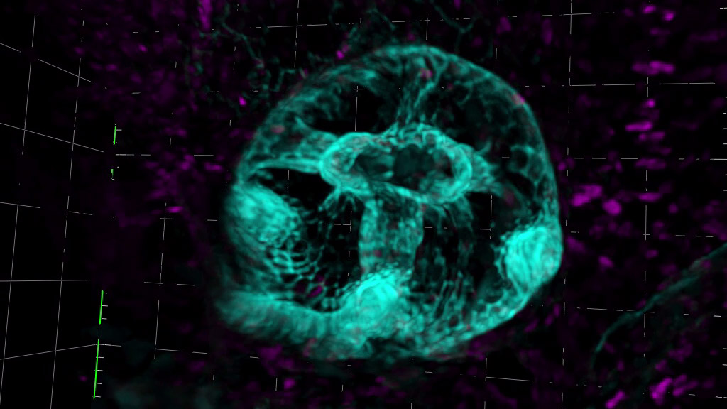
    Organoids are popular biological models for analyzing properties within cancer systems, such as responses to drug treatments, extracellular environments, and immune cell interactions. Image acquisition of such large 3D structures and screening through large sample sets is particularly time consuming. Lightfield 4D enables 3D image acquisition of organoids at a rate of several per second, dramatically increasing the throughput for screening through large numbers compared to traditional microscopy methods.

    Colorectal cancer organoids, actin cytoskeleton labelled with phalloidin (magenta), nuclei labelled with DAPI (blue). Captured with a 40× objective using 100 ms exposure time for each fluorophore.

    Courtesy of Nikki R. Paul, Cancer Research UK Scotland Institute, Glasgow. Data acquired at the Cellular Analysis Facility, University of Glasgow.

20% 할인된 금액으로 만나는

Lightfield 4D 특별 패키지
1 of 5

* 프로모션 기간: 2026년 6월 1일 - 2027년 5월 31일

  • 초고속 4D 이미징의 시작

    Lightfield 4D 업그레이드

  • 안정적인 성능을 자랑하는 최신 Light Source

    ZEISS Viluma 5 RGB-UV

  • 최적의 형광 이미징을 위한 Filter Set

    Filter Set 90 HE LED

  • 효율적인 멀티 채널 이미징을 위한 Reflector Module

    Reflector Module FL EC P&C

  • 새로운 가능성을 위한 첨단 워크스테이션

    최신 ZEN 소프트웨어와 NVIDIA RTX PRO4500 그래픽카드를 탑재한 신규 PC + 기존 PC를 분석용으로 전환시킬 수 있는 소프트웨어 업그레이드

First
Last

LSM 업그레이드로 새로운 연구의 가능성을 발견하세요!

아래 폼 작성하시면 자세한 상담 도와드릴게요.

Form is loading...

개인정보 수집·이용에 대한 동의

칼자이스㈜(이하 “칼자이스”)는 귀하의 개인정보 보호에 최선을 다하고 있습니다. 칼자이스는 개인정보보호법 및 관련 법률에 따라 고객의 개인정보를 다음과 같이 수집. 이용하고자 합니다.

1. 개인정보의 수집 및 이용 목적

마케팅/광고 활용(이벤트 등 광고성 정보전달 및 통계학 정보에 따른 서비스 제공 및 광고 게재)
고객의 요구사항을 이해하고 제품 및 서비스의 개선을 위해 개인정보를 칼자이스 코리아의 자회사(전세계 모든 자이스)와 공유할 수 있으며, 자회사는 이를 처리하고 저장할 수 있습니다.

2. 수집하는 개인정보

소속, 이름, 부서, 소속 유형, 연구 주제, 이메일, 연락처

3. 개인정보의 보유 및 이용기간

수집 및 이용에 관한 동의일로부터 (2) 년 또는 고객이 동의를 철회하는 때 개인정보를 보관합니다.

4. 개인정보의 제3자 제공
(1) 개인정보를 제공받는 자
- 칼자이스 독일 계열사 및 관계사
- 칼자이스의 대리점, 협력기관, 협력회사
(2) 개인정보 항목: 성명, 회사명, 부서, 이메일, 연락처, 회사 유형 및 지역, 연구 분야, 연구 주제
(3) 제공받는 자의 개인정보 이용목적: 칼자이스의 제품 및 서비스 제공, 영업활동, 고객관리
(4) 제공받는 자의 보유 · 이용기간: 제공의 목적이 달성된 때 또는 정보주체가 동의를 철회할 때까지

고객께서는 위 동의를 거부할 수 있으나, 만일 동의를 거부하시는 경우 칼자이스㈜와의 거래 및/또는 서비스가 이루어지지 않을 수 있음을 알려드립니다.

칼자이스코리아 ㈜