解鎖生命的架構

吳恭和 史丹佛大學生醫影像專家

ZEISS Cryo光電聯用技術深入解析生物結構看見真實生命樣貌
產品

ZEISS 關聯式冷凍工作流程

冷凍條件下的 TEM 薄片製備與3D影像分析

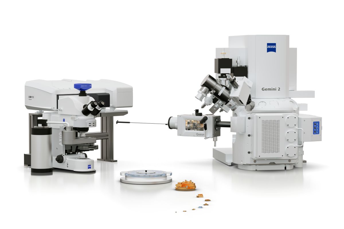
ZEISS 關聯式冷凍工作流程將寬場螢光顯微鏡、雷射掃描共軛焦顯微鏡,以及聚焦離子束掃描式電子顯微鏡(FIB-SEM)整合於一套順暢且易於操作的流程中。
此解決方案提供針對相關冷凍工作流程需求優化的軟硬體整合,涵蓋從螢光巨分子的定位,到高對比三維體積影像,再到用於冷凍電子斷層掃描的網格上薄片製備。

  • 無縫整合跨硬體冷凍工作流程
  • 高解析度螢光影像
  • 高對比3D影像與三維重建
  • 用於冷凍穿透式電子顯微鏡的網格上薄片製備
  • 同時支援冷凍與常溫樣品的多功能應用

冷凍寬場與共軛焦顯微成像

Cryogenic Widefield & Confocal Microscopy

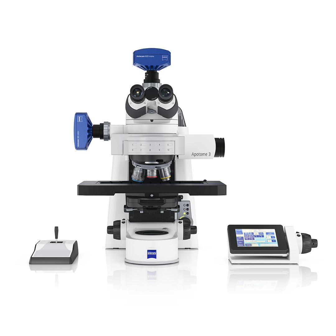
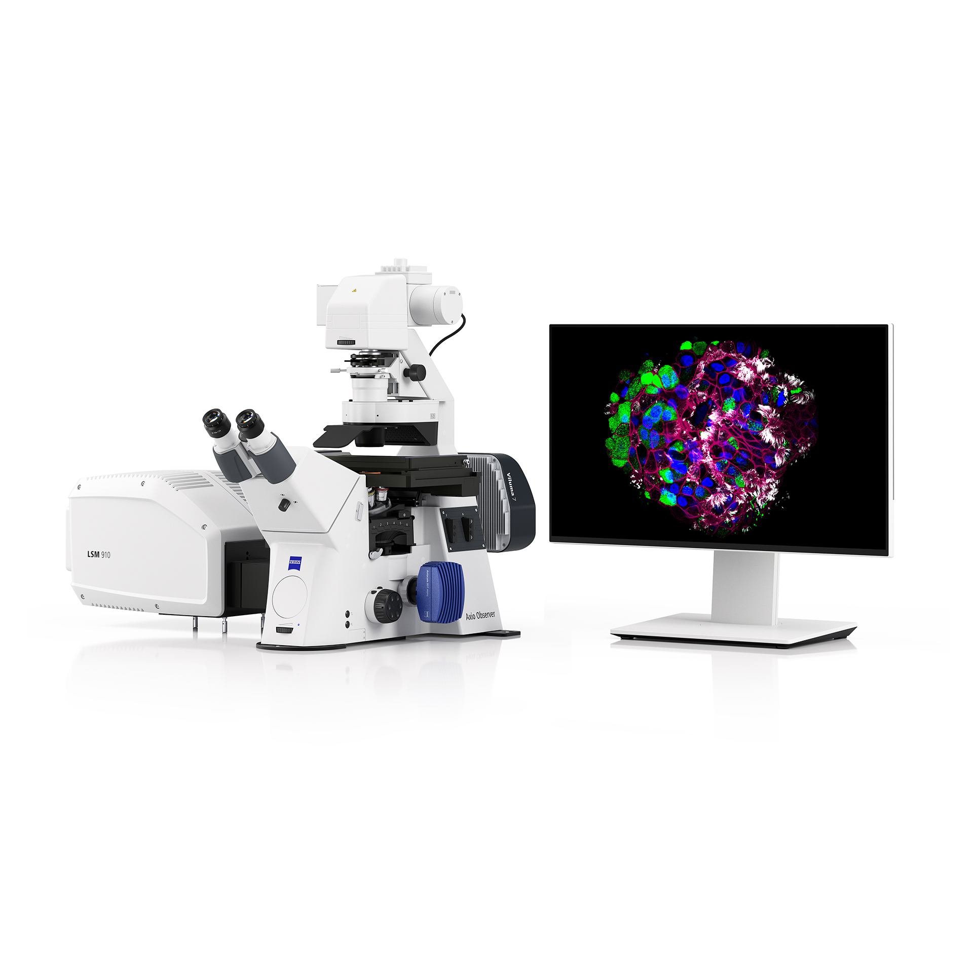
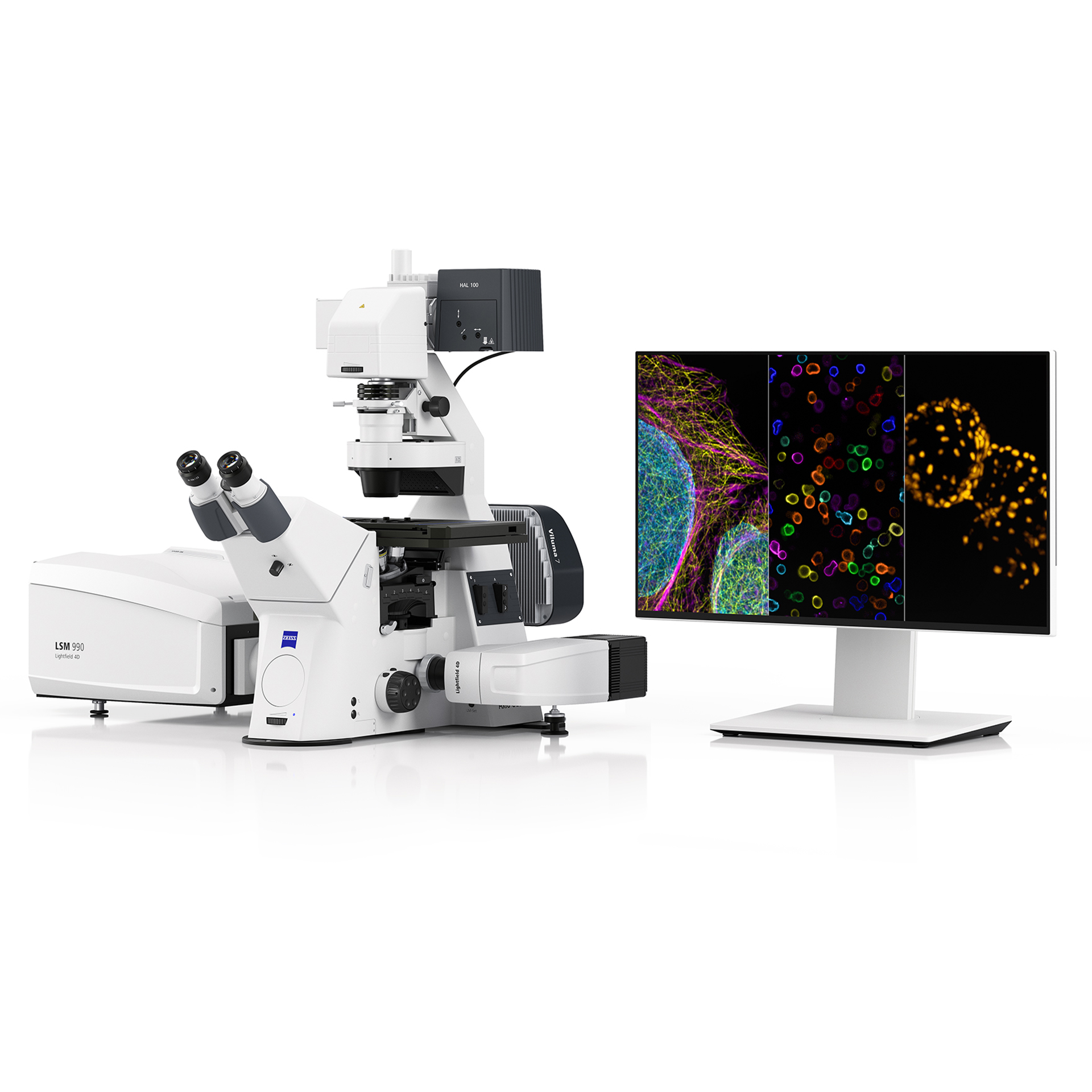
ZEISS Axio Imager

  • ZEISS Correlative Cryo Workflow 的核心光學顯微鏡平台
  • 可搭載 Linkam CMS196 V4 冷凍載台

靈活配置選項

  • 寬場顯微系統(Widefield)
    搭配 Apotome 3 → 可取得高品質 3D 影像
  • 共軛焦系統(Confocal)
    LSM 910 / 990 + Airyscan 2 → 提供高解析度成像

多功能應用

  • 寬場與 LSM 系統皆為多用途平台
  • 可快速於「冷凍(cryo)」與「常溫」實驗之間切換
  • 切換過程 不影響影像品質

冷凍 SEM 體積影像與 TEM 薄片製備

Cryogenic SEM Volume Imaging & TEM Lamella Preparation

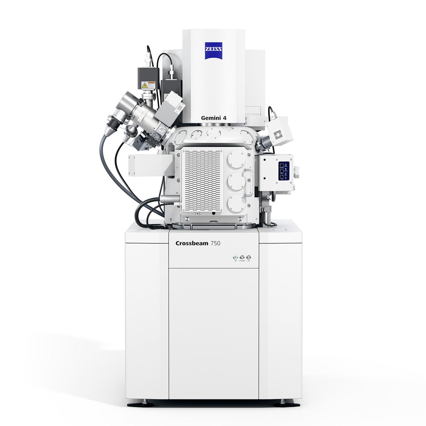
ZEISS Crossbeam FIB-SEM

  • 高使用便利性(usability)
  • 優異影像對比(contrast)

卓越低溫成像能力

  • 即使是未染色的冷凍玻璃化樣品(vitrified samples)仍可獲得高對比影像
  • 支援:細胞與組織超微結構觀測 + 清晰辨識細胞內區域

即時製程監控

  • 可同時觀察影像與 milling 過程
  • 精準控制 FIB 切削
  • 支援 TEM 薄片網格上薄片製備
  • 支援薄片 lift-out

多用途平台

  • 可於冷凍與常溫之間切換 
  • 可搭配元素定性分析 EDS

ZEISS Correlative Cryo Workflow 一覽

Advantage

  • HPF carriers with vitrified samples (click for more details); Image Courtesy: Anat Akiva & Nico Sommerdijk, Radboud University Medical Center, The Netherlands

    HPF carriers with vitrified samples (click for more details); Image Courtesy: Anat Akiva & Nico Sommerdijk, Radboud University Medical Center, The Netherlands

  • TEM grids with vitrified samples. (Click for more details)

    TEM grids with vitrified samples. (Click for more details)

樣品品質評估與損傷預防

冷凍顯微技術中常見的挑戰包括樣品流失、冰污染以及去玻璃化。ZEISS Correlative Cryo Workflow 專為保護珍貴的玻璃化樣品而設計,可有效減少在高精度流程中可能發生的各種風險。

結合 ZEISS LSM/Airyscan 與 ZEISS Crossbeam 的高解析及高對比影像能力,可在低溫條件下操作且同時大幅降低樣品流失或損壞的風險。

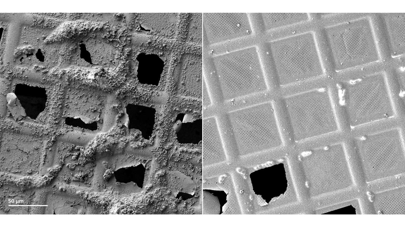
在進入工作流程之前,樣品的玻璃化本身即是一大挑戰。儘管玻璃化技術近年已有進展,樣品仍常被厚冰層覆蓋,或僅部分玻璃化,並出現結晶態的冰區域。不良的玻璃化會破壞細胞與組織的超微結構,而這些區域通常只能透過 TEM 才能辨識——除非在工作流程初期,光學顯微鏡或 FIB-SEM 已具備樣品品質評估能力。

ZEISS LSM 可透過螢光及反射光成像方式提供此類評估能力;同時,ZEISS Crossbeam 傑出的對比表現也能可靠地判斷樣品品質。這不僅能節省寶貴時間,更能提升整體實驗效率。

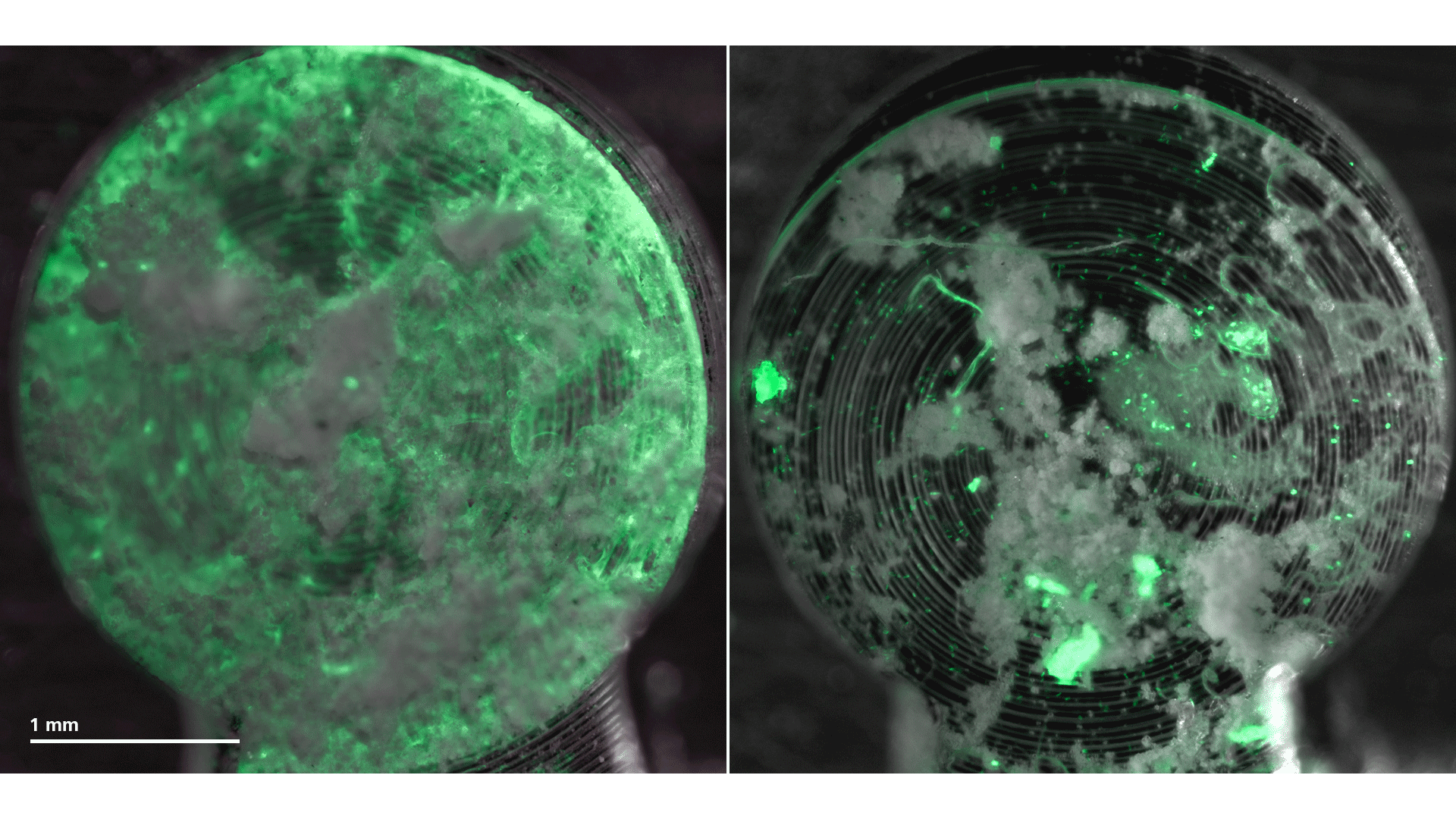
Plunge-frozen Hela cells (Histone 2-GFP labeled) illustrating ideal conditions for further imaging. The ice layer is around 6.8 μm thick and covers the vitrified cells. These cells are perfectly suited for further FIB-SEM analysis.

冰層厚度量測與高效率目標區域定位

冰厚量測對於評估樣品品質,以及在玻璃態冰包埋的樣本中定位目標細胞至關重要。透過光學顯微鏡,即可輕鬆完成樣品驗證;反射光與共軛焦螢光成像能提供初步的品質判斷,並協助精準定位具潛力的細胞區域。

當螢光訊號呈現如蜘蛛網狀模糊的圖案時,通常代表冷凍品質不佳。此外,即使是在同一個急速冷凍樣本中,也可能存在不同的冷凍品質與保存狀態。透過掌握冰層厚度與冰品質資訊,可在進入下一階段關聯式冷凍工作流程前,先行進行細胞的快速篩選,大幅節省後續時間與資源。

Application

  • LM and EM dataset – from the grid overview to the region of interest identified for further TEM tomography
  • FIB image of the prepared lamella; lamella thickness: 230 nm

  • Segmented and reconstructed tomogram

細胞生物學

稀有事件的辨識

紡錘極體(spindle pole bodies)在酵母細胞中相當難以定位。它們體積微小,且出現頻率低。ZEISS 的 Correlative Cryo Workflow 可讓您在接近原生狀態(near-to-native state)下,精準識別並成像這類細胞結構。搭配配備 Airyscan 檢測器 的 LSM,可使這些結構的辨識更加容易,並進一步取得更多細節影像。從整個細胞的大範圍總覽,到這些微小結構的高解析影像,所有資料皆整合於 ZEN Connect 專案中,提供後續在 ZEISS Crossbeam 中重新定位這些細胞結構所需的完整資訊。

透過 ZEISS Crossbeam,可對已識別的區域進行 TEM 薄片製備,以供cryo electron tomography (cryo-ET) 使用,同時也支援3D成像。此外,此工作流程允許在影像取得後串聯所有資料,例如將 Crossbeam 所獲得的影像或 TEM 的斷層影像(tomograms)與 LSM 多通道影像整合,並在三維空間中進行呈現。

Yeast cells labeled with NUP (nuclear pore complex)-GFP and CNM67-tdTomato.
Sample and tomogram courtesy of M. Pilhofer, ETH Zürich, Switzerland

  • Overlay of a high-resolution LSM/Airyscan image with a high-contrast Crossbeam image acquired under cryogenic conditions. The overlay was done with ZEN Connect.
  • 3D volume reconstruction of yeast cells and segmentation of the nucleus (dark blue) as well as several mitochondria.

細胞生物學

關聯式3D影像

當在 LSM 系統中辨識出紡錘極體(spindle pole bodies)等細胞結構後,ZEISS Crossbeam 憑藉其卓越的影像品質,可進一步進行目標定位,並透過冷凍3D成像分析其超微結構。即使在低加速電壓條件下,Crossbeam 仍能對未染色、玻璃化(vitrified)的樣品提供高對比影像,同時有效保護樣品免於損傷。LSM 所取得的高解析影像結合 Crossbeam 的高對比影像,使得影像能進行精準的疊合。透過 ZEN Connect 在 Crossbeam 系統中重新定位目標區域後,即可針對已識別的細胞取得 3D 影像。在此關聯式3D影像中,成功鎖定兩個紡錘極體。依據 FIB 切削方向,在高對比影像中可清晰辨識單一微管(microtubules)的排列方向。此外,3D 影像中亦可辨識其他細胞組成結構。

Sample courtesy of M. Pilhofer, ETH Zürich, Switzerland

癌症研究

癌細胞常呈現出明顯的粒線體分裂表型,這可能也是其對藥物產生抗性的原因之一。然而,傳統的化學固定方法往往會產生贗像 (artifact),例如粒線體的異常聚集,進而可能被誤判為分裂事件。冷凍固定則可避免這些贗像 (artifact),並能在接近原生的狀態下保存樣品。

本案例展示了藍寶石片上的腺癌細胞,經由投入式冷凍固定。LSM 影像已顯示出密集的粒線體網絡與增加的分裂現象,而這一結果進一步由 Crossbeam 影像確認。 在完成 LSM 與 Airyscan 成像後,玻璃化樣品被轉移至 Crossbeam。透過 ZEN Connect,研究人員可重新定位目標區域、在不同影像之間進行疊合,並有效整合與管理所有取得的影像。

  • Stomata and internalized plastids were identified with an LSM using the autofluorescence of the sample. The selected stoma was re-located and imaged with the Crossbeam.

    Stomata and internalized plastids were identified with an LSM using the autofluorescence of the sample. The selected stoma was re-located and imaged with the Crossbeam.

  • Stromules are clearly visible in the section planes acquired with the Crossbeam.

    Stromules are clearly visible in the section planes acquired with the Crossbeam.

  • Stromules are clearly visible in the section planes acquired with the Crossbeam.

    Stromules are clearly visible in the section planes acquired with the Crossbeam.

  • 3D reconstruction and segmentation of the FIB image stack reveals the morphology of the plastids. The reconstruction shows stromules closely interacting with mitochondria.

植物科學

植物對不斷變化的環境條件(例如鹽度升高)的反應,是植物科學中的一項重要研究課題。當植物適應這些變化時,通常會表現出壓力反應。其中一種可在超微結構層級觀察到的現象,是所謂的基質小管 (stromules),即葉綠體中延伸出的細長管狀結構。

ZEN Connect 專案展示了多模態的影像:首先利用 LSM 透過樣品的自體螢光來定位氣孔與內部的葉綠體。在成功重新定位目標區域後,將 LSM 影像與所選氣孔的 SEM 概覽影像進行疊加。接著取得該氣孔的 3D 影像。由 Crossbeam 取得影像顯示,在葉綠體中觀察到 stromule 形成增加的現象。

Sample courtesy: B. Franzisky, University of Hohenheim, Germany

  • Top: The worm was imaged under cryogenic temperature with an LSM / Airyscan system before freeze substitution. Bottom: The embedded and stained worm was then imaged with a Crossbeam.

    Top: The worm was imaged under cryogenic temperature with an LSM / Airyscan system before freeze substitution. Bottom: The embedded and stained worm was then imaged with a Crossbeam.

  • Reconstruction of cellular structures such as an autophagosome (AP) or the genome in different mitotic phases (*cell in metaphase, # cell in telophase).

    Reconstruction of cellular structures such as an autophagosome (AP) or the genome in different mitotic phases (*cell in metaphase, # cell in telophase).

  • 3D reconstruction of cellular structures

生物學研究

C. elegans 有絲分裂細胞的發育生物學研究

完整的 C. elegans 線蟲以高壓冷凍(HPF)固定,並利用冷凍螢光顯微鏡在原位(in situ)對處於中期分裂(metaphase)的胚胎細胞進行成像。篩選後的樣本再經由冷凍置換進行重金屬染色,隨後嵌入樹脂並切片,使相同體積的區域能夠被再次定位,並透過 Crossbeam 進行高解析度、高對比度的成像。透過此工作流程,成功地重建了目標的中期分裂細胞。此外,此方法也促成了意外的發現:在鄰近區域觀察到一個具有吸引力的點狀螢光訊號,經比對後推測可能對應於自噬體(autophagosome)。

因此,對經高壓冷凍處理的厚樣本進行冷凍螢光顯微成像,可在接近天然狀態下捕捉並觀察短暫存在的細胞結構;再配合適當的後續處理與關聯式體積電子顯微(correlative volume EM)技術,便能以高解析度與三維方式重建這些目標結構。

Courtesy of Kedar Narayan, National Cancer Institute / NIH and Frederick National Laboratory for Cancer Research, USA

Downloads

  • ZEISS Correlative Cryo Workflow

    Your Solution for TEM Lamella Preparation and Volume Imaging under Cryogenic Conditions

    7 MB


Contact us

Form is loading...

If you want to have more information on data processing at ZEISS please refer to our data privacy notice.