

Solutions and workflows built for the clinical lab
Discover ZEISS microscopy solutions designed around the realities of routine clinical work. From trusted optics and efficient digital documentation to ergonomic viewing and application-focused configurations, Axiolab 5 and Axioscope 5 help laboratories build microscopy workflows around the way their teams work.


A clinical microscope has to get a lot right.
Hours at the eyepiece. Detailed observation. Consistent illumination and color. Documentation, consultation, and teaching. These are the everyday demands that shape the way ZEISS clinical microscopes are designed.


Two smart microscopes, matched to how you work.
Both platforms bring Smart Microscopy and ergonomic design to routine clinical microscopy. Axiolab 5 provides a compact, flexible platform across six clinical disciplines, while Axioscope 5 extends the portfolio with premium optics, a larger field of view, and expanded co-observation capabilities for pathology and histology workflows.


Explore products by discipline
Learn more about ZEISS Axiolab 5 and Axioscope 5 configurations designed around the needs of different clinical workflows. Select your application to explore recommended microscopy solutions, compare platform capabilities, and find the configuration that fits your laboratory.Axiolab 5 • Axioscope 5
Configured for routine pathology review, consultation, collaboration & teaching.- Brightfield imaging for routinely stained tissue sections.
- Smart Microscopy for streamlined digital documentation.
- Ergonomic viewing options for extended microscope use.
- Co-observation configurations for consultation, collaborative review & teaching.


Axiolab 5 · Axioscope 5
Stained tissue-section review, documentation & extended microscope use.- Premium optics.
- True-color brightfield imaging for stained tissue sections.
- Smart Microscopy for streamlined digital documentation.
- Ergonomic viewing options for extended microscope use.
- Larger field of view available with Axioscope 5.


Axiolab 5
From brightfield to fluorescence.- Brightfield, phase contrast, darkfield & fluorescence options.
- Digital imaging compatibility for routine documentation.
- Oil-immersion objective options for detailed specimen observation.
- Ergonomic viewing options for extended microscope use.


Axiolab 5
Blood smears and bone marrow, viewed in true color.- True-color brightfield imaging for stained blood and bone marrow preparations.
- 10× and 100× oil objectives in the Standard configuration; 10×, 50× oil, and 100× oil in Advanced configurations.
- Smart Microscopy for streamlined digital documentation.
- Ergonomic viewing options available for extended microscope use.


Axiolab 5
See urinary sediment in a different light.- Brightfield & phase contrast for urinary sediment review.
- Polarized light with lambda plate functionality for crystal evaluation.
- Ergonomic viewing options available for extended microscope use
- Smart Microscopy for streamlined digital documentation.


Four things every clinical setup is built on
Faithful visualization, streamlined documentation, ergonomic operation, and shared viewing when collaboration matters. These are the capabilities designed into ZEISS clinical microscopy solutions.


Smart Microscopy
Document without leaving the standFrame your specimen, press Snap, and capture an image directly from the microscope with scale information and metadata.


Ergonomics
Designed for comfortable microscope operationAdjustable viewing options and thoughtfully positioned controls help users adapt the microscope to their preferred working position during extended microscope sessions.


Visualization
See your specimen in true colorZEISS optics, LED illumination, and imaging components are designed to support consistent visualization across magnifications and documentation workflows.


Co-Observation
See the same field, togetherFor consultation, teaching, and collaborative review, co-observation configurations allow multiple viewers to observe the same specimen field with consistent orientation and illumination.
Image capture in three simple steps
Frame, focus, and capture directly from the microscope stand.


Microscopes designed around the way you work.
Extended microscope sessions place demands on more than optics. Adjustable viewing options, accessible controls, and intelligent illumination help users configure the microscope around their working position and routine.


Faithful visualization from eyepiece to image
True-color illumination, consistent brightness across magnifications, and ZEISS optical systems support faithful specimen visualization, with additional contrast and fluorescence options available for specific workflows and configurations.


Compatibility with staining and contrast techniques
Clinical specimens may be transparent or nearly colorless in transmitted light. Staining adds color and contrast to cellular and tissue structures, while optical contrast techniques, including phase contrast, polarization, and fluorescence, provide additional ways to visualize unstained specimens, birefringent materials, and fluorescently labeled targets. ZEISS Axiolab 5 and Axioscope 5 support a range of contrast methods depending on configuration. Available methods vary by platform and setup, talk with an imaging specialist to match the appropriate capabilities to your workflow.
-
Brightfield Staining
Brightfield microscopy is widely used with stained clinical preparations. Staining provides color and contrast to cellular and tissue structures, while ZEISS true-color LED illumination supports natural-color visualization and documentation across commonly used stains.


Wright / Giemsa (Romanowsky)
Commonly used with blood smear and bone marrow preparations for visualization of cellular morphology.

Courtesy of Annekatrin Richter, Leibniz Institute on Aging, Fritz Lipmann Institute e.V. (FLI), Jena, Germany. Hematoxylin & Eosin (H&E)
Provides characteristic contrast between nuclei, cytoplasm, and surrounding tissue structures for visualization of tissue architecture and cellular morphology.

Papanicolaou (Pap)
Multi-dye cytology stain used to visualize cellular morphology and detail in cytological preparations.

Gram Staining
Provides characteristic staining contrast for visualization of Gram-stained bacterial preparations.
-
Phase Contrast
Phase contrast converts differences in refractive index into visible contrast, supporting visualization of transparent, unstained specimens that can be difficult to see in brightfield.

Urinary sediment
Unstained urinary sediment viewed with phase contrast and brightfield (400x). Drag to compare.

Courtesy of Gerhard Ey Praxis Dr. Holthaus und Partner Lemförde, Germany Microorganisms
Supports visualization of unstained microorganisms, including bacteria, fungi, and protozoa.
-
Fluorescence
LED fluorescence provides bright, stable excitation without lamp warm-up or cool-down. Depending on configuration, Axiolab 5 and Axioscope 5 support fluorescence imaging across a range of excitation wavelengths and fluorophores. Examples used in clinical laboratory workflows include:

Dr. med. Harald Hoffmann, WHO - Supranationales Referenzlabor IML Gauting, Germany Auramine (acid-fast fluorescence)
Fluorescence visualization of auramine-stained acid-fast bacilli.

FITC-labeled antibodies
Fluorophore-conjugated antibodies provide fluorescent contrast for labeled targets in immunofluorescence workflows.
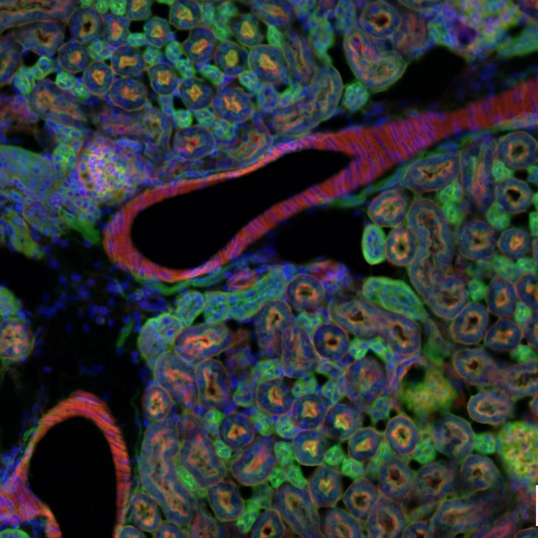
DAPI
Fluorescent nuclear counterstain used to visualize cell nuclei in tissue and cell preparations.
-
Polarization
Polarized light provides contrast for birefringent materials that may be difficult to distinguish in brightfield alone. Depending on configuration, Axiolab 5 supports polarized-light observation for specimens including crystals and stained tissue deposits.

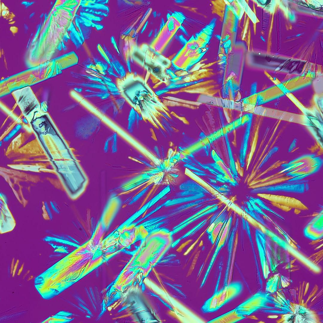
Urine crystals
Polarized-light observation provides contrast for birefringent crystals in urine sediment.
Birefringent tissue deposits
Congo red-stained tissue can be observed under polarized light to visualize birefringent structures.


One field of view. Every observer, together.
Both clinical stands support ZEISS multidiscussion configurations for shared observation. Setups range from face-to-face viewing to larger rows, U-form, or square configurations, depending on the platform and number of observers.


Axiolab 5 vs Axioscope 5, side by side
Both platforms share ZEISS Smart Microscopy, ergonomic design options, digital documentation, and a broad range of contrast techniques. Compare the features, optical capabilities, and clinical configurations available for each platform.


|


|
|
|---|---|---|
|
Compare |
ZEISS Axiolab 5 |
ZEISS Axioscope 5 |
|
Clinical Portfolio |
Configured for: pathology, histology, microbiology, Cytology, hematology, urology |
Configured for: pathology and histology |
|
Configured Workflows |
15 |
5 |
|
Contrast Methods *Available capabilities depend on system configuration. |
Brightfield, phase contrast, darkfield, polarization, fluorescence |
Brightfield, phase contrast, DIC, darkfield, polarization, fluorescence |
|
Fluorescence *Available capabilities depend on system configuration. |
LED fluorescence available depending on configuration |
LED fluorescence available depending on configuration; multichannel options available |
|
Documentation |
Smart Microscopy with integrated Snap controls, automatic scaling and metadata; operation without a PC supported |
Smart Microscopy with integrated Snap controls, automatic scaling and metadata; operation without a PC supported |
|
Field of View |
Up to 23 mm |
Up to 25 mm |
|
Ergonomics |
Ergonomic viewing and operating options, including adjustable tubes and accessible controls |
Ergonomic viewing and operating options, including adjustable tubes and accessible controls |
|
Co-observation |
Active two-position co-observation and multidiscussion configurations for up to 20 co-observers |
Active two-position co-observation and multidiscussion configurations for up to 20 co-observers |
Not sure which fits? Start from your discipline in the portfolio above, or talk it through with an imaging representative.


