

Microscopy solutions for Cytology
Prepared, stained, ready to review.From Papanicolaou and Romanowsky-stained preparations to exfoliative cytology, fine-needle aspirates, and fluid specimens, cytology microscopy supports visualization and morphological review of prepared cytology specimens across diverse sample types. ZEISS microscopy solutions combine high-quality brightfield imaging, ergonomic viewing options, and digital documentation capabilities to support daily cytology workflows.
Cellular detail across diverse specimens and stains
Cytology workflows encompass varied specimen types, preparation methods, stains, and magnifications. Microscopy supports visualization and morphological review of prepared cytology specimens while meeting the demands of routine slide examination.


Configure Axiolab 5 for your cytology workflow
Two configurations, one Axiolab 5 platform. Choose Standard for routine brightfield cytology or Standard Ergo for the same imaging capabilities with an ergonomic viewing setup for extended microscope use. Both combine ZEISS optics, a 2.5×–100× objective range, and digital imaging compatibility.


One Axiolab 5 across your cytology workflows
From Papanicolaou-stained preparations to fine-needle aspirates and fluid specimens, Axiolab 5 delivers trusted ZEISS optics across your routine cytology workflows, with Standard and Standard Ergonomic configurations to match the preparations, stains, and working needs in front of you.
Built around your cytology workflow
Find the cytology workflow that sounds like yours and explore the Axiolab 5 configuration designed to support it.


Examine stained preparations
True-color brightfield imaging supports visualization of nuclear and cytoplasmic morphology across Papanicolaou-stained cytology preparations.


Visualize Romanowsky-stained preparations
Brightfield imaging supports visualization of cellular morphology and characteristic staining in Romanowsky-stained cytology preparations.
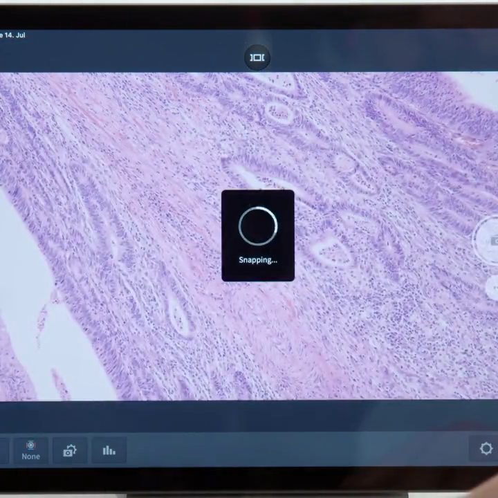

Streamline image documentation
Smart Microscopy supports streamlined image capture with automatic imaging functions and scaling for routine cytology documentation.
Explore the science behind cytological staining
Download the white paper to learn more.

A Quick Guide to Cytological Staining Cytology is the science of the interpretation of cells removed from the human body through clinical procedures or exfoliation. One of the most widely used groups of stains in cytology, the Papanicolaou series of stains, was developed by Dr. George N. Papanicolaou. These stains impart a characteristic color to nuclei and cytoplasmic components.
Clinical cytology — frequently asked questions
Answers to help you configure the right setup for your organisms and stains.
-
Which Axiolab 5 configuration should I choose for cytology?
Both configurations provide the same core brightfield imaging capabilities and 2.5×–100× objective range. Choose Axiolab 5 Standard for routine cytology microscopy, or Standard Ergo when you want an adjustable ergonomic viewing setup for extended microscope use.
-
What types of cytology preparations can I examine with Axiolab 5?
Axiolab 5 supports brightfield examination of stained cytology preparations, including Papanicolaou and Romanowsky-stained specimens, across a range of cytology sample types.
-
What is different about the Standard Ergo configuration?
Standard Ergo provides the same core imaging capabilities as Standard with the addition of an ergonomic phototube. This allows the viewing position to be adjusted to accommodate different users and working positions during extended microscope sessions.
-
How does Smart Microscopy support cytology documentation?
Smart Microscopy integrates image capture into the microscopy workflow, with automatic imaging functions and scaling that help streamline digital documentation of cytology preparations.
-
Can I capture images without using a PC?
Depending on the camera and system configuration, Axiolab 5 supports stand-alone image capture without operating a PC, providing another option for routine digital documentation.


