

Microscopy solutions for Hematology
Blood smears and bone marrow, viewed in true color.From peripheral blood smears to bone marrow preparations, hematology microscopy depends on clear visualization of cellular morphology and subtle color differences. ZEISS microscopy solutions combine high color-fidelity brightfield imaging, a broad range of magnifications, ergonomic viewing options, and digital documentation capabilities to support routine hematology workflows.
Routine hematology depends on true color and the right magnification
Peripheral blood smears and bone marrow preparations bring different cellular details into view across a range of magnifications. Faithful color, clear brightfield imaging, efficient movement from specimen overview to cellular detail, and a comfortable working setup all matter in routine hematology microscopy.


Configure Axiolab 5 for your hematology workflow
Three configurations on one Axiolab 5 platform. Choose Standard for routine brightfield hematology, Advanced to add a 50× oil objective for greater magnification flexibility, or Advanced Ergo for the same expanded objective range with an ergonomic viewing setup. All three support true-color brightfield imaging and digital documentation.


One Axiolab 5 across your hematology workflows
From peripheral blood smears to bone marrow preparations, Axiolab 5 delivers trusted ZEISS optics across routine hematology workflows, with configurations matched to the stains, magnifications, and working needs in front of you.
Built for the way hematology labs actually work
Find the hematology workflow that sounds like yours and explore the Axiolab 5 configuration designed to support it.


Examine peripheral blood smears
True-color brightfield imaging supports visualization of cellular morphology across routinely stained peripheral blood preparations, with magnification options for moving from specimen overview to cellular detail.


Explore cellular detail with 50× oil
The Advanced configuration adds a 50× oil objective, providing high-resolution observation with a larger field of view than 100× for detailed examination of stained hematology preparations.


Adjust your setup for extended microscope use
The Advanced Ergo configuration combines the expanded objective range with an ergonomic phototube, providing adjustable viewing options for extended microscopy sessions.

Streamline image documentation
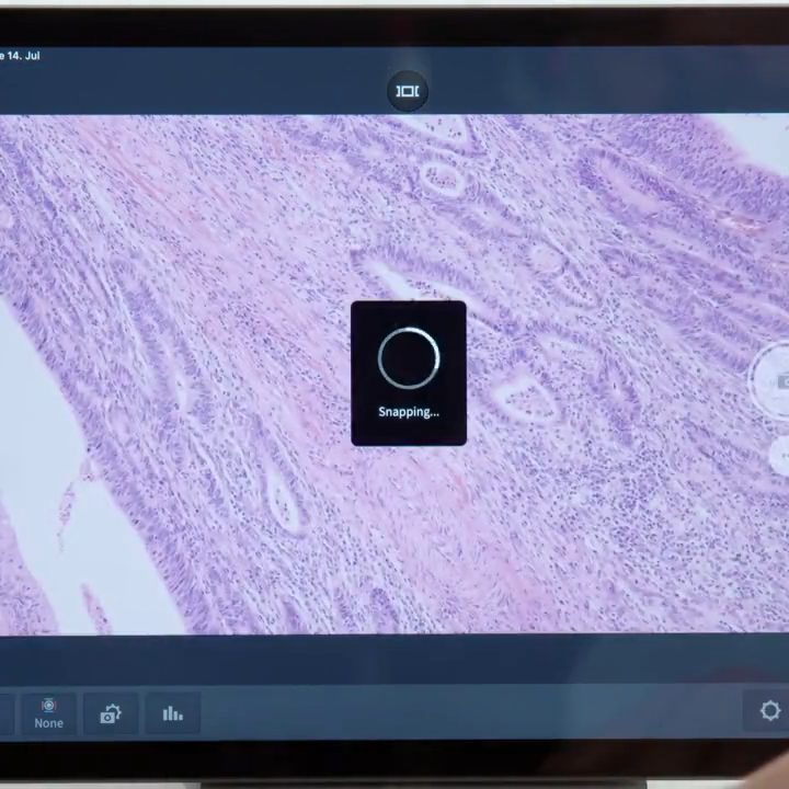
Smart Microscopy integrates image capture into the microscopy workflow, with automatic imaging functions and scaling to support routine hematology documentation.
Explore the science behind microscopy in hematology
Download the white paper to learn more.

Explore the fundamentals of hematology microscopy, from peripheral blood and bone marrow preparations to staining, magnification, and cellular morphology. This practical guide also explores the role of color fidelity, objective selection, ergonomics, and digital documentation in the hematology microscopy workflow.
Clinical hematology— frequently asked questions
Answers to help you choose the right Axiolab 5 configuration for your hematology preparations, magnification needs, and working setup.
-
Which Axiolab 5 configuration should I choose for hematology?
Choose Axiolab 5 Standard for routine brightfield hematology with 10× and 100× oil objectives. Advanced adds a 50× oil objective for greater flexibility across magnifications, while Advanced Ergo combines the same expanded objective range with an ergonomic phototube for adjustable viewing during extended microscope use.
-
Why would I choose the 50× oil objective?
The 50× oil objective provides high-resolution observation with a larger field of view than 100×, offering an additional magnification option for detailed examination of stained hematology preparations.
-
Can I use Axiolab 5 for both peripheral blood and bone marrow preparations?
Yes. Axiolab 5 supports true-color brightfield imaging of routinely stained peripheral blood smears and bone marrow preparations. Its high color-rendering LED illumination supports faithful visualization of the characteristic colors produced by hematology stains.
-
Which hematology stains can I view in brightfield?
Brightfield microscopy supports commonly used Romanowsky-type stained preparations, including May-Grünwald-Giemsa, Giemsa, and Wright-stained specimens.
-
What does the Advanced Ergo configuration add?
Advanced Ergo provides the same 10×, 50× oil, and 100× oil objective range as Advanced, with an ergonomic phototube that allows the viewing position to be adjusted for different users and working positions.
-
How does Smart Microscopy support hematology documentation?
Smart Microscopy integrates digital image capture into the microscopy workflow. With a compatible camera configuration, automatic functions support image capture and scaling for routine documentation while reducing the number of manual imaging steps.


