Improve Success Rates for Packaging Failure Analysis
with 3D X-ray Microscopy
While investigating the root cause of defects in semiconductor advanced packages, 3D XRM can help you…


Non-destructive 3D Imaging
ZEISS 3D X-ray microscopy (XRM) provides non-destructive, high-resolution visualization of embedded defects, preserving the integrity of both sample and defect structures. This solution integrates seamlessly into standard failure analysis (FA) workflows, enhancing the efficiency of subsequent physical FA for root cause determination.
- Image diverse samples at high-resolution from systems to package to interconnect levels
- Obtain 3D insights by viewing virtual XRM cross-sections from any angle
- Enable accurate physical cross sections with 3D navigational data


Breakthrough Resolution with Cutting-edge Imaging Technology
Advanced packaging techniques such as heterogeneous integration and chiplets are used in larger package architectures, pushing the resolution limit of traditional microCT systems.
ZEISS X-ray microscopy extends the frontiers of submicron-resolution 3D imaging and analysis techniques, enabling unprecedented 450 nm spatial resolution to image 3D interconnects, redistribution layers (RDLs), through silicon vias (TSVs), embedded bridges, hybrid bonds, solder bumps, and more.


Superior Contrast across Diverse Materials
Diverse materials such as polymers, resins, adhesives, thermal interface materials, as well as metal wires, traces and solder can be difficult to visualize due to their low contrast.
ZEISS X-ray microscopes unique architecture design employs advanced scintillator coupled optics, providing superior contrast to enable visualization of these materials.


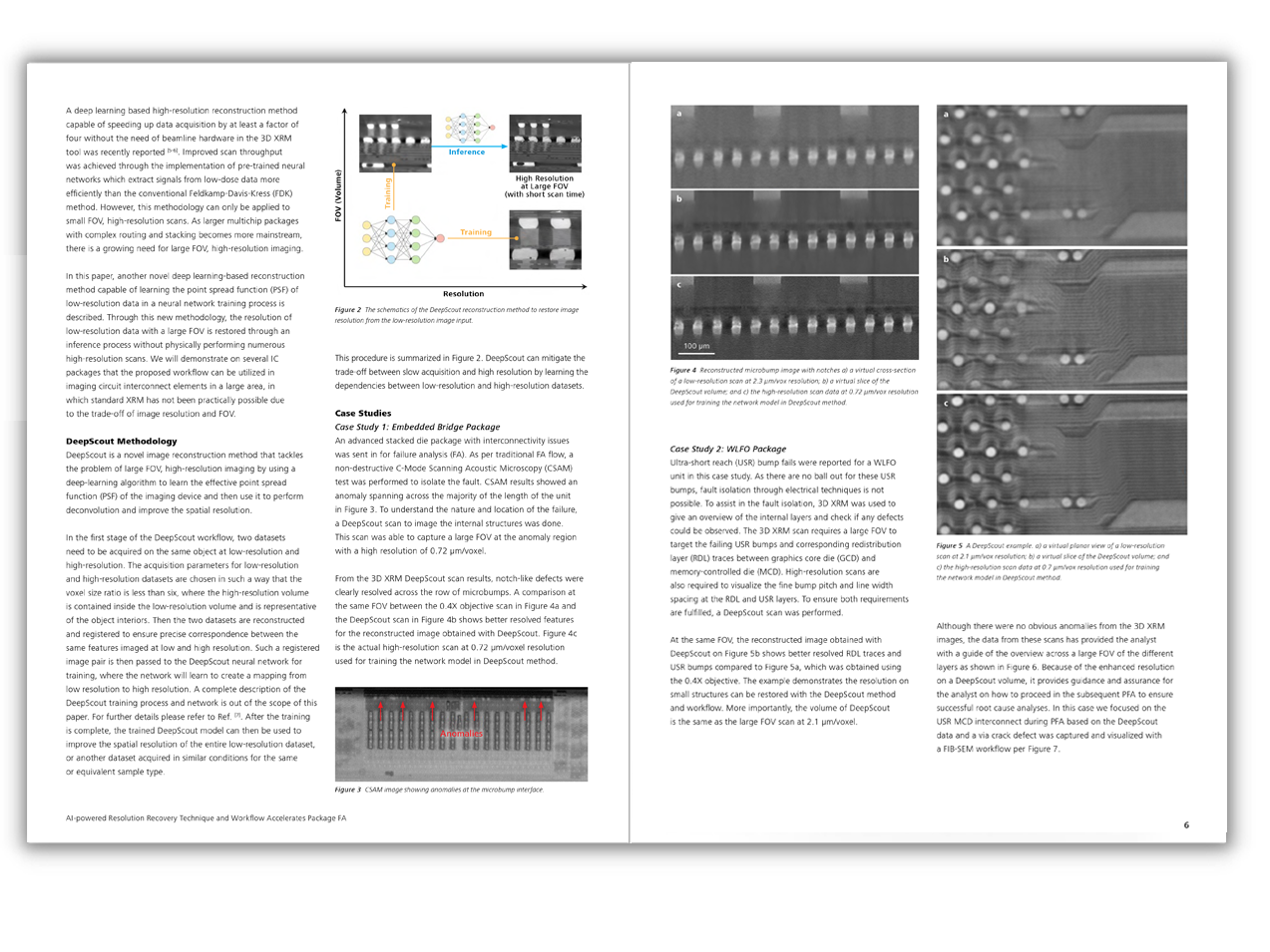
Award-winning AI-powered Reconstruction for High-throughput Imaging
- Deep learning-based reconstruction technique, ZEISS DeepRecon Pro delivers 4X faster scan times and improved image quality.
- ZEISS DeepScout enables high-resolution recovery at large FOV, significantly reducing the numerous long scans required to cover the entire FOV.


Integrated User-centric Interface for Effortless Scan Set up
Simplify and optimize X-ray scan set ups with ZEN navx XRM user interface, which
- Guides users through embedded workflows with system intelligence
- Delivers results easily and efficiently for novice users and allows experienced users to explore full system versatility
- Enables optimized scans for all users with on-screen guidance
- Enhances data management and archiving with automated file transfer utility (FTU)
Download the free semiconductor packaging failure analysis eBook.
What will you receive in your download?


Download the Article Compendium here ↓
After completion of the form, the compendium will be available for download.
Introducing ZEISS VersaXRM 730
Discover limitless potential with the exclusive 40x-Prime objective and award-winning ZEN navx.