Dr. James Schiffbauer customer profile
Little things. big impact.

Dr. James Schiffbauer

Unraveling the origins of animal life

Associate Professor of Geology  |  University of Missouri

Imaging life’s earliest blueprint.

Dr. James Schiffbauer focuses his work on our earliest animal ancestors that evolved during the Ediacaran-Cambrian transition, and he employs analytical microscopy to gain insight into the role of soft tissue preservation during this evolutionarily important interval.

About Dr. Shiffbaeuer

  • Years of experience

  • Awards

  • Peer-reviewed papers published

Breakthroughs by Dr. Shiffbauer

Earth’s earliest animals

The dawn of animal life.

The evolutionary origin of animals remains shrouded in mystery, though the fossil record provides exceptional clues. Tiny but ornate spherical fossils from the Ediacaran Period (635 541 million years ago) mayrepresent the first evidence of Earth's earliest animals.

These beautiful fossils preserve several features suggesting that they are animal embryos, including their cellular arrangement and volumetrically reductive division. In addition, some examples preserve subcellular structures, interpreted as nuclei and lipid vesicles -- revealed by advanced 3D microscopic imaging.

Dr. James Schiffbauer's work on these fossils has detailed the preservation pathway that allowed for capturing such fine biological details in such old fossils.

From Dr. Schiffbauer's lab

  • Cellular phosphatization

    Cellular phosphatization

    This electron microscopy image shows the fractured surface of a 600-million-year old animal embryo from south China, illustrating phosphatic crystal growth/replication of cellular material.

  • An electron microscopy image of a fossil embryo from China that shows distinct and ornate surface textures, and an apparent spiral structure.

    Spiral cleavage

    An electron microscopy image of a fossil embryo from China that shows distinct and ornate surface textures, and an apparent spiral structure.

  • Embryo preservation

    Variation in embryo preservation

    Another electron microscopy image of a fossil embryo from China with distinct cellular shrinkage and surface ornamentation.

  • Spiny spheres

    Spiny spheres

    An electron microscopy image of another fossil from the same geological unit hosting the embryos, although the nature and affinity of this fossil are
    unresolved.

Breakthroughs by Dr. Shiffbauer

Ancient biomineralization

Which came first: the predator or the shell?

During the last 10 million years of the Ediacaran period, cloudinomorphs begin to evolve from simple walled organisms to those that produce a series of cone-shaped shells. That act of making a protective covering, biomineralization, represents a vital step in the development of bones,including our own.

It raises important questions. Because no sooner do these early shells appear in the fossil record than bite marks of predators appear as well. So, did the shells predate predators? Or did the animal develop a shell to protect itself?

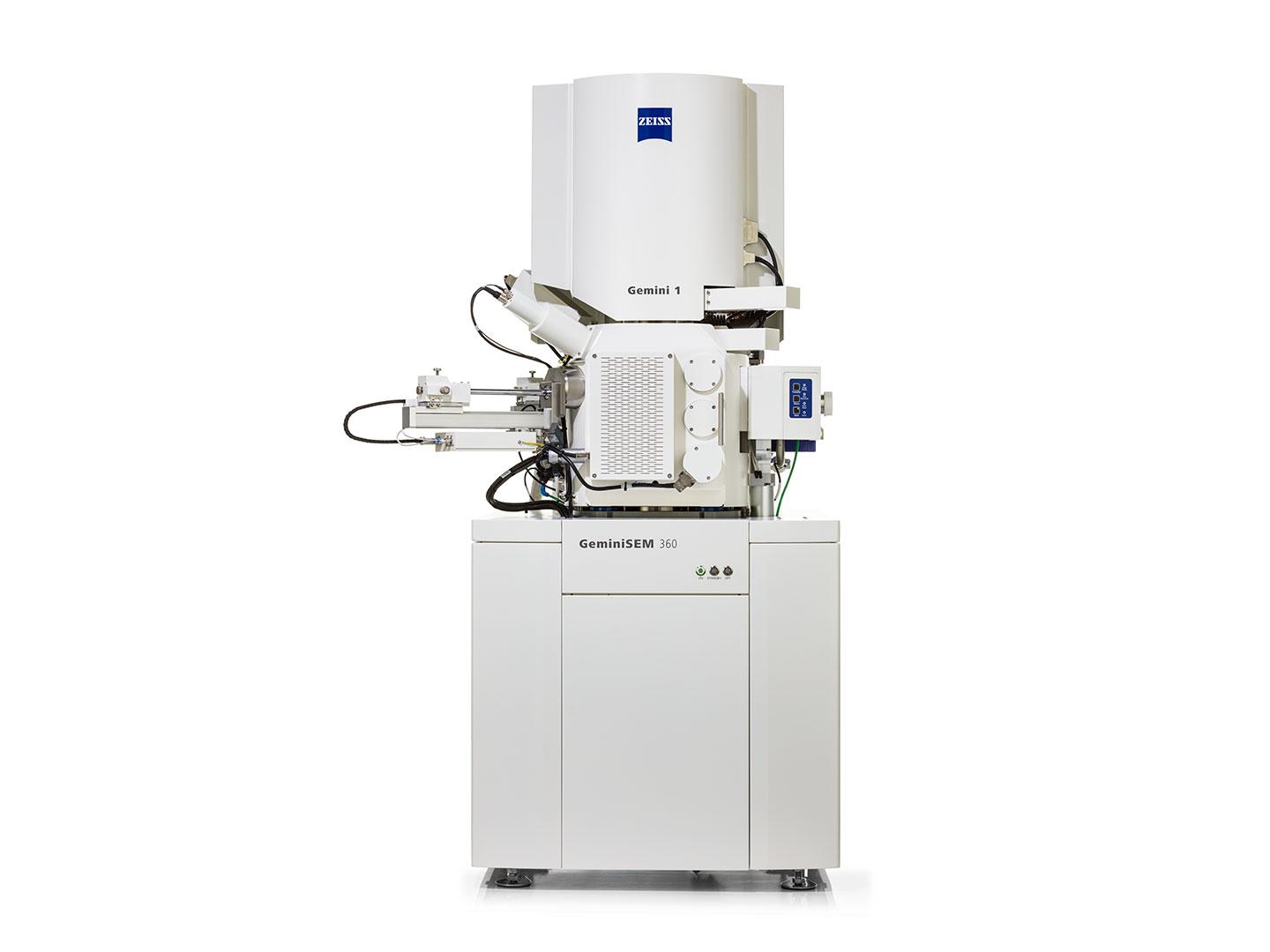
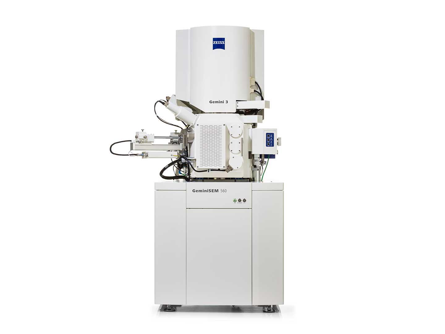
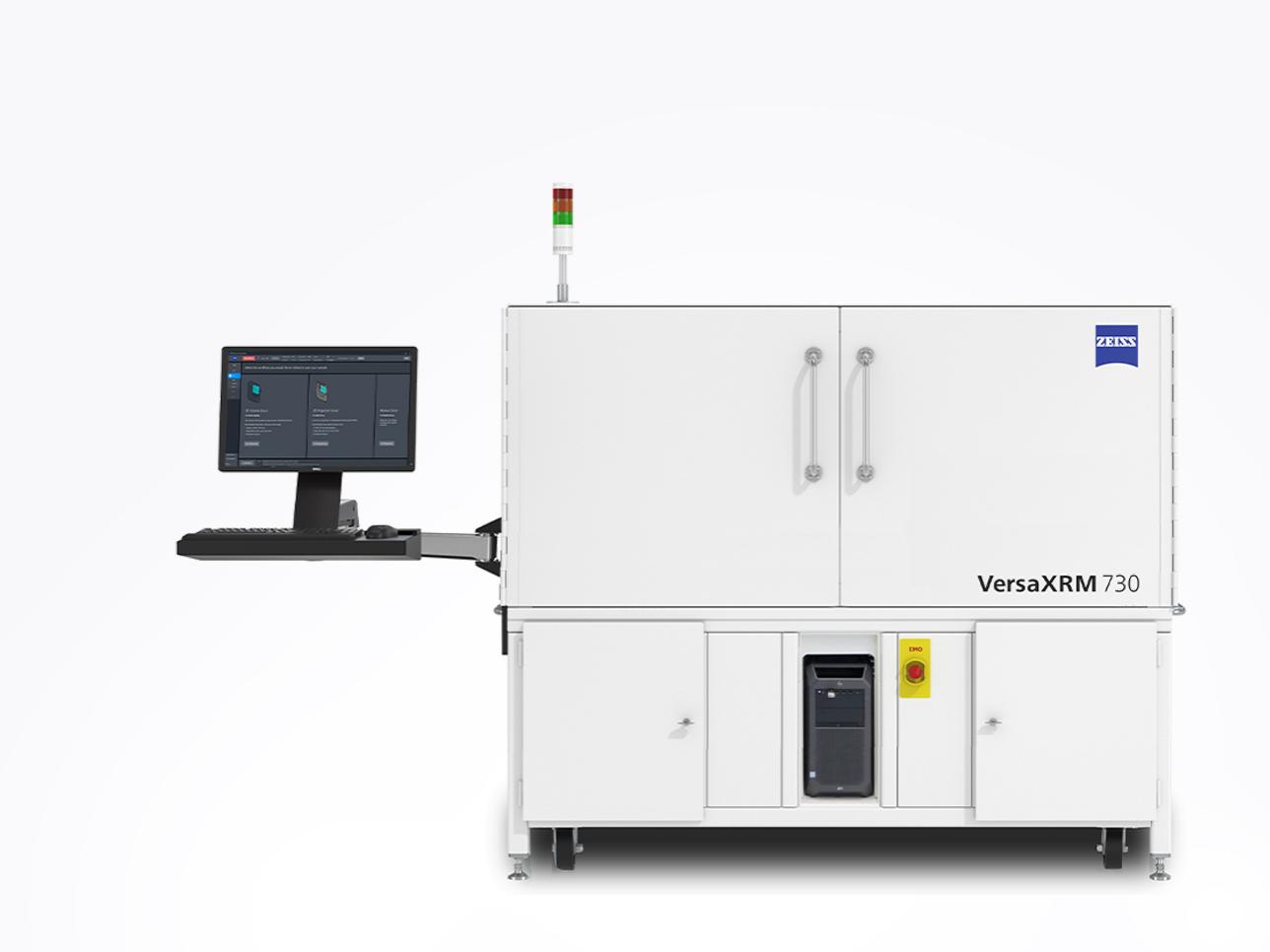
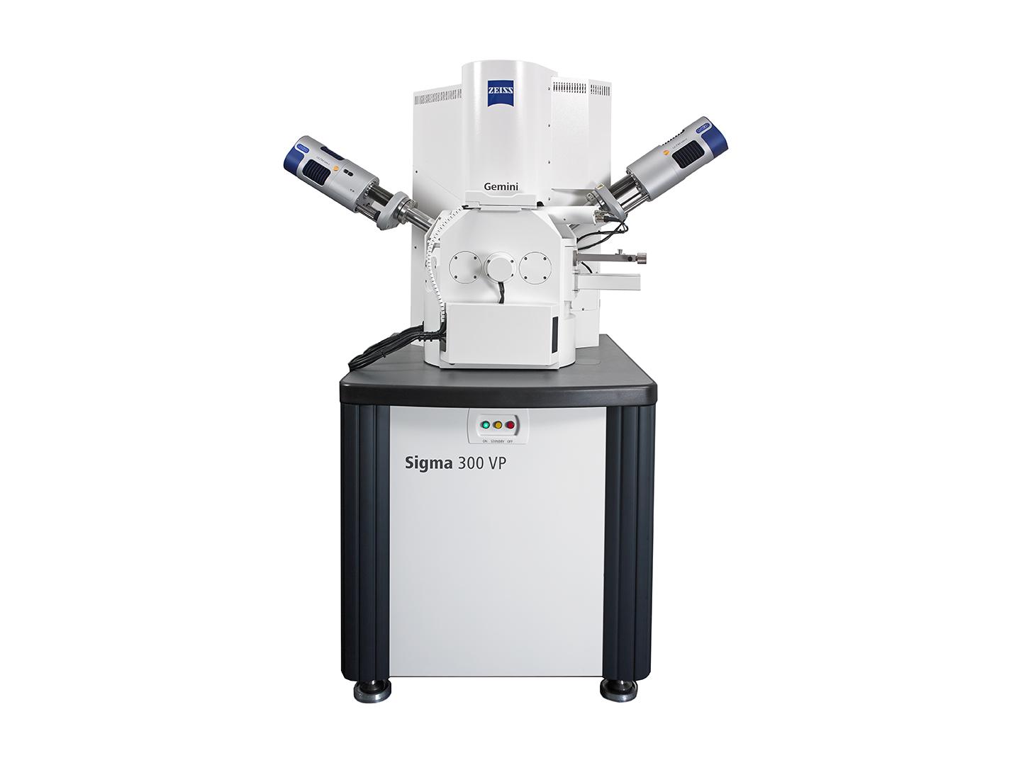
These kinds of questions help motivate Dr. James Schiffbauer's work in the lab. Together, with advanced microscopy methods powered by ZEISS, he hopes to learn even more about animal evolution.

From Dr. Schiffbauer's lab

  • Pyritized cloudinomorph fossil

    Pyritized cloudinomorph fossil

    This cloudinomorph tube, viewed here using backscatter electron imaging in a Zeiss Sigma 500VP, shows characteristic preservation via pyrite growth and replacement. This suite of fossils from southern Nevada dates to approximately 550M years ago.

  • Nevada cloudinomorph fossil

    Nevada cloudinomorph fossil

    Another pyritized cloudinomorph fossil tube from Nevada showing rusty coloration from oxidative weathering.

  • Nevada Saarina fossil

    Nevada Saarina fossil

    A well-defined cloudinomorph tube assigned to the genus Saarina. Typically known to come from Russia, the one shown here is from Nevada.

Animal origin stories are elusive. Microscopy is helping to connect
previously unconnected dots.

Dr. James Schiffbauer University of Missouri

Breakthroughs by Dr. Shiffbauer

A gutsy discovery

The gut at the heart of the debate.

Dr. James Schiffbauer s team used ZEISS X-ray microscopy to examine one of their cloudinomorph fossils. When evidence of soft tissue appeared, Dr. Schiffbauer quickly realized he was looking at a primitivedigestive tract.

A straight digestive tract indicates a distinct flow of nutrient intake and waste output. Not only does this differ from the way corals process nutrients, but it suggests more efficient eating, and thus a clear evolutionary advantage.

This tiny discovery was huge. Dr. Schiffbauer discovered the first known example of a digestive tract in animal life and gained valuable insight into the worm vs. coral debate. Said Dr. Schiffbauer, “…we can now say that their anatomical structure appears more worm-like than coral-like.

From Dr. Schiffbauer's lab

  • Preserved soft tissues

    Preserved soft tissues

    This image shows a segmented view of x-ray microscopy data, collected with a Zeiss Xradia 510 Versa, of a Nevada cloudinomorph with welldefined soft tissue (gold) preserved within the
    skeletal tube (red). This soft tissue has been interpreted as the digestive tract of the animal that lived within the tube.

  • Cross section of preserved soft tissues

    Cross section of preserved soft tissues

    The preserved soft tissues were physically sectioned in order to view their cross-sections, shown here. The exterior skeletal tube and interior soft tissue cylinder are distinct, and are preserved via oxidized pyrite.

Shop Microscopes Online

Precision at your fingertips.

Accelerate your research with ZEISS top-class microscopes, designed to meet your research needs.

Comprehensive Solutions and Capabilities

Discover Our Application Hub

Explore applications to discover tailored solutions for your unique laboratory needs and elevate your research capabilities.