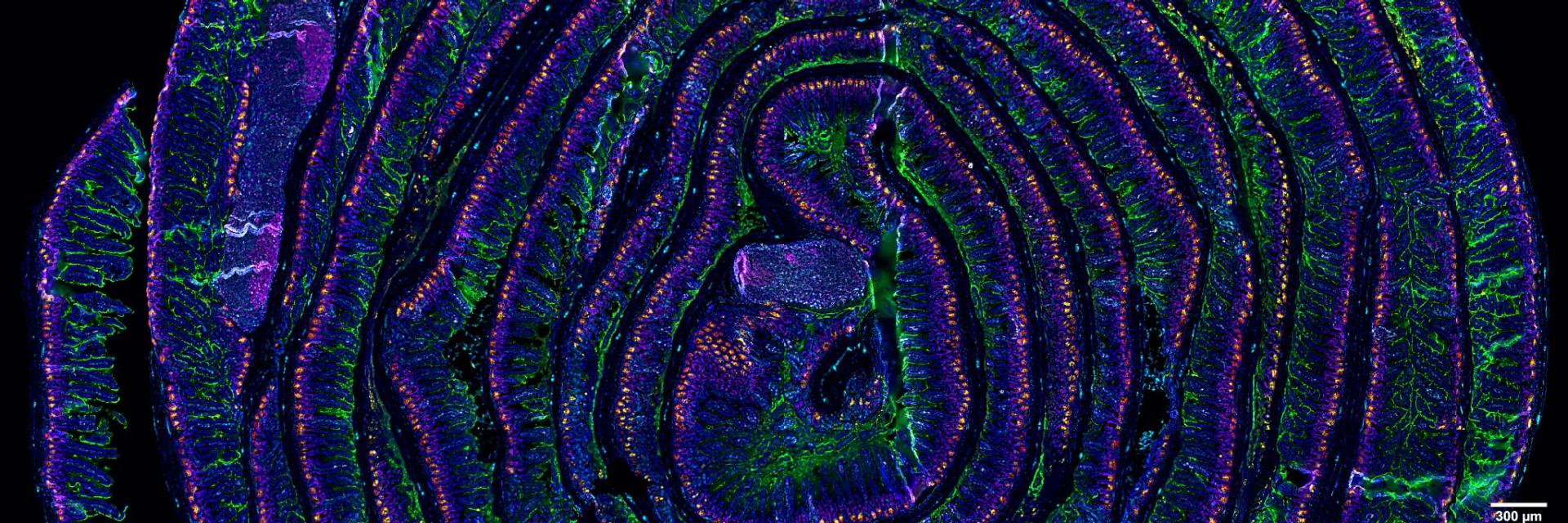
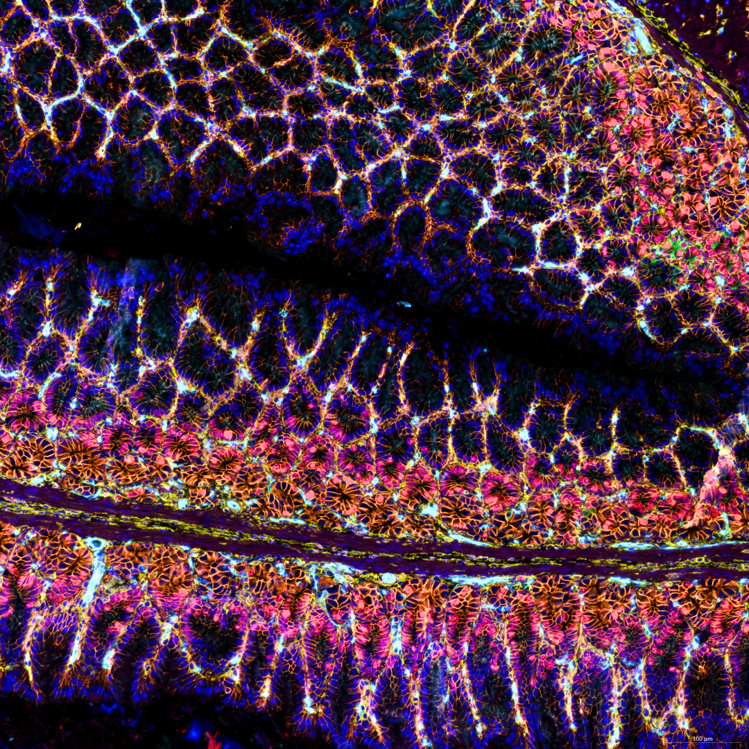
5 µm section of a formalin fixed paraffin-embedded mouse small intestine swiss roll. Section was stained with a 14 CODEX antibody panel designed to visualize intestinal stem and enteroendocrine cells (Lysozyme in red, villin in green, MHCII in gray, vimentin in cyan, Ki67 in magenta, and Olfm4 in yellow). Blue signals correspond to DAPI nuclear staining. Acquisition was done with a ZEISS Axio Observer microscope and processing with the Akoya CODEX Processor.
Courtesy of D. Bejarano, Life and Medical Sciences Institute (LIMES) of the University of Bonn, Germany Courtesy of D. Bejarano, Life and Medical Sciences Institute (LIMES) of the University of Bonn, Germany
Use Case

Tissue Imaging

Break through traditional imaging barriers with ZEISS solutions that combine deep penetration, minimal photodamage, and automated workflows for consistent results.

Unlock Advanced Tissue Imaging Solutions ZEISS microscopy solutions empower researchers to image complex tissues and whole organs with exceptional clarity, speed, and scale. From deep 3D imaging of cleared samples to high-resolution multiplexed analysis and advanced spectral unmixing, ZEISS systems address key challenges in tissue microscopy—minimizing photodamage, enabling accurate marker separation, and delivering scalable data analysis. Whether you're studying development, disease, or spatial biology, ZEISS delivers the precision and performance to push your research further.
  • Preserve structural detail without photodamage

    Fast, gentle 3D imaging of optically cleared tissues—such as whole brains, lungs, or kidneys.

  • Enhanced sensitivity and resolution

    Visualize and separate up to 5+ markers—even in autofluorescent or challenging samples—supporting spatial biology, immunophenotyping, and detailed cellular mapping.

  • Scalable analysis tools for large tissue datasets

    AI-driven segmentation, batch processing, and cloud collaboration to streamline analysis and insight generation.

Courtesy of Ultivue, Inc.

New Dimensions in Tissue Imaging

ZEISS Spatial Biology Systems

From high-speed, high-resolution imaging and non-destructive 3D visualization, to real-time analysis, discover advanced microscopy techniques that capture both intricate and dynamic details of plant specimens.

  • Using ZEISS Axioscan, we can image whole tissue sections with up to 7 fluorophores. Combined with powerful software, this allows us to assess spatial phenotyping of tumor microenvironments and is now applied to a wide range of projects.

    Dr. Caroline Bouzin Facility Manager, 2IP Imaging Platform at the Institute of Experimental and Clinical Research at the UCLouvain, Brussels, Belgium
  • We would like to exploit the high dimensionality of multiplex tissue microscopy to study the diversity of myeloid cells in multiple tissues and identify non-described tissue-specific populations. There is so much we still don’t understand and we see great potential in leveraging such findings to eventually lead to new and better targeted treatments for multiple health conditions.

    Dr. David Bejarano Postdoctoral Researcher, Schlitzer Lab at the Life and Medical Sciences Institute (LIMES), University of Bonn, Germany
  • The light sheet microscope shows us extremely complex neural networks without spending hours trying to piece the images back together.

    Dr. Kent Willis University of Alabama at Birgminham, USA

From Complexity to Clarity

Workflow Automation for Spatial Biology at Scale

ZEISS solutions for spatial biology facilitate the seamless integration of spatial profiling into your workflows, ensuring efficient and consistent multiplexed spatial profiling at scale.

Tissue Imaging FAQs

  • ZEISS Axioscan 7 creates high-resolution digital maps that can be registered with spatial omics data, while arivis Pro provides powerful tools for overlaying and analyzing multi-modal datasets. This combination enables seamless correlation between morphology and molecular profiles.

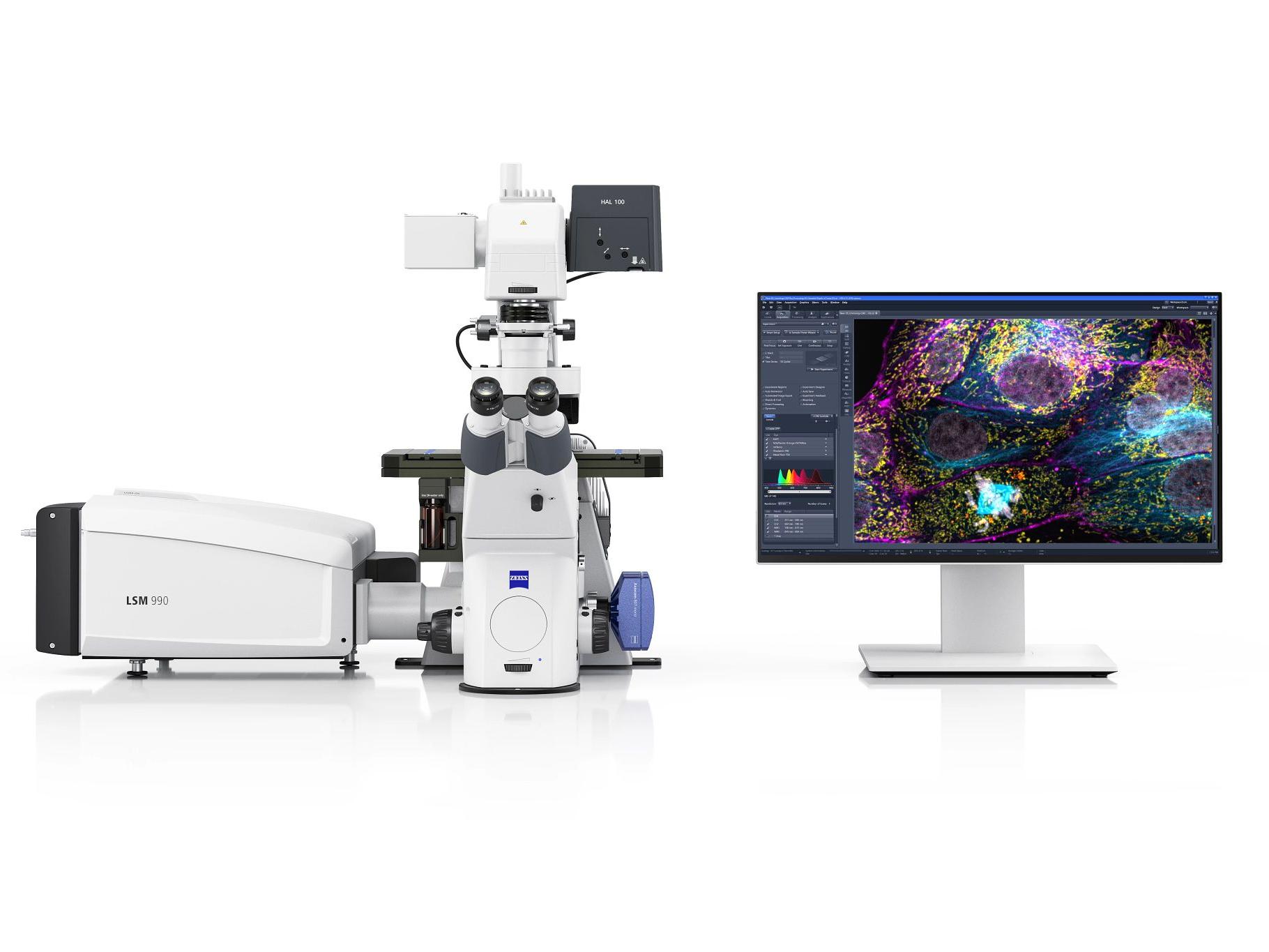
  • Yes. ZEISS LSM 990 with Airyscan and Spectral Multiplexing provides high-resolution imaging of multiple fluorescent markers, even in autofluorescent tissues. LSM 990 Spectral Multiplex covers a wavelength range from 380-900 nm and can separate 10 or more labels simultaneously.

  • The ZEISS Axioscan 7 is designed for high-throughput whole slide scanning in both brightfield and fluorescence. It's ideal for digitizing tissue libraries, supporting spatial omics workflows, and integrating with analysis platforms..

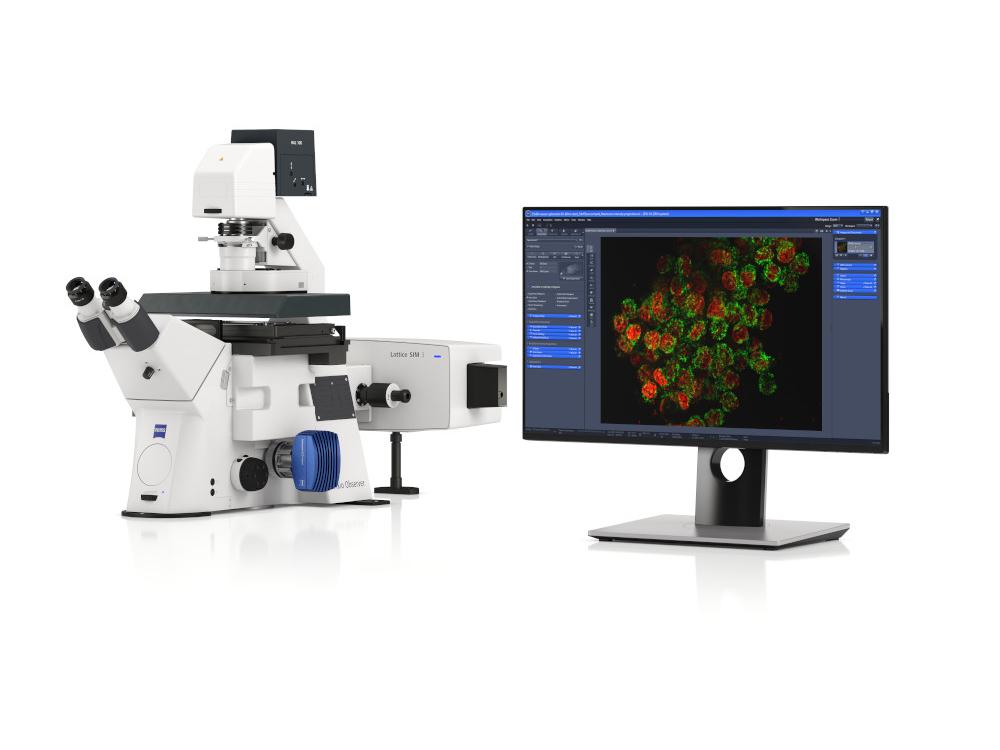
  • Yes. ZEISS Lattice Lightsheet 7 and Lightfield 4D systems allow for fast, long-term 4D imaging under physiological conditions with minimal phototoxicity. These are ideal for developmental biology, immune cell tracking, and organoid dynamics.

  • ZEISS arivis Pro and arivis Cloud provide scalable 3D/4D analysis for large datasets. You can segment, track, and quantify features like glomeruli, neurons, or vasculature—even across full organs or whole-slide images.

  • Lattice SIM 3 provides 120nm lateral resolution with structured illumination that works effectively in tissue samples. Unlike other super-resolution techniques, it maintains compatibility with standard fluorophores and provides excellent performance in scattering tissue environments.

Comprehensive Solutions and Capabilities

Discover Our Application Hub

Explore applications to discover tailored solutions for your unique laboratory needs and elevate your research capabilities.