

Zebrafish Research
From development to disease models, explore the full potential of zebrafish using tools for deep, dynamic, and reproducible imaging.
Advanced Microscopy for Zebrafish Development, Disease, and Discovery
ZEISS microscopy systems empower researchers to explore zebrafish biology with exceptional resolution, speed, and flexibility. Whether you're tracking cardiac dynamics, mapping neural activity, analyzing cell migration, or screening drug responses, ZEISS tools support gentle live imaging, deep-tissue visualization, and high-throughput workflows. From whole-organism development to targeted disease models, our systems and software—like Lightfield 4D, Airyscan, Celldiscoverer 7, and arivis Pro—enable reproducible, data-rich imaging at every scale of zebrafish research.
Unveiling Zebrafish Research Potential


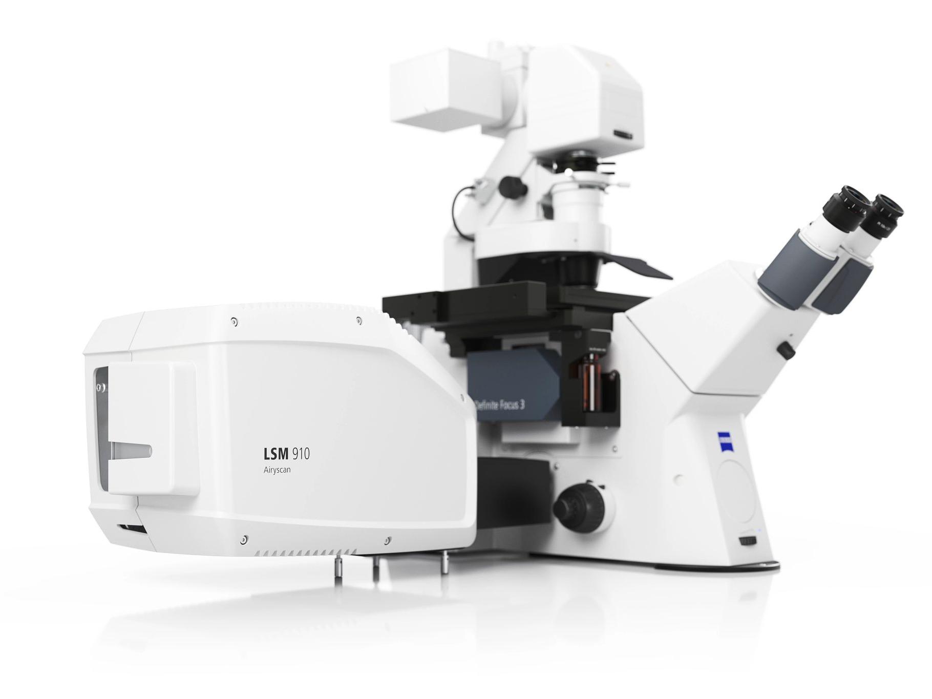
Recommended Products for Zebrafish Research


ZEISS Lab Essentials


Zebrafish Research FAQs
-
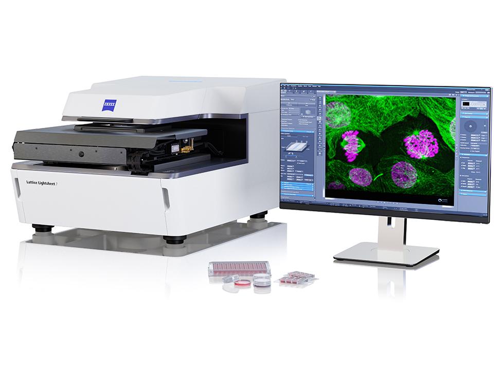
Zebrafish embryos are optically transparent, making them ideal for live imaging—but prolonged exposure to light can still lead to phototoxicity and photobleaching. ZEISS Lightsheet 7 and Lightfield 4D systems use gentle illumination strategies that minimize light exposure while still providing high-speed, volumetric data. These systems are ideal for tracking morphogenesis, organ development, and cell migration across extended time-lapse experiments, even over multiple days.
-
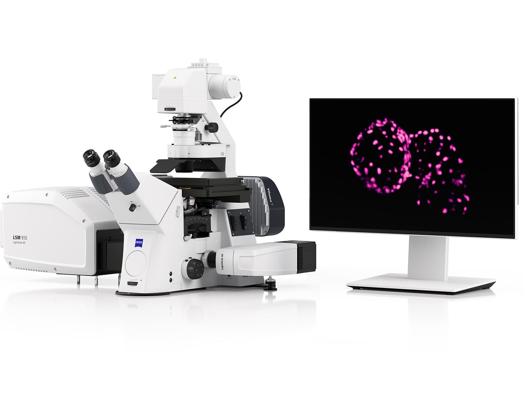
Capturing rapid motion like cardiac contractions or twitching tissues requires both speed and stability. ZEISS Lightfield 4D enables instantaneous 3D volume capture—perfect for recording every beat of the zebrafish heart without motion artifacts. For fluorescent reporters or biosensors in dynamic tissues, Airyscan Fast provides exceptional temporal resolution with enhanced signal-to-noise, making it ideal for imaging fast biological events in developing zebrafish.
-
Yes. Zebrafish are widely used for functional imaging due to their transparency and genetic accessibility. ZEISS Airyscan Fast and Lightfield 4D systems allow researchers to image biosensor activity—such as calcium, cAMP, or voltage indicators—with high sensitivity and speed. These systems support volumetric imaging of the brain or spinal cord in real time, making them powerful tools for studying neural circuits, sensory processing, and pharmacological effects in vivo.
-
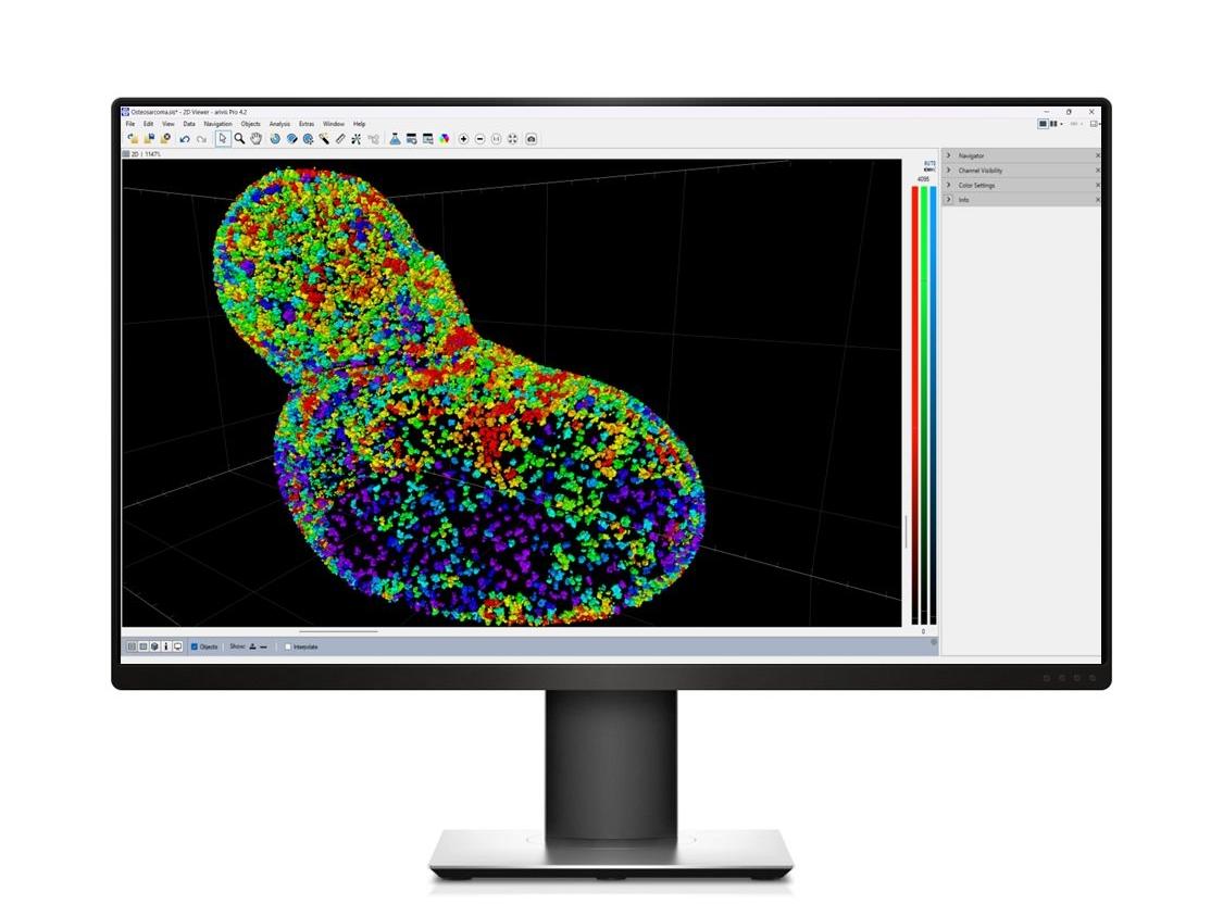
Zebrafish studies often generate complex 4D datasets across multiple samples, conditions, or time points. ZEISS arivis Pro is built to handle these challenges, offering robust tools for stitching, segmentation, cell tracking, and batch quantification. Whether you’re analyzing a single embryo over 12 hours or hundreds of wells from a screening run, arivis Pro provides scalable, reproducible workflows that help you extract actionable data from even the largest experiments.
-
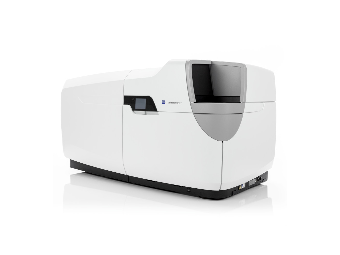
For large-scale experiments like toxicity testing, phenotype analysis, or drug discovery, automation and consistency are critical. ZEISS Celldiscoverer 7 and Axioscan 7 provide streamlined, high-content imaging of embryos and larvae in standard multi-well plates or slides. These platforms combine automated acquisition with standardized optics, enabling reproducible results across replicates and conditions. Pairing them with arivis Pro adds automated quantification, making them ideal for both academic and industrial screening pipelines.