Turn the intricacies of development into data
From early embryos to complex organoids, ZEISS solutions support live-cell imaging, tissue clearing, and long-term time-lapse acquisition. Capture morphogenesis with lattice light sheet, resolve fine structures with Airyscan and SIM, and image intact samples in 3D using light sheet and X-ray microscopy. With AI-powered analysis and automation, you can quantify structure, track lineages, and study function across scales. These workflows accelerate discovery in stem cell biology, neurodevelopment, regeneration, and more.
Scroll animation items
When imaging Becomes Insight
Transforming Developmental Biology with Quantifiable Visualization
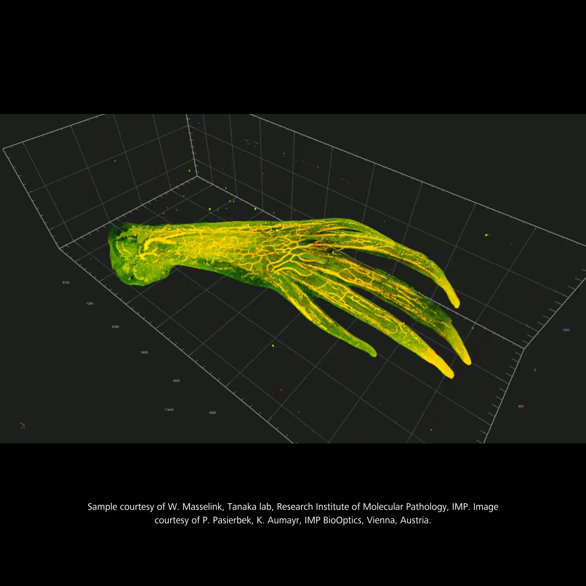
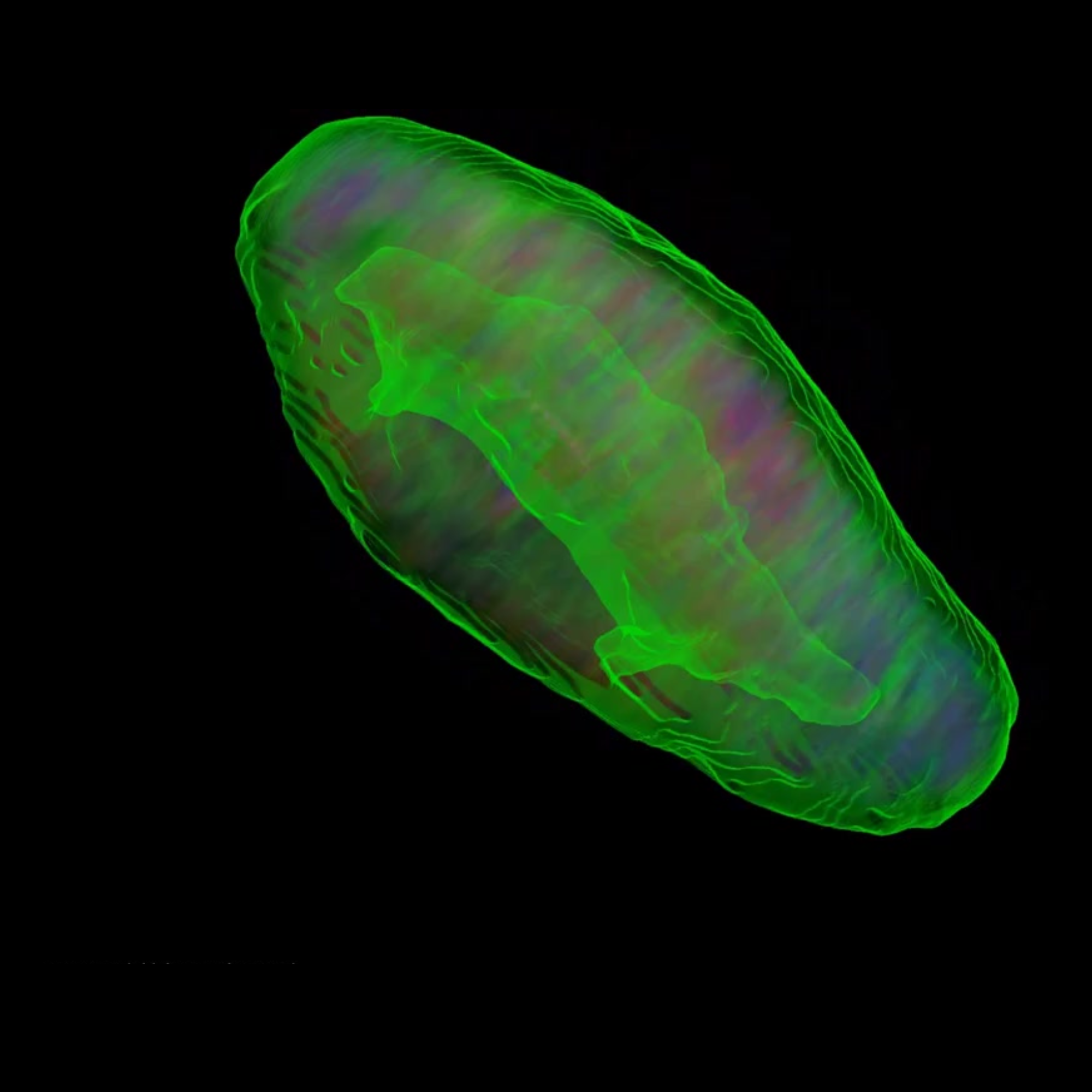
Decoding the architecture of regeneration
3D imaging of a regenerating axolotl forelimb reveals the re-establishment of complex tissue networks during limb regrowth. By capturing intact structure at cellular resolution, researchers can study spatial patterning, connectivity, and developmental reprogramming in regenerative systems.
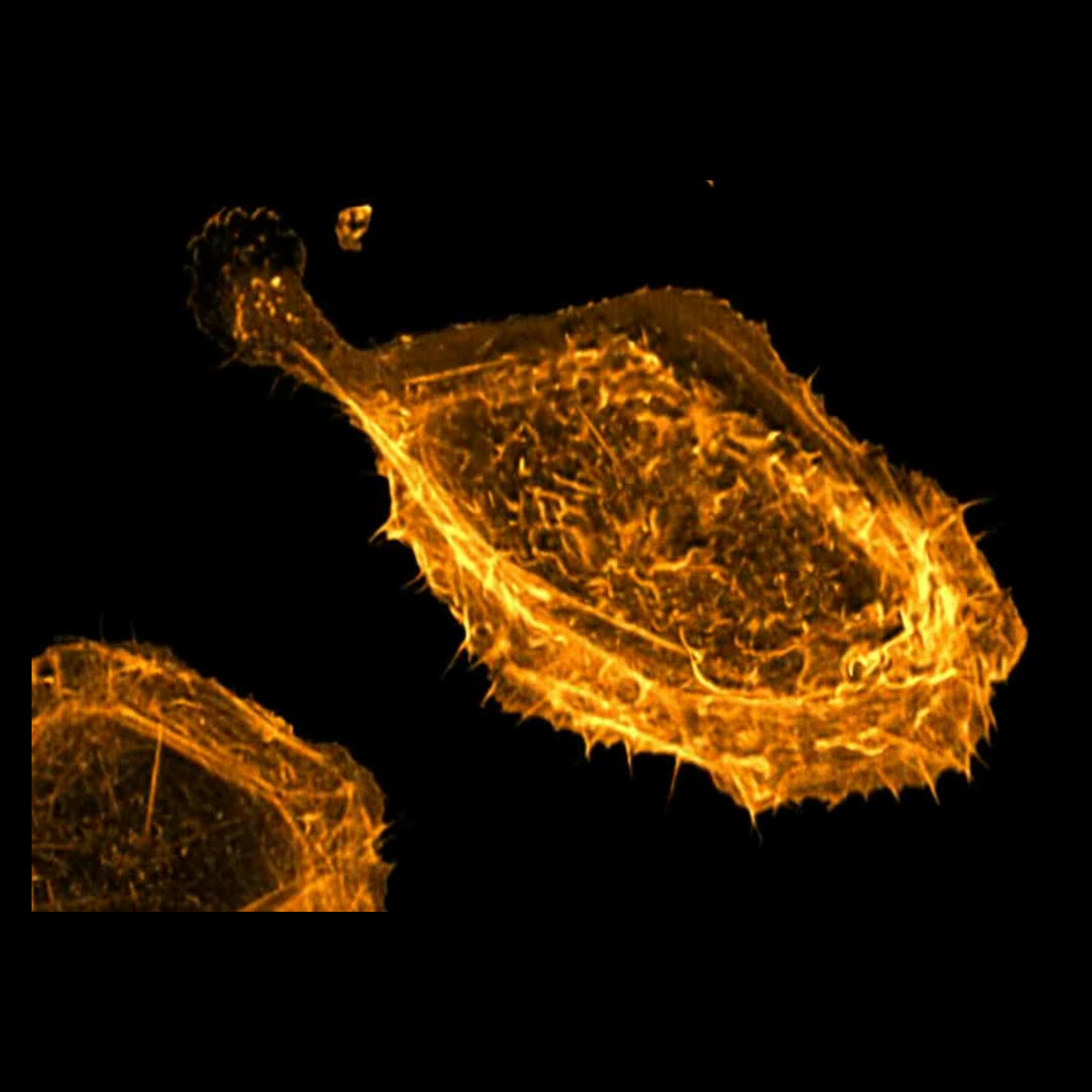
High-resolution volumetric imaging reveals changes in cell architecture during mitosis. From cytoskeletal reorganization to spindle dynamics, these structural transitions contribute to the symmetry, polarity, and spatial organization that shape developing tissues.
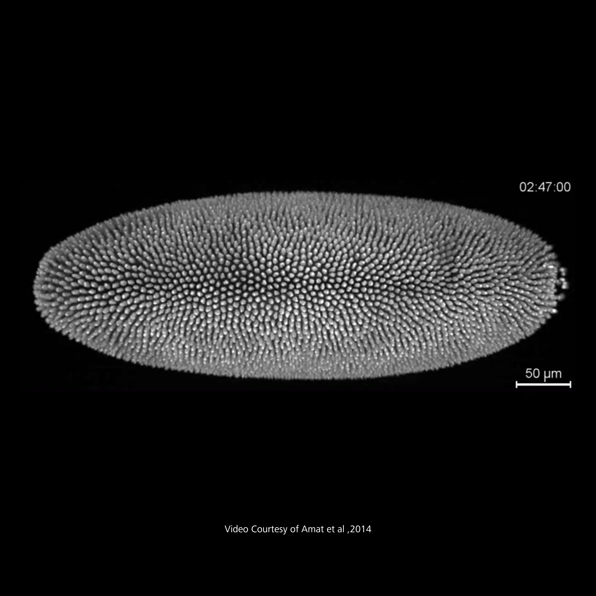
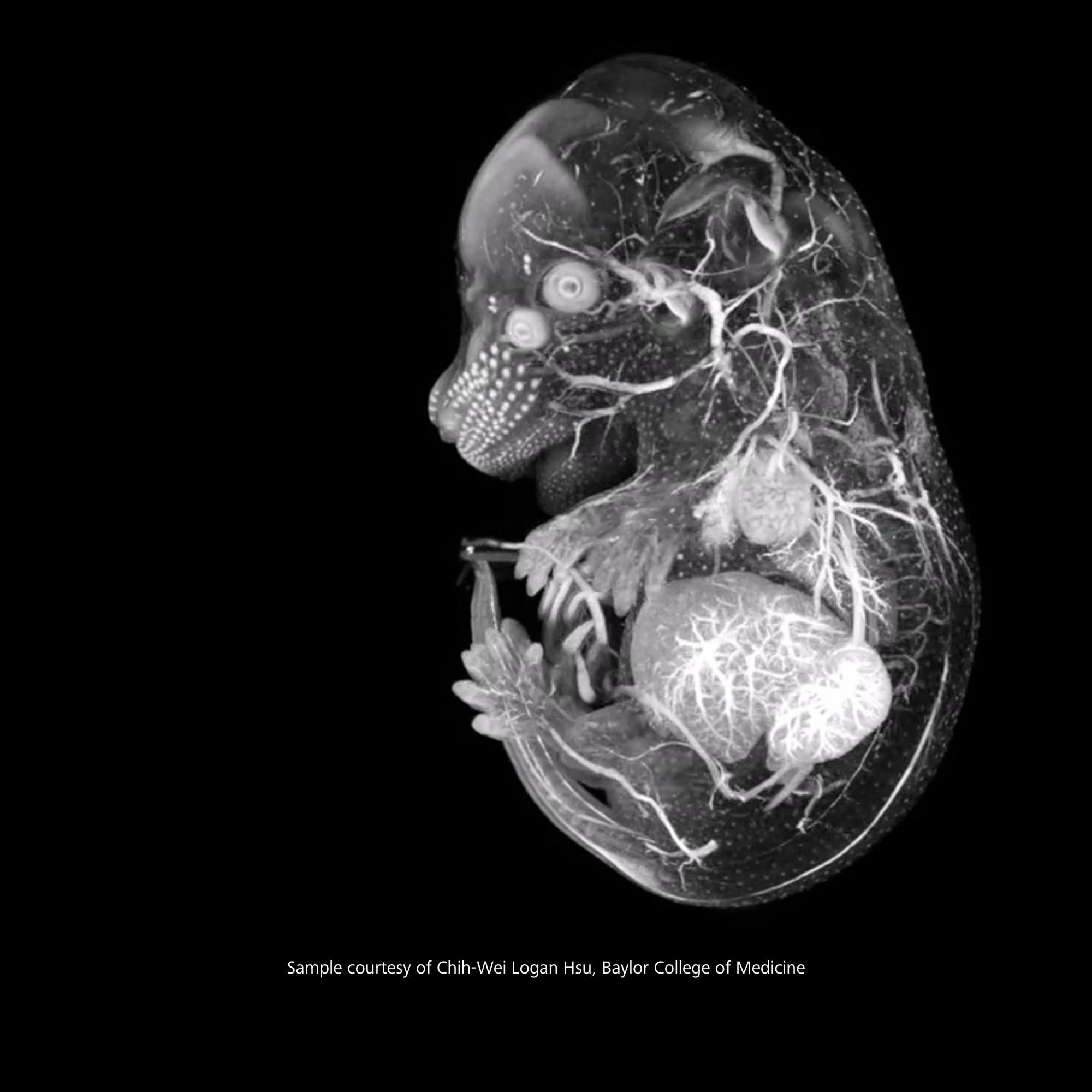
Visualize whole-embryo anatomy during critical stages of organogenesis. Segment and analyze developing tissues—such as the brain, heart, and spinal cord—to understand structural relationships and growth trajectories with deep learning-enabled volumetric analysis.
Real-time volumetric imaging reveals cardiac development as it happens, capturing the emergence of rhythmic beating and coordinated tissue motion. This dynamic view of organogenesis offers insight into morphogenesis, function, and developmental timing in vivo.
Multiplexed imaging and analysis reveal spatial relationships between nuclei, membranes, and enterocyte-specific markers in a developing intestinal organoid. Integrated segmentation and 3D visualization enable researchers to measure differentiation, architecture, and cell-type composition in stem cell–derived models of tissue development.
The ZEISS light sheet system gives us the resolution we need to identify and analyze individual cells.... We aim to analyze and understand the accumulation of senescent cells during aging and investigate their impact on life- and health span.
From whole embryos to single cells, in live or fixed samples
Capture detailed structural and molecular information across stages of development—from early morphogenesis to late-stage differentiation—in both live and cleared tissue preparations.
Volumetric time-lapse for development in motion
Explore dynamic processes like cell migration, lineage formation, and organogenesis with high-speed, high-resolution 3D imaging. Ideal for live imaging workflows across model organisms and organoids.
AI-powered analysis and reproducible results
Automated segmentation, quantification, and image-based measurements reduce variability and accelerate discovery. Scalable tools support reliable, high-throughput developmental biology research.
Courtesy of Dr. Zhao Xinying | Shandong Agricultural University | China
Courtesy of Dr. Zhao Xinying | Shandong Agricultural University | China
ZEISS Microscopy as a Service
Not every Developmental Biology breakthrough requires new equipment.
From live volumetric imaging of embryos to cleared-tissue 3D reconstruction and quantitative image analysis, our experts design and execute end-to-end workflows that reveal spatiotemporal patterns of gene expression, morphogenesis, and cellular organization. This enables researchers to generate robust, publication-ready data without the need for in-house capital equipment.
Recommended Products for Developmental Biology Research
See the Whole Embryo. Miss Nothing.
ZEISS Lightsheet 7
ZEISS Lightsheet 7 lets you follow it all: whole-organism imaging of living and cleared specimens at subcellular resolution, with multiview acquisition that eliminates shadowing artifacts and dead angles. From the first cell division to late-stage organogenesis, see development as it actually unfolds.
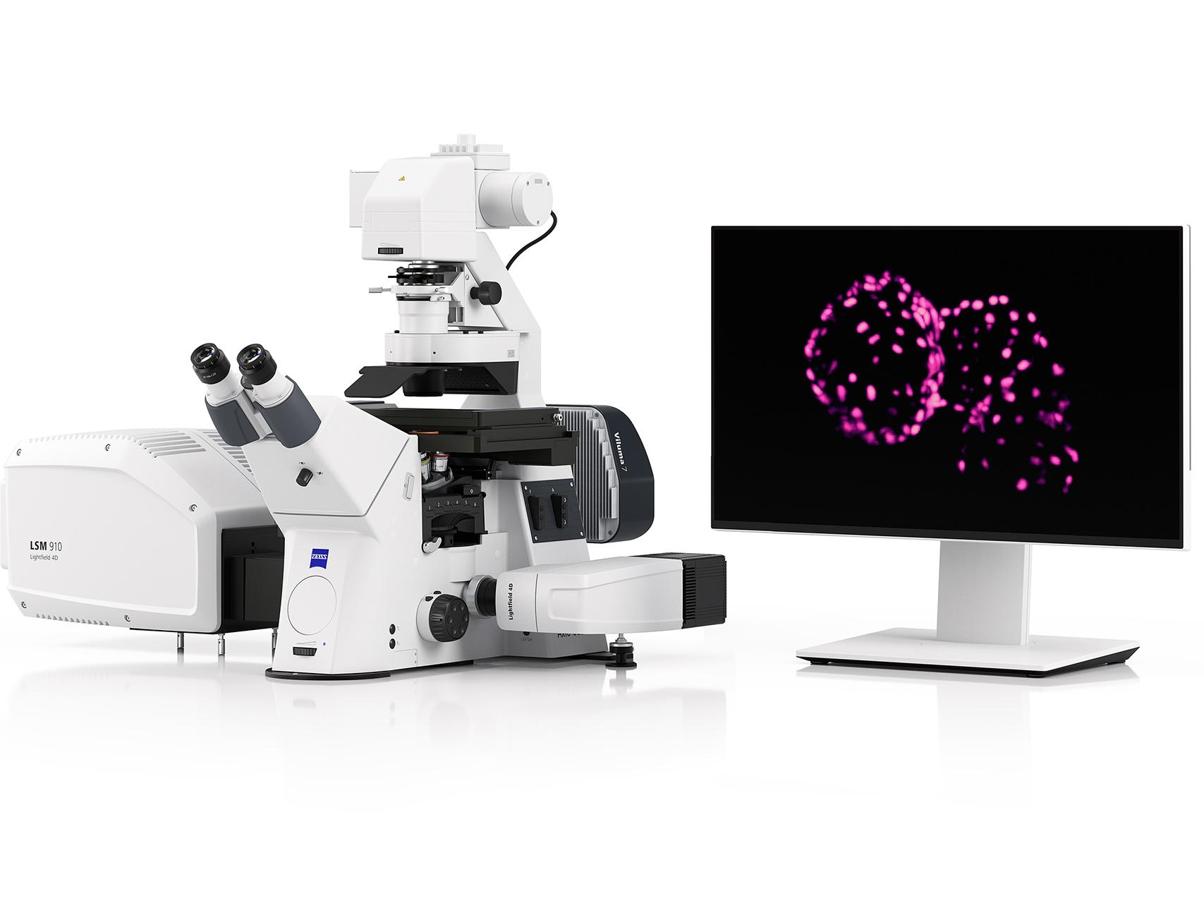
Catch Every Heartbeat, Every Cell Division — 80 Volumes Per Second
ZEISS LSM Lightfield 4D
ZEISS LSM Lightfield 4D captures complete 3D volumes with a single snap at up to 80 volumes per second, with all spatiotemporal information intact. No more choosing between speed and depth. Watch whole organisms move, beat, and develop in real time, for as long as your experiment demands.
The Dissection Bench Workhorse Every Developmental Biology Lab Relies On
ZEISS Stemi 508
ZEISS Stemi 508 is designed for heavy workloads and suitable for almost any observation task in developmental biology, with apochromatic zoom optics and efficient stray light suppression delivering a crisp, three-dimensional, distortion-free image without color fringes. From zebrafish staging to Drosophila dissection to mouse embryo selection, the Stemi 508 is the reliable foundation your workflow is built on.
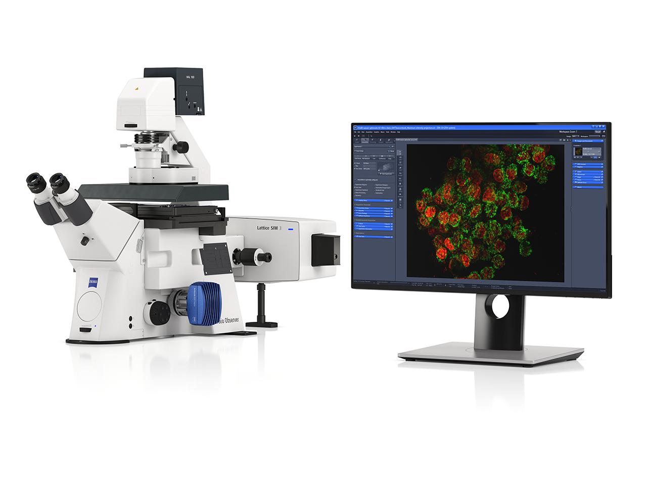
Resolve Tissue Architecture Across the Whole Organism — As Fast As Widefield
ZEISS Lattice SIM 3
ZEISS Lattice SIM 3delivers super-resolution imaging across entire model organisms and tissue sections at widefield speed and light dose, with no trade-off between field of view and resolution quality. From Drosophila wing discs to mouse brain sections, capture the spatial context that gives subcellular observations their meaning.
Super-resolution at widefield speed and gentleness
Whole model organism coverage in a single acquisition
Seamless zoom from tissue overview to subcellular detail
3D/4D Visualization and AI-Driven Analysis
ZEISS arivis Pro
arivis Pro is a modular desktop software platform designed for advanced analysis and visualization of multi-channel 2D, 3D, and 4D microscopy images regardless of dataset size, enabling developmental biologists to segment tissues, track cell lineages, quantify organoid architecture, and reconstruct morphogenetic movements — all without coding expertise.
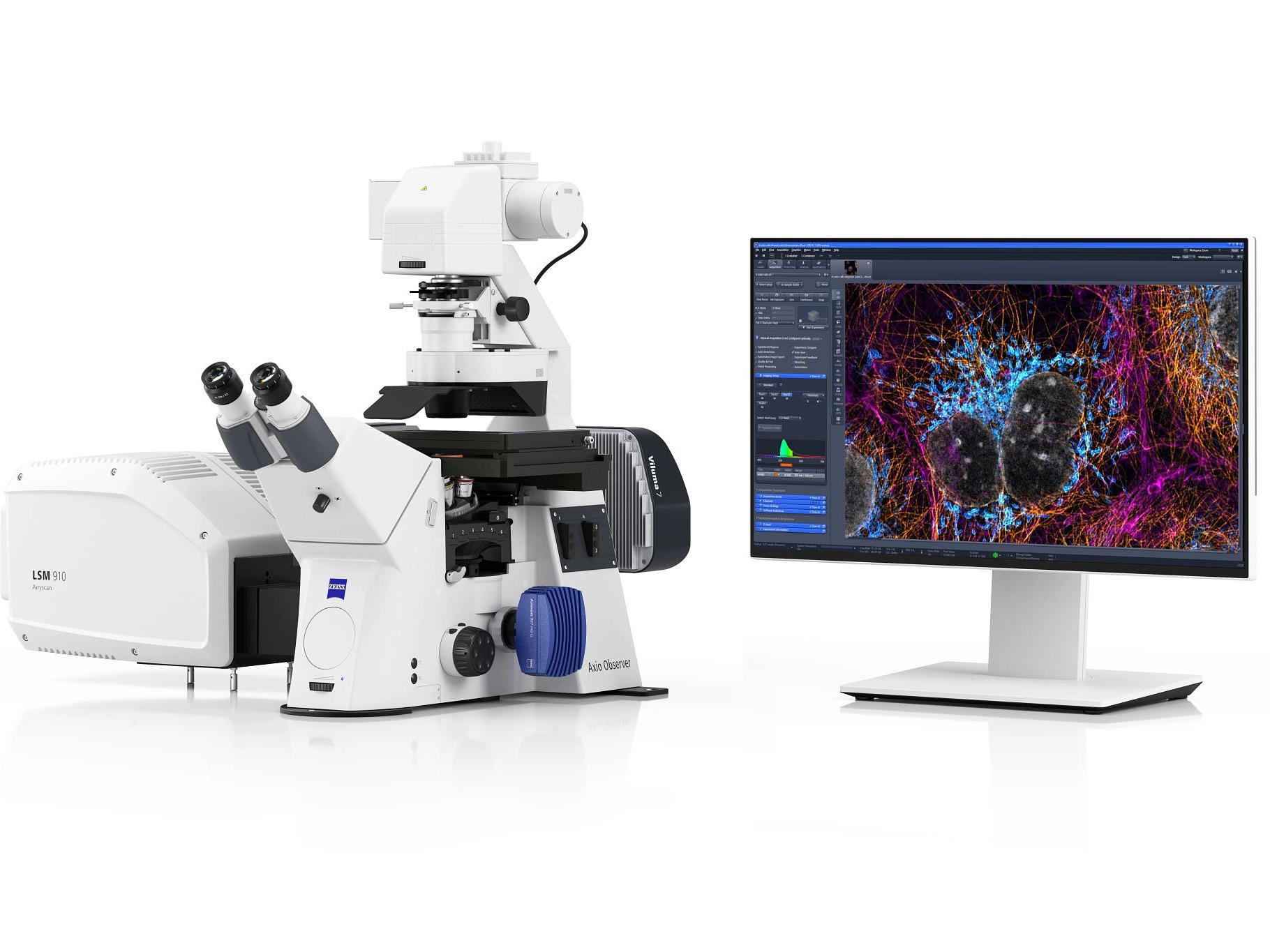
ZEISS LSM with Airyscan provides gentle super-resolution imaging for both fixed developmental specimens and live cells, combining the flexibility of a full confocal platform with the resolution enhancement needed to resolve the structural details that drive developmental decisions. From characterizing fluorescent reporter expression in embryo sections to resolving cytoskeletal dynamics during morphogenesis, the LSM Airyscan bridges the gap between routine confocal and dedicated super-resolution.
Super-resolution without specialized sample preparation
Simultaneous speed and resolution improvement
Full confocal flexibility on a single platform
Watch Morphogenesis Unfold — Without Disturbing the Biology You're Filming
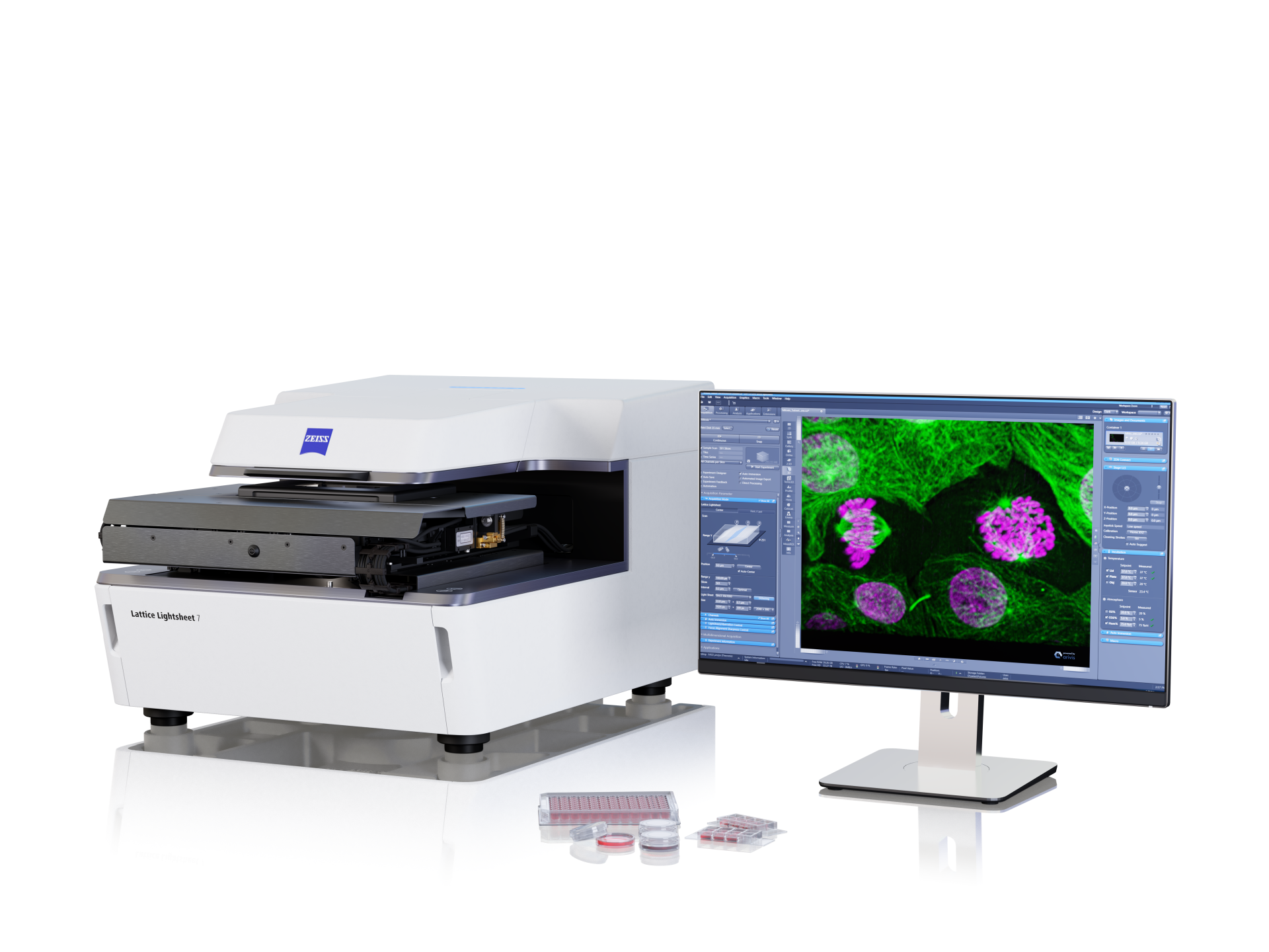
ZEISS Lattice Lightsheet 7
ZEISS Lightsheet 7 uses a thin sheet of light to image living cells and embryos with minimal energy deposition, capturing subcellular dynamics — cytoskeletal reorganization, vesicular trafficking, membrane remodeling — over hours without compromising cell viability or developmental trajectory.
ZEISS light sheet systems allow gentle, volumetric imaging of live or cleared embryos—ideal for capturing early development in model organisms like zebrafish, Drosophila, and mouse. For fixed, optically dense, or later-stage samples, ZEISS X-ray microscopy offers non-destructive 3D imaging with micrometer-scale resolution—perfect for staging, phenotyping, and structural analysis of embryonic organ systems.
For stem cell–derived organoids or cultured cells, the ZEISS Celldiscoverer 7 offers integrated environmental control and high-resolution confocal imaging in multi-well formats—ideal for tracking growth, differentiation, and morphology over days. For dynamic imaging of developing embryos or delicate 3D cultures, the ZEISS Lattice Lightsheet 7 provides ultra-fast, low-phototoxicity volumetric imaging—perfect for capturing rapid events like mitosis, migration, or morphogenesis in real time.
ZEISS offers super-resolution imaging tools designed for developmental biology. Airyscan enhances sensitivity and resolution while remaining gentle enough for live imaging of dynamic processes like spindle formation or junction remodeling. For even finer structural detail, Lattice SIM enables precise visualization of cytoskeletal and polarity structures—ideal for fixed or live samples where spatial organization is critical to understanding developmental mechanisms.
Yes. ZEISS imaging systems support fast, volumetric acquisition for tracking cell lineage and migration in whole embryos, organoids, and cultured systems. Tools like Lattice Lightsheet and Lightfield 4D enable real-time 4D imaging with minimal photodamage, ideal for capturing dynamic behaviors over hours or days. Paired with analysis platforms like arivis Pro, you can trace trajectories, segment cells, and quantify fate decisions across space and time.
Regenerative models like axolotl, zebrafish, and mouse require deep imaging of complex, often thick tissues. ZEISS light sheet systems are optimized for gentle, volumetric imaging of large samples, including cleared or live regenerating tissue. Their low phototoxicity and deep penetration enable repeated imaging over time, ideal for studying tissue re-patterning, stem cell activation, and structural regrowth—without compromising sample viability or resolution.
 Spinning Volvox Algae Sample 2) Adult Drosophila Ovary")
 Spinning Volvox Algae Sample 2) Adult Drosophila Ovary")