

Drosophila Research
Image, screen, and analyze Drosophila to uncover mechanisms driving biology and behavior
Microscopy Solutions for Drosophila Development, Genetics, and Behavior
Unlock deeper insights into Drosophila development, morphogenesis, neural circuits, and genetic function with ZEISS microscopy solutions. Whether you're imaging live embryos, tracking cell migration, or screening genetic variants, ZEISS systems—like Lightfield 4D, Lattice Lightsheet 7, and Airyscan-enabled LSMs—offer high-speed, low-phototoxicity 4D imaging with batch-mode efficiency and unmatched clarity. From subcellular detail to full-organism structure, ZEISS empowers Drosophila researchers with scalable, reproducible workflows for every stage of discovery.
Model Systems for Developmental Biology & Genetic Research
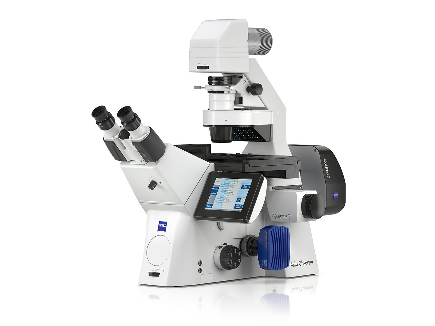
Recommended Products for Drosophila Research


ZEISS Lab Essentials


Drosophila Research FAQs
-
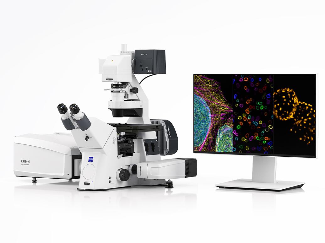
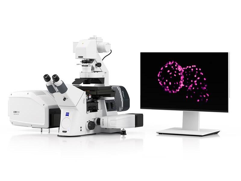
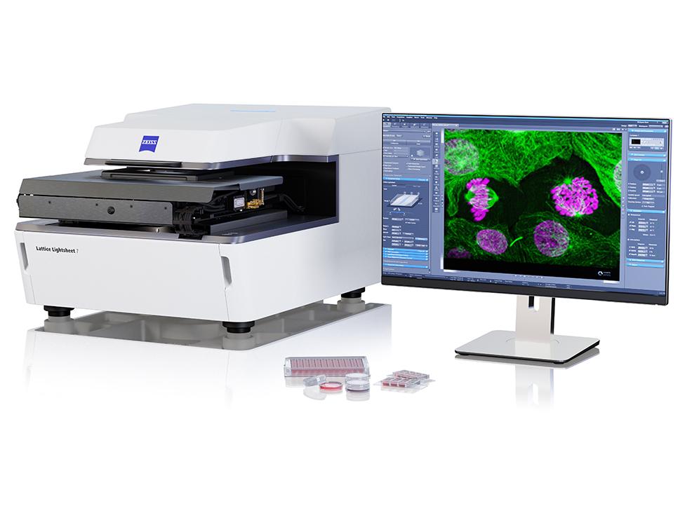
Long-term imaging of Drosophila embryos and pupae can be limited by light exposure. ZEISS Lightfield 4D enables instantaneous volumetric imaging using multiple sensors, capturing entire 3D datasets without mechanical z-scanning. This high-speed approach reduces the total light dose, helping to preserve viability while capturing dynamic events like morphogenesis, cell migration, or signal transduction in real time. For slower or gentler acquisitions, Lattice Lightsheet 7 provides complementary low-phototoxicity imaging with selective plane illumination.
-
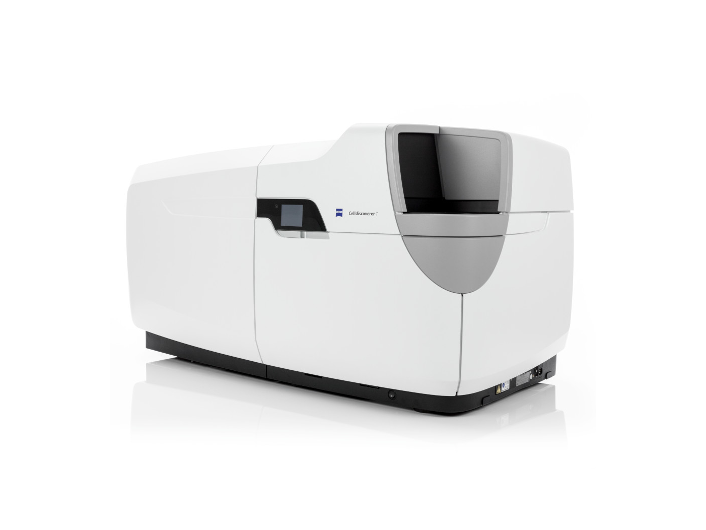
For screening experiments involving large numbers of embryos, imaginal discs, or tissue samples, ZEISS Celldiscoverer 7 and Axioscan 7 offer automated, high-throughput imaging with consistent quality and reproducibility. These systems are ideal for CRISPR or RNAi phenotype screens, biosensor reporters, or drug-response assays. When paired with arivis Pro, you can streamline analysis with batch quantification, classification, and visualization tools built for scale.
-
Mapping fly brain circuits requires balancing coverage with resolution. ZEISS Lightsheet 7 enables full-volume imaging of the intact adult brain and connected sensory structures. For higher-resolution neuroanatomy or cellular localization, LSM 990 with Airyscan 2 provides exceptional sensitivity and spatial detail. Tools like arivis Pro allow for 3D reconstruction, region annotation, and integration with anatomical reference atlases.
-
ZEISS arivis Pro provides powerful tools for cell tracking, lineage reconstruction, and 3D quantification. Whether you're imaging progenitor migration, tissue morphogenesis, or structural changes across development or disease models, arivis Pro scales seamlessly with lightsheet, confocal, and high-content screening data. Built-in machine learning and AI models help extract meaningful features from complex datasets.
-
Yes. ZEISS offers a full correlative imaging workflows to support connectomics and structural neuroscience. Start by identifying neurons or regions of interest non-destructively using X-ray imaging with ZEISS Xradia Versa or fluorescence microscopy with systems like LSM 990 or Lightsheet 7. These datasets guide targeted volume imaging with ZEISS Crossbeam FIB-SEM, enabling nanometer-scale 3D reconstruction of neuronal architecture and synapses. This multiscale approach allows researchers to link light-based molecular signals with detailed ultrastructure in the same specimen.
-
Biosensors like GCaMP, cAMP, or ROS indicators require sensitive, fast imaging. ZEISS Lightfield 4D allows for high-speed volumetric capture of dynamic signals across tissues without z-scanning delays. For biosensors expressed in localized brain regions or imaginal discs, LSM 990 with Airyscan Fast Mode offers rapid frame rates with excellent resolution and sensitivity, enabling detailed analysis of pathway dynamics or neural activity.