ZEISS microscopy solutions empower pathology researchers with cutting-edge tools designed to enhance accuracy, efficiency, and insights in disease analysis. By combining advanced imaging technologies, such as high-resolution light and electron microscopy, with intuitive software for image analysis and data management, ZEISS enables researchers to visualize and quantify cellular and tissue structures with unparalleled clarity. These solutions support a wide range of pathology applications, from routine diagnostics to translational research, facilitating deeper understanding of disease mechanisms and accelerating the development of targeted therapies. With a commitment to precision and innovation, ZEISS helps pathology professionals achieve reliable results while streamlining workflows to meet the demands of modern research.
Microscopy applications for advanced pathology & digital diagnostics
Courtesy of Dr Kumiko Isse, Department of Pathology | University of Pittsburgh & Thomas E. Starzl |Transplantation Institute
Overall, whole-slide imaging is essential for the study of the pancreas and of type 1 diabetes. Understanding and characterizing the large heterogeneity and the changes that we observe in the pancreas during disease progression, even within an individual, is greatly needed in order to move towards successful therapeutic and preventive strategies in diabetes.
Exceptional image quality & resolution
With technologies like advanced digital slide scanning and super-resolution confocal microscopes, ZEISS enables precise imaging for complex pathology applications, such as tumor microenvironment studies or biomarker localization.
Advanced digital and multiplex workflows
Whole-slide scanning, spectral unmixing, and multiplex fluorescence imaging enable AI-ready, quantitative pathology across large tissue sections.
Correlative & live-cell capabilities
From live 3D organoid imaging to correlative light and electron microscopy, ZEISS systems bridge dynamic processes and ultrastructure.
Recommended Products for Pathology Research
Slide Scanning Solutions for Your Field of Application
ZEISS Axioscan 7
Discover high-performance digital slide scanning tailored to your application needs. Whether your focus is spatial biology at scale, life science research, clinical applications or geology, the Axioscan 7 slide scanning microscope brings you advanced automation capabilities and exceptional image quality.
Multi-Fluorescence Imaging for In-Depth Understanding of Spatial Biology
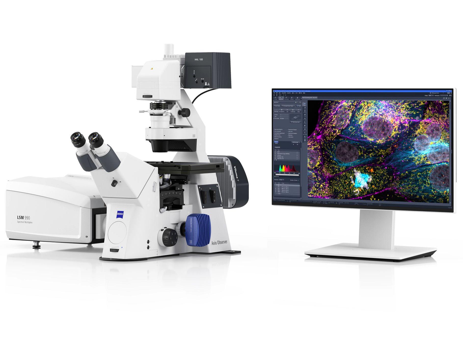
ZEISS LSM 990 Spectral Multiplex
ZEISS LSM 990 Spectral Multiplex excels in the spectral separation of fluorescent labels. Optimize your advanced spectral multiplexing experiments with numerous protein markers and clear separation of fluorescence signals while reliably eliminating autofluorescence.
Your Upright Microscope Platform for Life Science Research
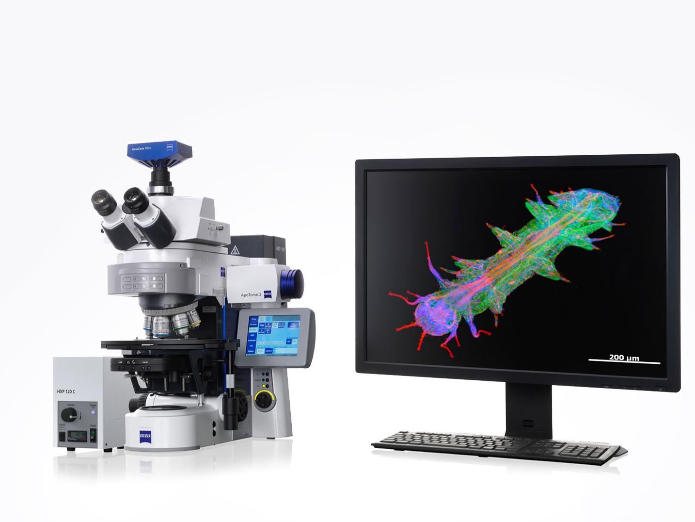
ZEISS Axio Imager
Combine the best for life science research in a single upright platform. Whether you are simply observing and recording or performing highly complex imaging experiments: It's easy to customize system components to meet the needs of your applications.
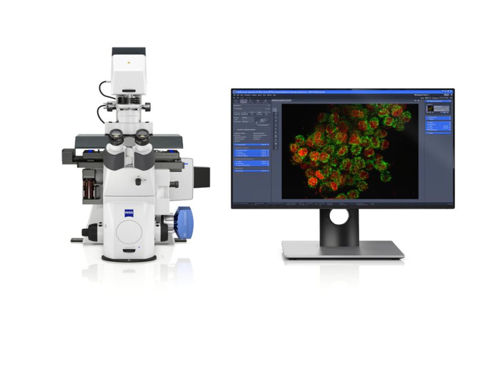
Inverted Microscope Platform with AI Assisted Experiment Startup
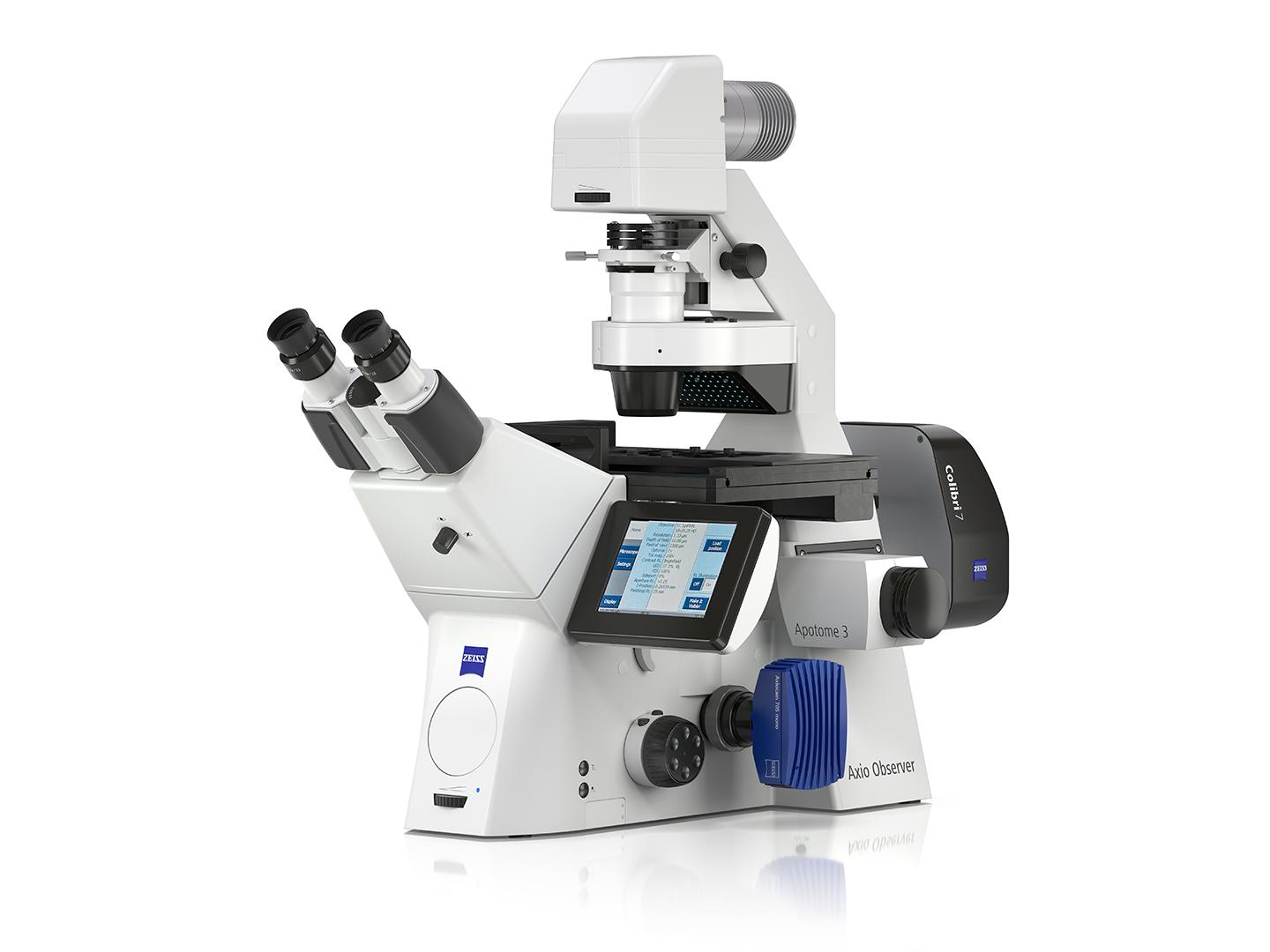
ZEISS Axio Observer
ZEISS Axio Observer is your inverse platform for demanding multimodal imaging of living and fixed specimens. Combine Axio Observer with a wealth of technologies and refine it to support your experiments precisely.
Your Smart Microscope for Cell Culture and Research
ZEISS Axiovert 5
ZEISS Axiovert 5 is a versatile inverted microscope designed for life science and biomedical research labs. It features high quality long working distance optics for phase contrast and powerful LED illumination.
Fast Optical Sectioning of Developing Organisms and Tissue Microstructures
ZEISS Lattice SIM 3
ZEISS Lattice SIM 3 is specifically designed to meet the imaging requirements of multicellular organisms and tissue sections. Discover fast optical sectioning at superior quality, large fields of view with access to smaller regions of interest, near-isotropic resolution, and the gentlest super-resolution imaging possible.
ZEISS solutions for spatial biology facilitate the seamless integration of spatial profiling into your workflows, ensuring efficient and consistent multiplexed spatial profiling at scale.
For high-volume digital pathology and whole-slide scanning, ZEISS Axioscan 7 is the flagship choice. It offers fully automated, high-resolution scanning of brightfield and fluorescence slides up to 100×, fast batch loading (up to 100 slides), and seamless integration with ZEN and arivis analysis software for AI-ready image management.
ZEISS automation solutions are specifically designed to eliminate workflow bottlenecks and reduce operator dependency in pathology labs. Our Axioscan 7 Spatial Biology with SlideStream Workflow Manager delivers a truly hands-off experience.
Yes. Platforms like LSM Airyscan, Axio Observer and the Axioscan 7 support high-plex fluorescence imaging, while ZEN software’s spectral unmixing accurately separates overlapping markers.
Systems such as LSM 990 or Elyra 7 capture sub-cellular details and multiple biomarkers in thick tissue, enabling precise tumor microenvironment analysis and molecular pathology.
ZEISS arivis software suite can handle very large 2D, 3D, and 4D datasets—such as multiplexed whole-slide scans—without down-sampling. They provide AI-driven segmentation, quantitative biomarker analysis, and interactive 3D visualization, making it easy to measure cell populations, map tumor microenvironments, and integrate results with other research or clinical data systems.
Comprehensive Solutions and Capabilities
Discover Our Application Hub
Explore applications to discover tailored solutions for your unique laboratory needs and elevate your research capabilities.
; GFAP (astrocytes)")
; GFAP (astrocytes)")