By integrating advanced optical technologies, AI-powered image analysis, and seamless digital workflows, ZEISS enables researchers to achieve faster, more accurate diagnoses and deeper insights into tissue structures. With a commitment to innovation, reliability, and user-centric design, ZEISS empowers histopathologists to push the boundaries of biomedical research and improve patient outcomes. Its comprehensive portfolio, including light, electron, and X-ray microscopy, supports multi-scale imaging and correlative workflows, ensuring a holistic understanding of complex biological systems. Backed by decades of expertise and global support, ZEISS remains a trusted partner for advancing histopathology research.
Examine Tissue Morphology with Precision & Clarity
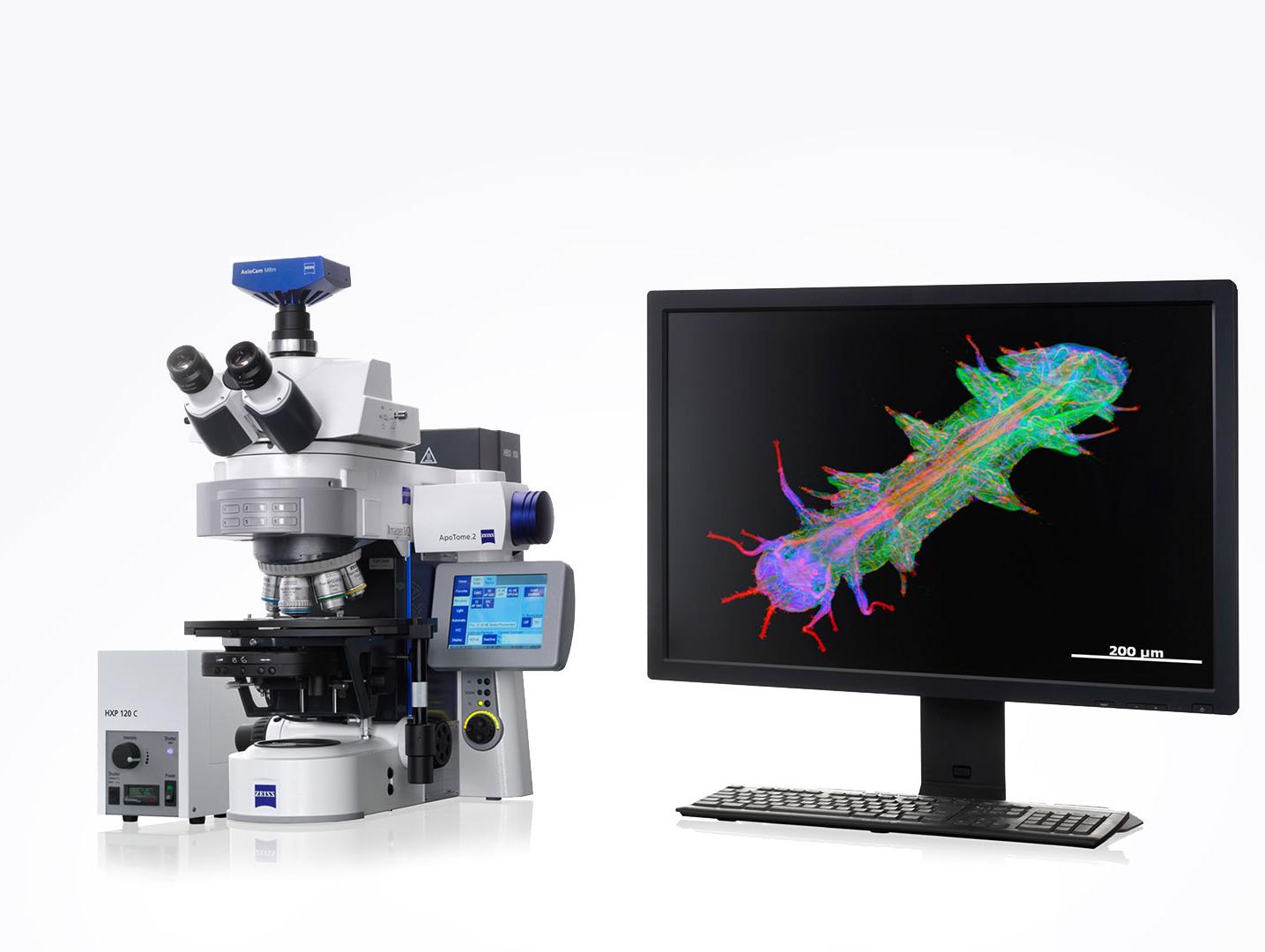
Courtesy of K. Ono and T. Horie, Kyoto University Hospital, Japan
From upright and inverted microscopes to slide scanners, lightsheet, and SEM, ZEISS' portfolio offers unmatched flexibility to your imaging and sample needs.
Software solutions that provide automated AI segmentation, measurement, and analysis, drastically reducing manual analysis time and ensuring reproducibility.
Thanks to our sample preparation methods and advanced light-sheet microscopy, we can visualize entire tissue samples in 3D without physically sectioning them. Our ability to perform rapid optical sectioning and obtain thousands of digital images in seconds or minutes has significantly increased our throughput.
Recommended Products for Histology
High-Performance Digital Slide Scanning
ZEISS Axioscan 7
The ZEISS Axioscan 7 is a high-performance slide scanner designed to support histology applications by enabling fast, high-resolution, and automated digitization of histological slides. It is particularly suited for research, pathology, and education, offering precise imaging and advanced analysis capabilities.
Your Upright Microscope Platform for Life Science Research
ZEISS Axio Imager 2
Combine the best for life science research in a single upright platform. Whether you are simply observing and recording or performing highly complex imaging experiments: It's easy to customize system components to meet the needs of your applications.
Inverted Microscope Platform with AI Assisted Experiment Startup
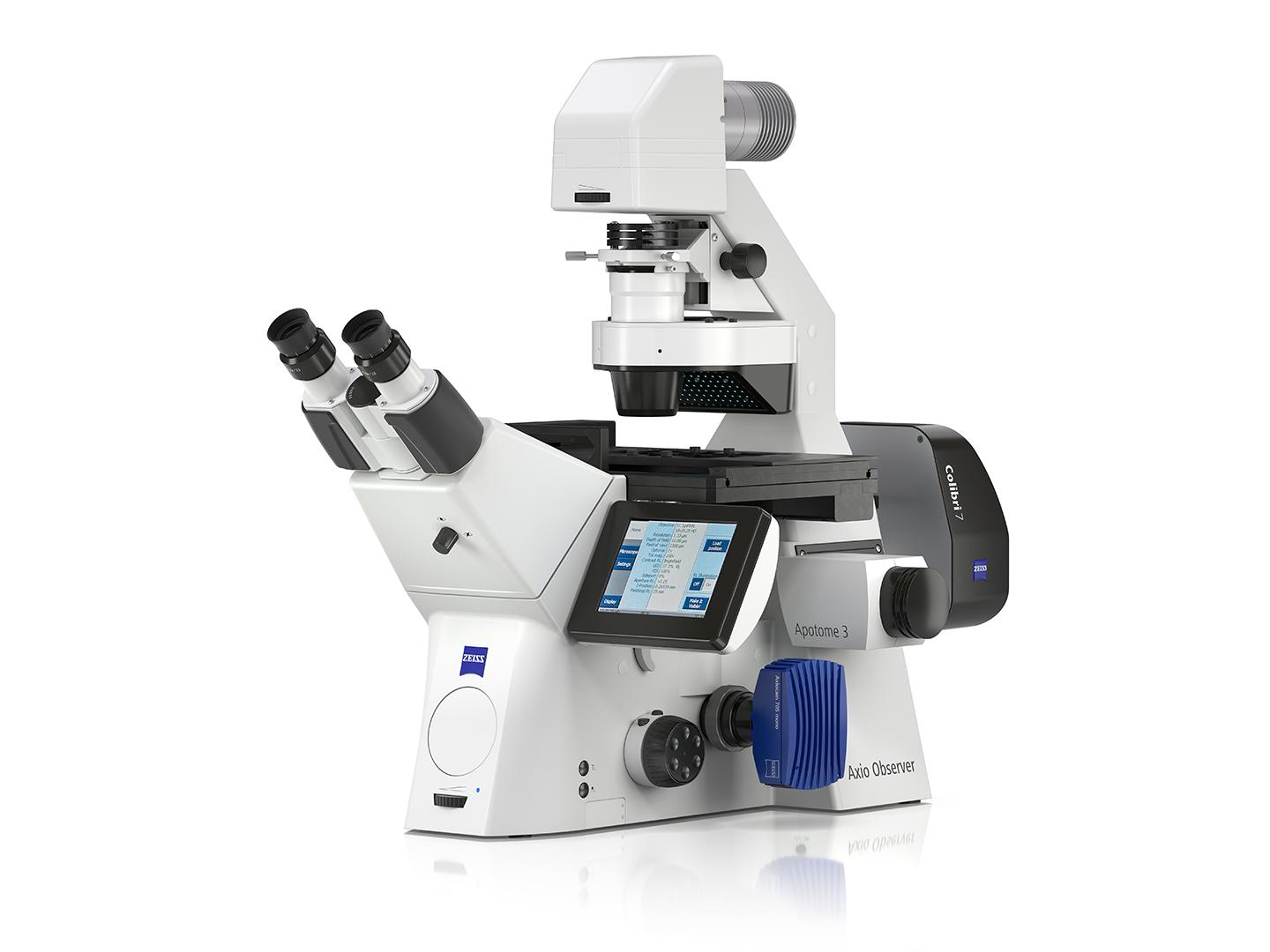
ZEISS Axio Observer
The ZEISS Axio Observer is an advanced inverted microscope platform that supports histology research by enabling high-resolution imaging, live-cell observation, and advanced analysis of tissue samples. Its modular design and compatibility with various contrast techniques make it ideal for diverse histological applications.
Light-Sheet Multiview Imaging of Living and Cleared Specimens
ZEISS Lightsheet 7
ZEISS Lightsheet 7 enabling fast, gentle, and high-resolution 3D imaging of large, cleared tissue samples. It preserves the spatial context of tissues, allowing researchers to study cellular and molecular structures in their native environment without the need for extensive sectioning.
ZEISS arivis advanced image analysis ecosystem offers a family of software products that help you make the most of your valuable datasets. We know you face the challenges of the ever-increasing volume, amount and speed in which microscopy data is generated. Limited infrastructure and inconvenient storage silos further impede progress.
ZEISS provides a wide range of microscopy solutions tailored for histology, including:
Light Microscopes: For routine histological analysis, such as ZEISS Axioscope and ZEISS Primostar.
Digital Microscopes: For documentation and sharing, like ZEISS Smart Microscopy systems.
Confocal Microscopes: For high-resolution imaging of tissue sections, such as ZEISS LSM 990.
Slide Scanners: For digitizing histological slides, like the Axioscan 7.
Yes, ZEISS microscopes are compatible with all standard histological stains, including:
Hematoxylin and eosin (H&E)
Periodic acid-Schiff (PAS)
Immunohistochemical (IHC) stains
Fluorescent dyes for advanced imaging
ZEISS systems are optimized to provide accurate color reproduction and fluorescence imaging, ensuring reliable results.
Here are the main fluorescence-imaging options ZEISS offers for histology and histopathology, covering everything from routine IHC to advanced multiplex and 3-D tissue analysis:
Upright Research Microscopes: ZEISS upright research microscopes, such as the Axio Imager series, deliver exceptional optical performance, modularity, and advanced imaging capabilities, making them ideal for demanding applications in life sciences, materials research, and histology.
Inverted Microscopes: ZEISS inverted microscopes, like the Axio Observer series, are designed for advanced live-cell imaging and materials research, offering superior stability, flexibility, and high-resolution imaging for dynamic and long-term studies.
Digital Slide Scanning: Systems like the Axioscan 7 provide automated fluorescence whole-slide imaging with multiple filter channels, autofocus, and high-throughput scanning—perfect for multiplex IHC and digital pathology workflows.
Laser Scanning & Super-Resolution Microscopy: Use a focused laser beam to scan samples point by point. The ability of confocal microscopy to eliminate out-of-focus light by employing a pinhole aperture results in excellent optical sections with high contrast and enables the high-resolution reconstruction of three-dimensional structures.
Lightsheet Microscopy: Systems like the Lightsheet 7 can enable fluorescence imaging of cleared whole organs or large tissue blocks with minimal photodamage, capturing intact 3D histological context.
Correlative & Volume Electron Microscopy: Combine fluorescence (widefield or confocal) with ZEISS GeminiSEM or Volutome for precise correlation of fluorescent markers to ultrastructure.
Specialized objectives and filters for unique imaging needs.
Tailored software workflows.
Personalized microscopy application services where we do the research for you.
ZEISS provides a full suite of software designed to support every stage of histology and histopathology workflows—from image capture to analysis, data management, and cloud collaboration. Here are the key offerings and how they help:
ZEISS ZEN:Core acquisition and analysis platform that streamlines multi-channel tissue imaging and quantitative assessment of stains or biomarkers with reproducible workflows.
Comprehensive Solutions and Capabilities
Discover Our Application Hub
Explore applications to discover tailored solutions for your unique laboratory needs and elevate your research capabilities.