
Skip to main content

Investigating the morphology and dynamic movement of developing embryos without compromise.
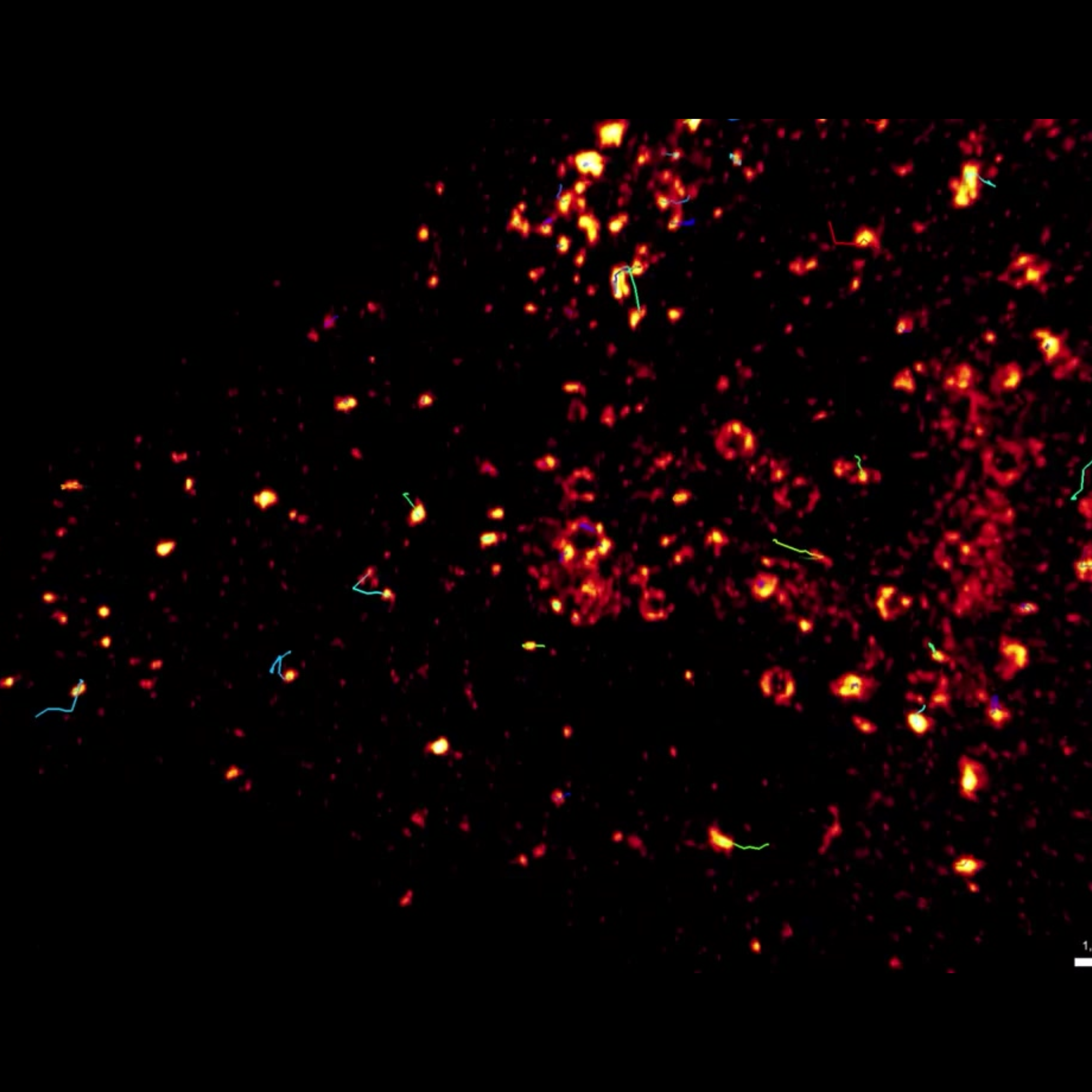

Investigate vesicular transport in live mammalian cells with the unique combination of gentle illumination, high speed, and super-resolution.
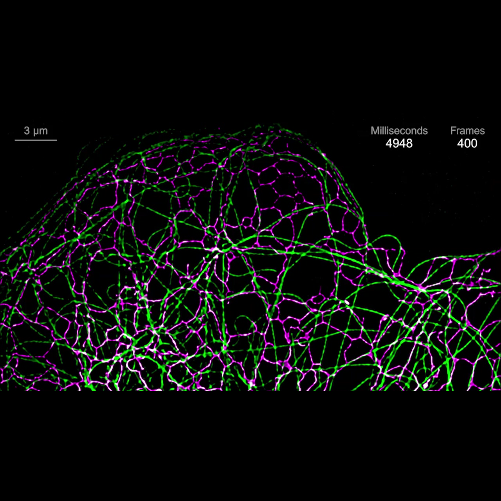

Combine high speed imaging with incredible light efficiency, low photon dosage and sensitivity to observe cellular, subcellular, and even sub-organelle structures in living specimens in 2D and 3D over time.


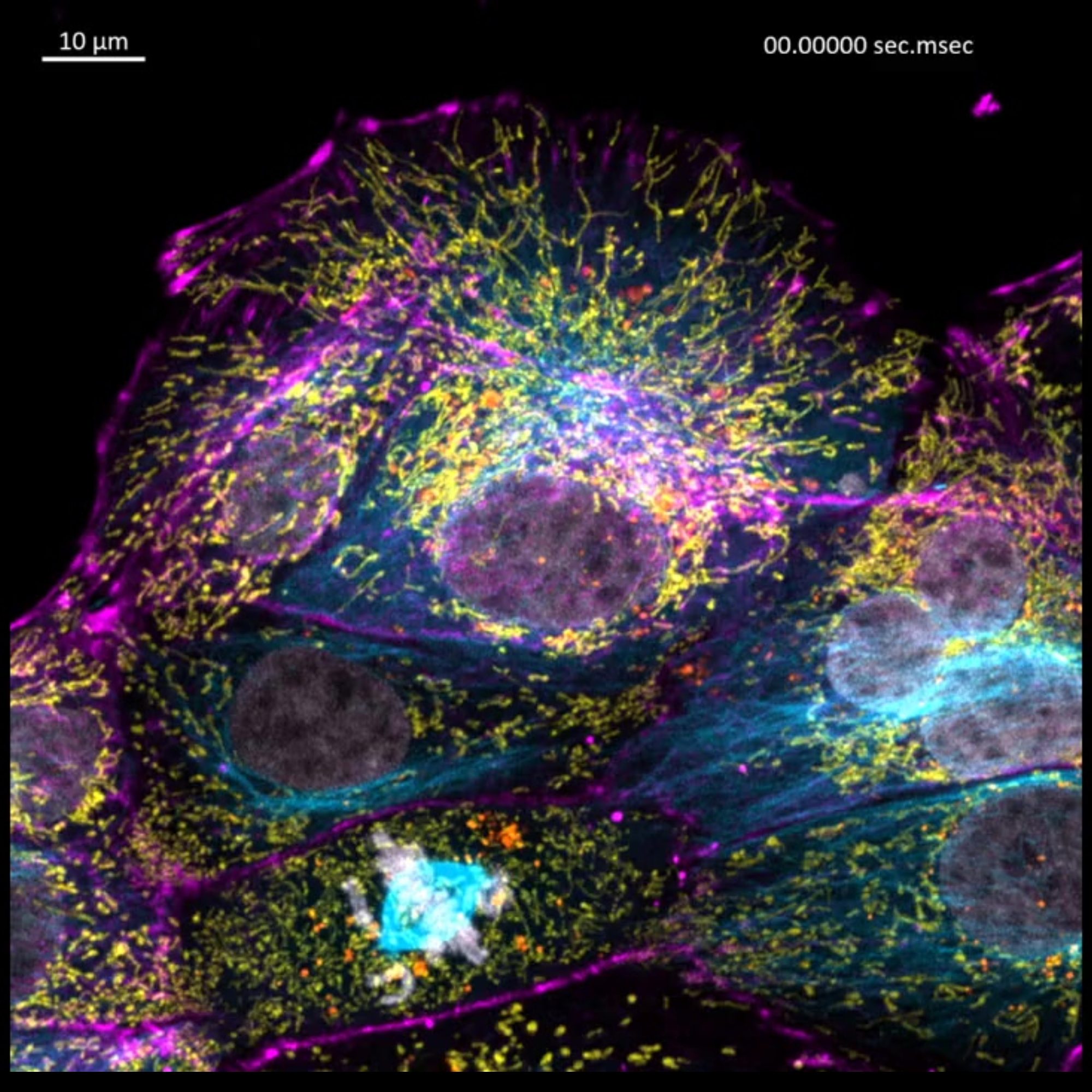
Image mitotic waves in live cells for days with next to no phototoxicity or bleaching.

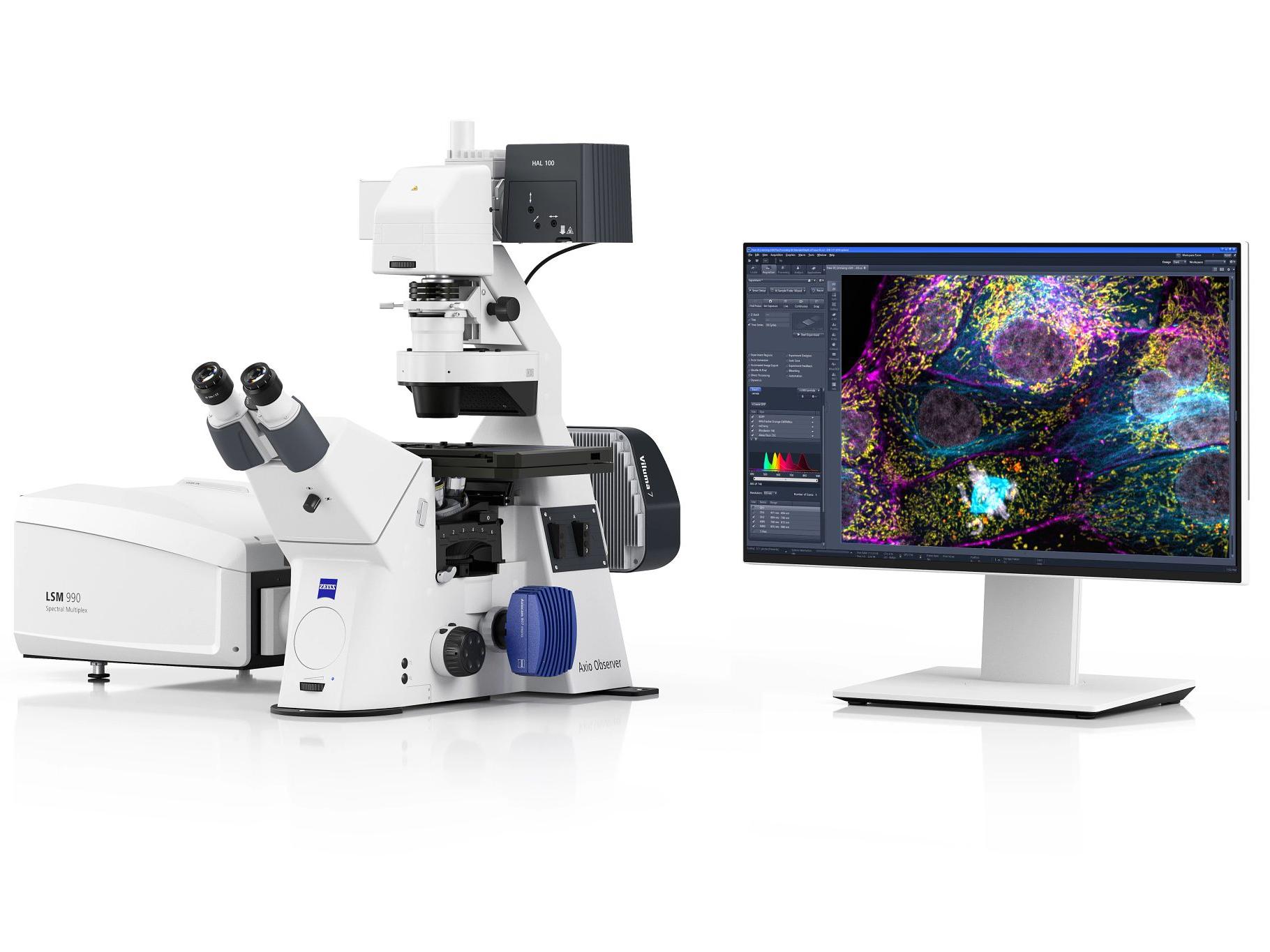
Unparalleled productivity for your demanding spectral imaging experiments, covering a wavelength range from 380 to 900 nm.

An intuitive, easy-to-use interface, providing effortless access to your data during confocal imaging experiments with low time and light investment.
ZEISS Microscopy offers a broad range of advanced microscopy techniques tailored for cell biology research, supporting both
qualitative and quantitative analysis across multiple scales. Here are the key types available:
Light Microscopy
Electron Microscopy (EM):
Correlative Microscopy
Digital and AI-Enhanced Imaging
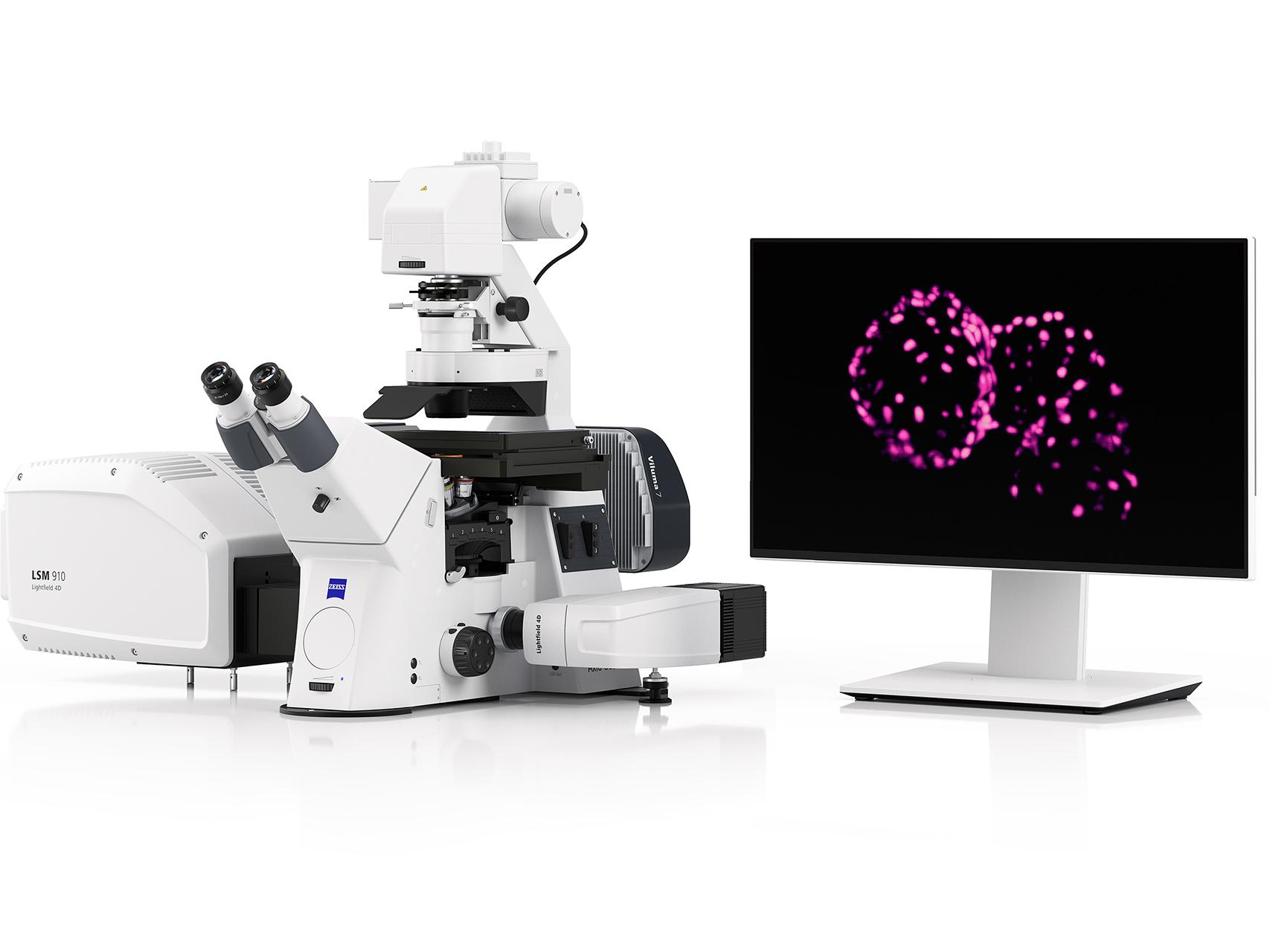
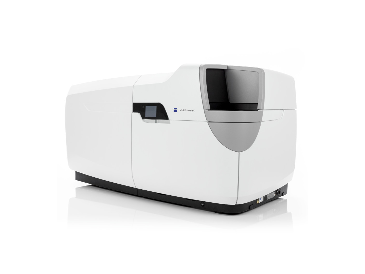
The ZEISS Celldiscoverer 7 and ZEISS LSM 980 and 990 with Airyscan 2 are optimized for live-cell imaging. They integrate incubation systems with precise control of temperature, CO₂/O₂ levels, and humidity, ensuring physiological conditions over long durations. The LSM 990 also offers gentle imaging with high sensitivity, making it ideal for time-lapse experiments with minimal phototoxicity.T
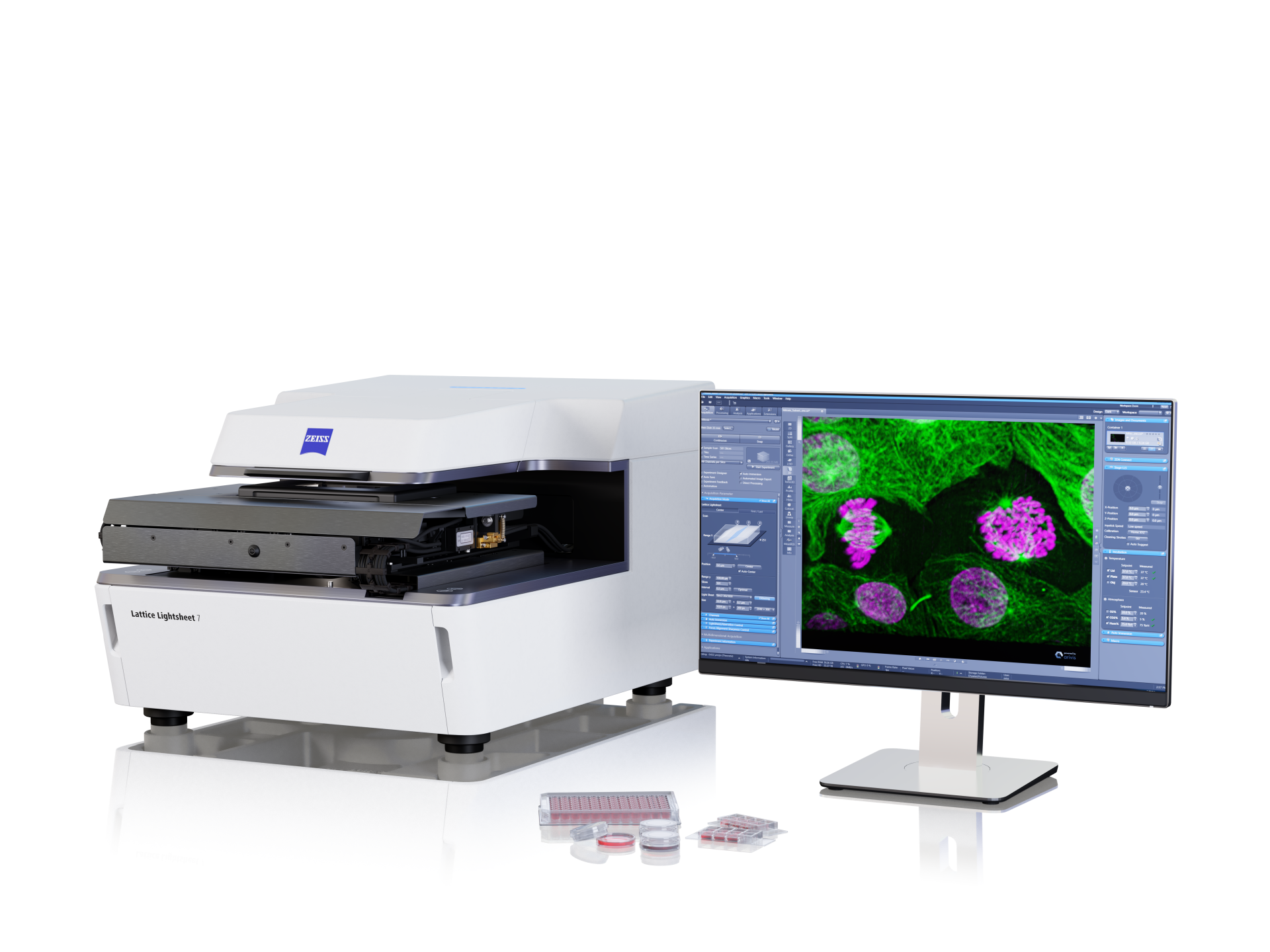
The ZEISS Lattice Lightsheet is ideal for long-term imaging of living cells due to its ability to provide high-resolution, three-dimensional images with minimal phototoxicity and photobleaching. This advanced imaging system uses lattice light-sheet illumination, which allows for gentle and continuous observation of cellular processes over extended periods, preserving cell viability and function while capturing dynamic events with exceptional clarity.
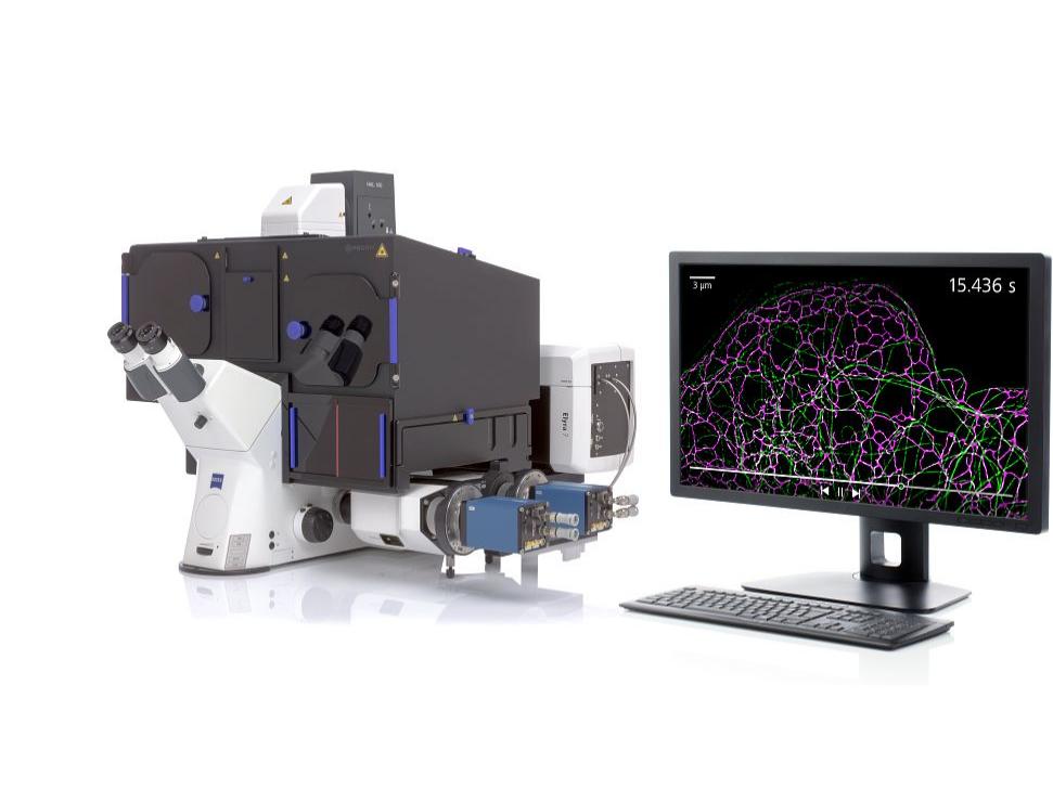
The Zeiss LSM 980 & 990 with Airyscan is ideal for long-term imaging of living cells because it utilizes Airyscan technology to achieve super-resolution imaging with enhanced sensitivity and reduced phototoxicity. This allows researchers to capture detailed, high-quality images of cellular processes over extended periods while maintaining cell health and viability. Additionally, the system's advanced environmental control features ensure optimal conditions for live-cell imaging, making it well-suited for prolonged studies of dynamic cellular events.
ZEISS provides multiple super-resolution modalities:
Yes. ZEISS offers several systems optimized for imaging 3D cell cultures:
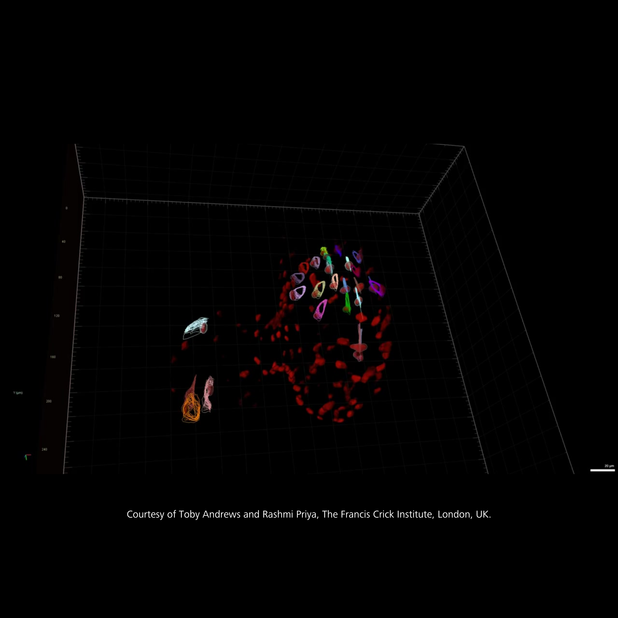
Image analysis tools (ZEISS arivis Pro, ZEN 3D) support full workflows from acquisition to 3D rendering and analysis.
Yes, ZEISS provides powerful AI-based tools for image segmentation, classification, and quantitative analysis, specifically designed to enhance cell biology workflows, including: