

Use Cases for Complex In Vitro Models
New Approach Methodologies (NAMs) High-resolution, scalable imaging and analysis workflows for 3D HCA imaging
New Approach Methodologies (NAMs) or Complex in vitro models (CIVMs), including organoids, spheroids, and organs-on-a-chip, require deep, gentle, and scalable 3D imaging. ZEISS microscopy systems combined with ZEISS arivis software connect image acquisition to quantitative 3D analysis for high-throughput screening and predictive drug discovery.
The challenge of imaging complex 3D cell cultures
Because organoids and other 3D cell cultures are thick, heterogeneous, and highly light-scattering, imaging workflows must address limitations in depth, resolution, sensitivity, speed, and throughput.|
Challenge |
How we address it |
|
Penetration Depth: Scattering and refractive mismatches limit light penetration |
Advanced optics and confocal and lightsheet modalities optimize depth without compromising resolution |
|
Resolution: Small subcellular details are lost in thick samples |
ZEISS Airyscan delivers super-resolution with high sensitivity |
|
Sensitivity: Gentle Imaging – Phototoxicity damages live samples |
ZEISS Lattice Lightsheet 7 and ZEISS Lightfield 4D modes minimize light exposure |
|
Speed: High-content workflows demand faster imaging |
ZEISS Celldiscoverer 7 with ZEISS LSM 910 module ensures rapid volumetric acquisition |
|
Throughput: Drug discovery requires scalability |
Integrated automation and ZEISS arivis analytics enable plate-based, high-volume workflows with 3D context and a multitude of 3D parameters |
 of the Austrian Academy of Sciences")
 of the Austrian Academy of Sciences")
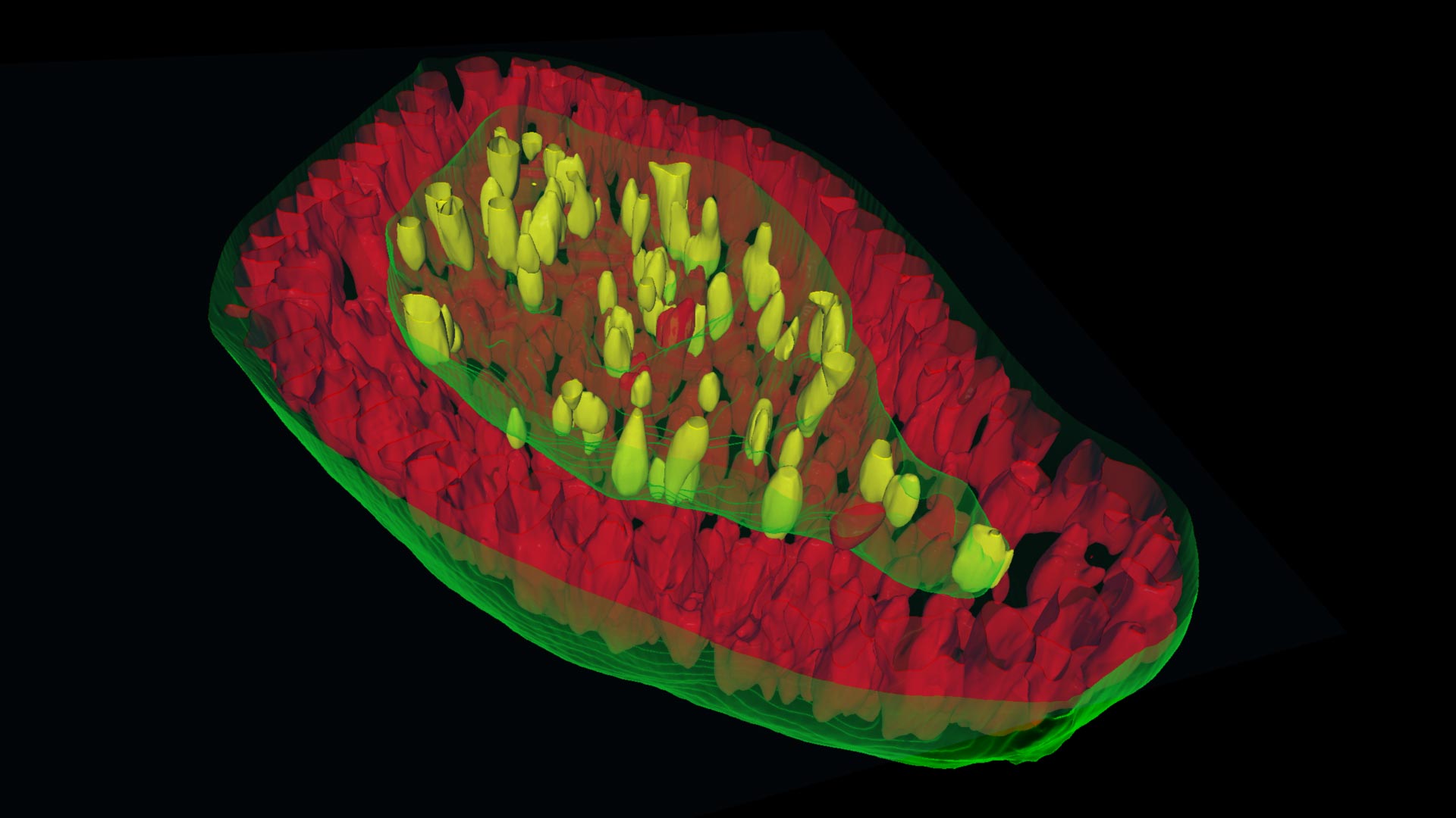
, membrane (mem9-GFP), enterocytes (Aldolase B-Alexa 647).")
, membrane (mem9-GFP), enterocytes (Aldolase B-Alexa 647).")
. Study the role of Wnt signaling in organoid formation.")
. Study the role of Wnt signaling in organoid formation.")
 of the Austrian Academy of Sciences")
 of the Austrian Academy of Sciences")


ZEISS arivis software has allowed us to extract crucial insights from our high-content imaging experiments, leading to better-informed decisions in drug discovery and development.
Advanced 3D imaging for new approach methodologies: organoids, spheroids, and organ-on-a-chip models
CIVMs are reshaping how the pharmaceutical and biotechnology industries study disease, evaluate drug efficacy and safety, and develop new therapies. By more closely replicating the structure and function of human tissues compared to traditional 2D cultures, these advanced 3D systems deliver greater translational relevance, reduce reliance on animal testing, and accelerate predictive drug discovery and personalized therapeutic development.
As adoption of 3D systems accelerates across pharma, biotech, and CROs, new challenges have emerged: thick, scattering, and heterogeneous samples require imaging systems that combine depth, sensitivity, resolution, and throughput, while handling the massive datasets generated in volumetric studies.
ZEISS 3D Imaging Solutions, integrated with ZEISS arivis ecosystem, deliver a unified workflow for 3D tissue imaging and analysis, enabling deep, gentle imaging, and reproducible results that scale from single organoids to plate-based screening, supported by Copilot-guided acquisition and powerful 3D segmentation algorithms powered by advanced AI-driven analysis.
Sample courtesy of Dr Shengping Xiao and Dr Sen Ye, Xellar Biosystems
3D images of the organ chips developed by Xellar Biosystems (with gastrointestinal tumor organoids on OC-Plex 32). Acquisition: Imaging system: LSM 990 LF4D.
Sample courtesy of Dr Shengping Xiao and Dr Sen Ye, Xellar Biosystems
ZEISS Microscopy solutions enable rapid, high-resolution 3D imaging of organ-on-chip models, capturing complex cellular architecture within physiologically relevant environments. With Lightfield 4D, uneven and volumetric samples can be imaged in a single acquisition—eliminating time-intensive tiling and z-stacks. Paired with ZEISS arivis, researchers can precisely visualize and quantify structures such as vascular networks, accelerating insights in disease modeling and drug response.
Scroll animation items


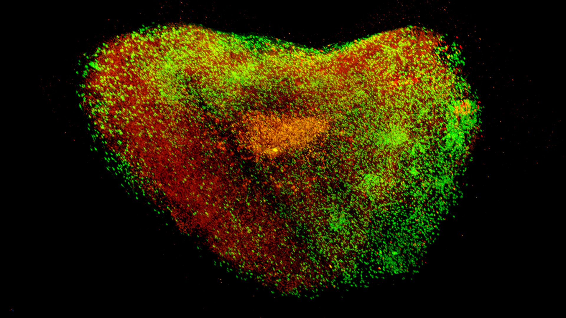
Human breast cancer organoids in matrigel
Capture deeper, clearer insights from live organoids—without compromising viability or throughputSolution: ZEISS Celldiscoverer 7 with LSM / Airyscan + ZEISS arivis
Key outcomes:
- Gentle long-term acquisition of live tumor organoids
- High-throughput image capture and analysis across multiwell plates
- AI-poewred segmentationf ro rapida nd precise quantification


Human embryonic stem cell-derived spinal cord organoids
Track complex tissue development in real time while enabling seamless, large-scale analysis.Solution: ZEISS Lattice Lightsheet 7 + ZEISS arivis
Key outcomes:
- Fast, gentle, high-resolution imaging of developing organoids
- Phototoxicity-free visualization of real-time tissue dynamics
- Seamless cloud-based analysis and data sharing for multi-sample studies
- Scalable handling of large datasets with ZEISS arivis


Cleared spheroid of a co-culture of HCT-116-GFP (colon cancer)
Advance spheroid research with fast, sensitive imaging and robust 4D analysis at scale.Solution: ZEISS LSM 910/990 with Lightfield 4D + ZEISS arivis
Key outcomes:
- Deep and fast imaging
- High sensitivity for short exposure times
- High-throughput acquisition of numerous Spheroids in multiwell plates
- Scalable 4D analysis, efficient segmentation and tracking for quantitative toxicology and metabolism studies


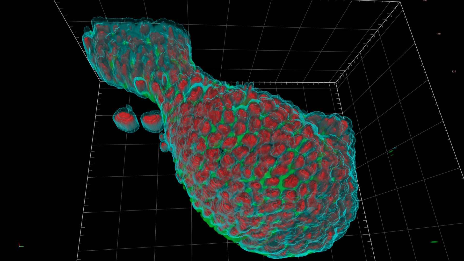
3D rendering of an intestinal organoid
Accurate 3D segmentation, quantification, and tracking of cellular dynamicsSolution: ZEISS arivis Pro Software + Celldiscoverer 7
Key outcomes
- Cell-level organoid growth analysis
- Organoid volume quantification


3D image acquisition of colorectal cancer organoids
Enhanced drug response analysis and cellular interactionSolutions: ZEISS LSM Lightfield 4D
Key outcomes:
- Increased throughput for screening large sample sets
- Fast, high-content 3D imaging
- Scalable, quantitative analysis for drug response studies
Imaging systems for New Approach Methodologie, including complex in vitro models (CIVMs)


Organoids Research FAQs
-
The thickness of organoids is often challenging for traditional confocal imaging due to light scattering, tissue absorption, and phototoxicity. ZEISS Lattice Lightsheet 7 and ZEISS Lightfield 4D provide gentle, rapid volume imaging with minimal photodamage, enabling extended time-lapse studies. The lightsheet approach illuminates only the focal plane being imaged, while lightfield offers one volume for each snapshot. The result is dramatically reduced photobleaching while maintaining exceptional image quality throughout the entire organoid volume.
-
High-throughput organoid screening requires limited user input combined with high-quality imaging to adequately survey each sample. The ZEISS Celldiscoverer 7 is fully automated and ensures reproducible results at scale. With its integrated workflows intended for high content screening, the Celldiscoverer 7 enables researchers to monitor development and response to treatment in either fixed or live samples.
-
This requires balancing resolution with field of view – a common challenge in organoid research. The ZEISS LSM 990 Lightfield 4D combines fast volume acquisition with enhanced sensitivity for deep confocal imaging. For comprehensive workflows, arivis Pro offers precise 3D analysis allowing you to correlate molecular-level events with tissue-scale organization patterns.