Lattice SIM² 3D image of a C. elegans larvae.  Image shows a maximum intensity projection.
Sample courtesy of InSphero AG. Schlieren, Switzerland Courtesy of Mango Lab (University of Basel, Switzerland)
Use Case

C. Elegans

From neural circuits to developmental biology, discover how ZEISS empowers high-resolution imaging and data-driven insights in Caenorhabditis elegans research.

Dynamic Insights from Development to Behavior in C. elegans

ZEISS microscopy solutions empower researchers to unlock insights from the C. elegans model across developmental biology, neuroscience, and genetics. Whether you are tracking cell lineages in embryos, imaging neuronal activity, or screening genetic variants, ZEISS systems—such as Lightsheet 7, Lattice Lightsheet, Airyscan-enabled LSMs, Celldiscoverer 7, and Crossbeam EM—deliver the resolution, speed, and versatility needed for every stage of discovery. From whole-organism dynamics to ultrastructural detail, ZEISS provides scalable, reproducible workflows supported by arivis Pro for advanced analysis, segmentation, and quantification. Together, these tools enable consistent, data-rich imaging that accelerates discoveries in behavior, aging, infection biology, and beyond.

  • High-Resolution Imaging is Essential for Small, Transparent Organisms

    C. elegans are optically clear but require exceptional contrast to resolve fine structures like neurons, germline cells, and organelles. ZEISS Airyscan and Lattice Lightsheet systems deliver the sensitivity and resolution needed to capture these details without compromising viability.

  • Dynamic Biological Processes Are Easy to Miss in Live Worms

    From embryogenesis to locomotion, critical events unfold rapidly in vivo. ZEISS Lightfield 4D and Airyscan Fast combine speed and gentleness to capture organism-wide and cellular dynamics in 4D, preserving biological function while revealing processes in unprecedented detail.

  • Large-Scale Genetic Screens Generate Complex Datasets

    C. elegans are a cornerstone of high-throughput genetics, producing vast datasets that demand reproducibility. ZEISS Celldiscoverer 7, Axioscan, and arivis Pro streamline population imaging, automated analysis, and multi-sample workflows, enabling consistent insights across screens, mutants, and drug assays.

A Valuable Model Organism for Translational Research

  • Research conducted by Andre Rajoo, Florida Atlantic University

    Andre Rajoo, Florida Atlantic University discusses his research highlighting the remarkable similarities between C. Elegans and human

Shop Microscopes Online

Precision At Your Fingertips

Accelerate your C. Elegans studies with our bundled solutions.

Imaging Large Model Organisms

Fixed or Living
  • Nuclear tracking in the beating heart of a zebrafish embryo. High-speed volumetric imaging enables zebrafish heartbeat data collection in real time for the first time in 3D.

    Courtesy of Toby Andrews and Rashmi Priya, The Francis Crick Institute, London, UK.

    Zebrafish

    From development to disease models, explore the full potential of zebrafish using tools for deep, dynamic, and reproducible imaging. Explore zebrafish biology with exceptional resolution, speed, and flexibility. Whether you're tracking cardiac dynamics, mapping neural activity, analyzing cell migration, or screening drug responses.

  • Drosophila embryo imaged with Multiplex Mode for LSM 900.

    Sample courtesy of J. Sellin, LIMES, Bonn, Germany

    Drosophila

    Image, screen, and analyze Drosophila to uncover mechanisms driving biology and behavior. Whether you're imaging live embryos, tracking cell migration, or screening genetic variants, ZEISS systems offer high-speed, low-phototoxicity 4D imaging with batch-mode efficiency and unmatched clarity.

  • mouse_hippocampus

    Mouse

    Investigate mouse models with unmatched clarity, speed, and flexibility. Whether you are mapping neural circuits, studying tumor microenvironments, tracing immune cell dynamics, or exploring organ development, ZEISS tools support gentle live imaging, deep-tissue visualization, and high-throughput analysis.

  • 3D segmented result after the starch granules have been segmented using Dragonfly Pro. Subsequent quantification of size and distribution of start granules can be performed from the high-resolution data

    Sample Courtesy of Jason (Chunjie Cao)

    Arabidopsis

    Discover a comprehensive toolkit for Arabidopsis thaliana research, supporting everything from basic plant development studies to advanced cellular and molecular analyses. Whether you're imaging whole seedlings, scanning tissue sections, or tracking gene expression at the single-cell level, ZEISS helps you visualize plant biology with clarity and confidence.


  • Human Distal Lung Organoid showing club cells and ciliated cells, everted for 10 days.

    Courtesy of Prof. C. Kuo, Department of Medicine, Hematology Division, Stanford University, USA.

    Organoids & Spheroids

    Streamline organoid analysis and spheroid screening with high-content 3D imaging solutions for faster research outcomes. Transform your organoid workflows with imaging solutions designed specifically for the unique demands of 3D cell culture research.

C. Elegans Research FAQs

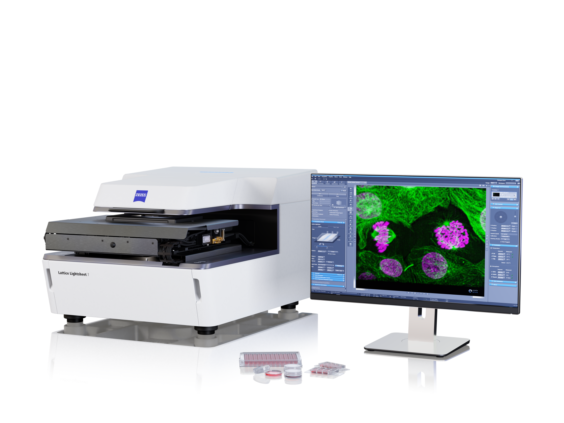
  • C. elegans embryos are sensitive and require gentle imaging to follow rapid developmental events. ZEISS Lightsheet 7 and Lattice Lightsheet systems enable long-term, volumetric imaging with minimal phototoxicity, while arivis Pro tracks cell lineages and divisions to capture the full developmental trajectory.

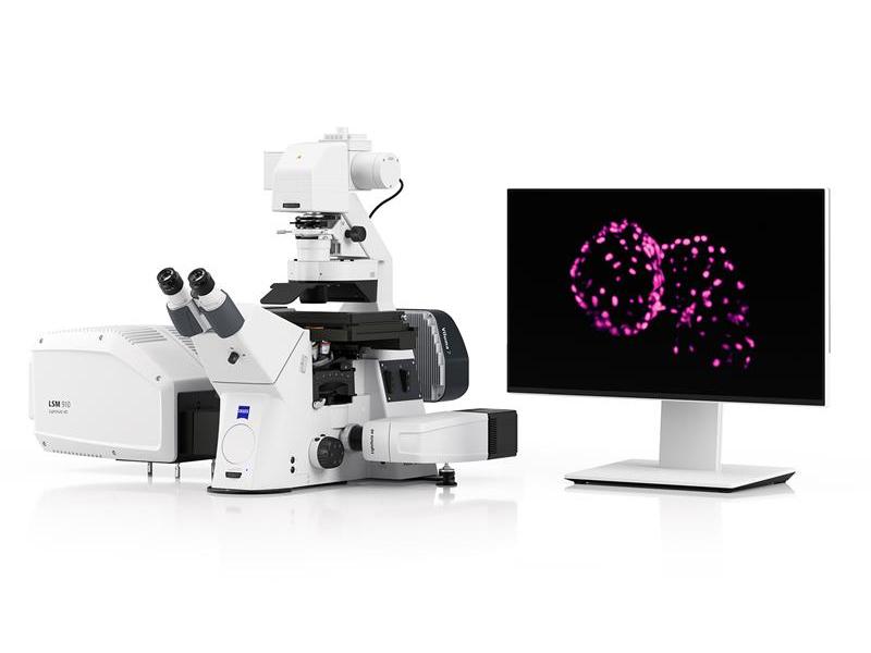
  • Functional imaging in C. elegans benefits from high speed and sensitivity. ZEISS Lightfield 4D and Airyscan Fast capture real-time calcium or sensor dynamics in intact animals, while Crossbeam EM provides ultrastructural detail for connectomic reconstruction. Together, these tools link neural activity with circuit architecture.

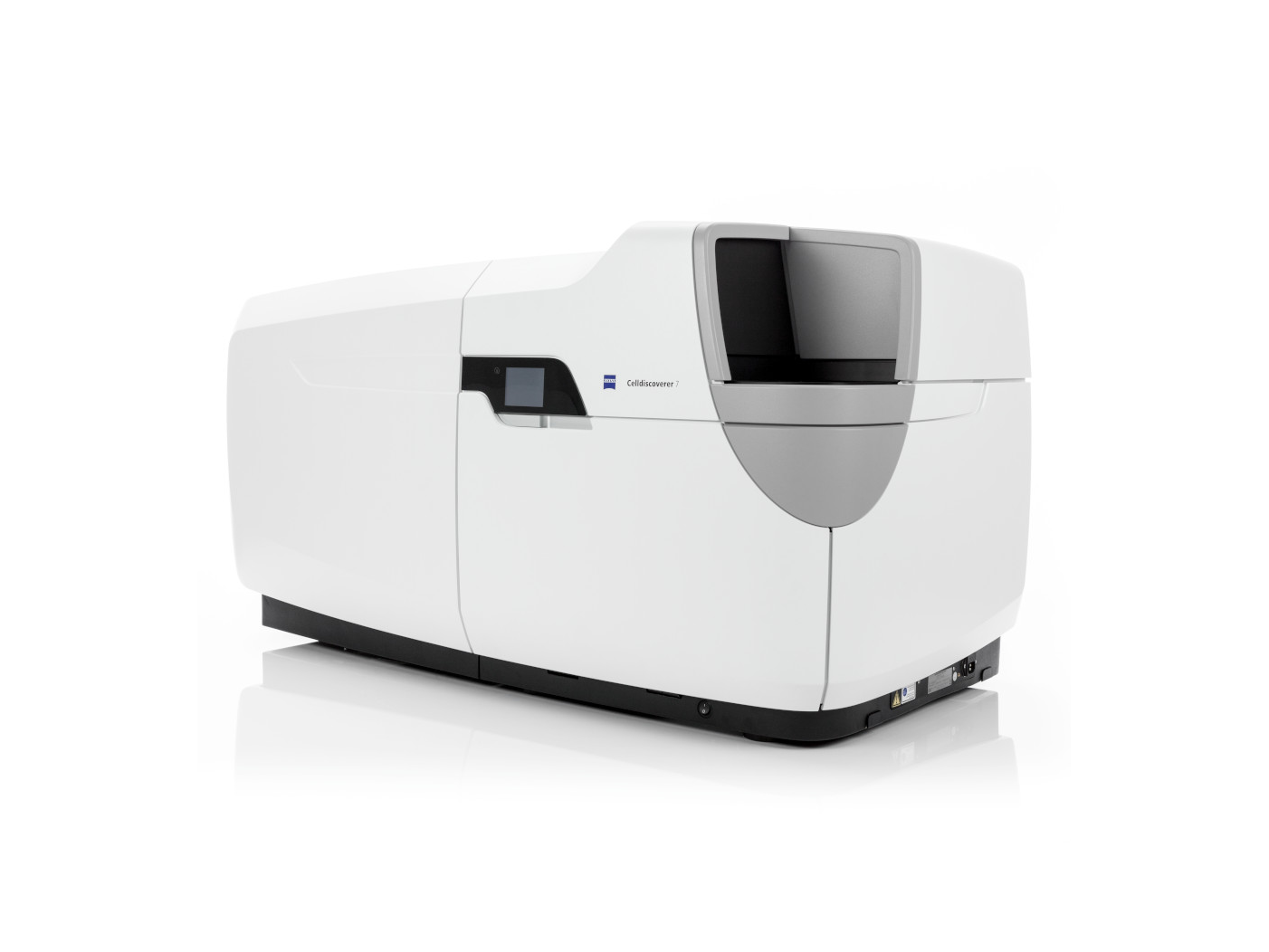
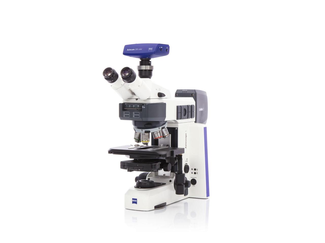
  • From locomotion to sensory-driven responses, behavior studies often require multi-worm tracking over time. ZEISS Celldiscoverer 7 and Axioscope systems allow standardized, high-throughput acquisition, while arivis Pro automates segmentation and trajectory analysis for consistent, reproducible results.

  • C. elegans screens can involve hundreds of conditions or genetic variants. ZEISS Celldiscoverer 7 and Axioscan streamline high-content imaging in multi-well formats, while arivis Pro provides automated analysis pipelines to quantify phenotypic changes, accelerating drug discovery and functional genomics.

  • C. elegans is increasingly used to model infection biology, from fungal invasion to bacterial colonization. ZEISS Airyscan confocal systems deliver the resolution and sensitivity to visualize host–pathogen interactions in vivo, revealing cellular responses and pathogen dynamics in real time.

  • Imaging whole animals, long-term time-lapse experiments, or multi-sample screens can generate terabyte-scale datasets. ZEISS arivis Pro provides scalable infrastructure for stitching, segmentation, and quantification, ensuring reproducible insights across experiments and enabling data sharing across teams.

Comprehensive Solutions and Capabilities

Discover Our Application Hub

Explore applications to discover tailored solutions for your unique laboratory needs and elevate your research capabilities.