ZEISS at Cell Bio 2026
Booth #713 | Dec 13-15 | San Diego, CA


Discover what's new at ZEISS
Stop by Booth #713 to see what's new at ZEISS for cell biology imagingZEISS LSM Lightfield 4D
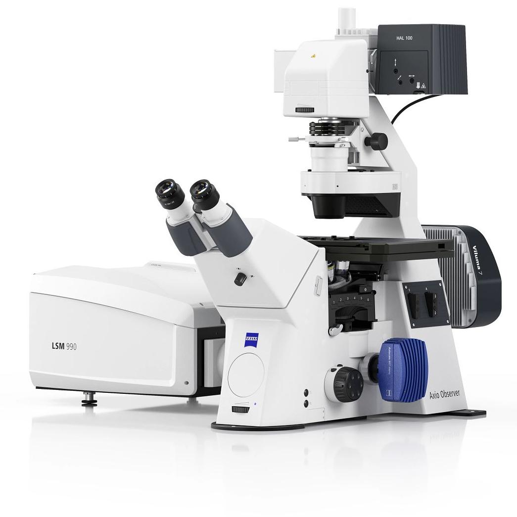
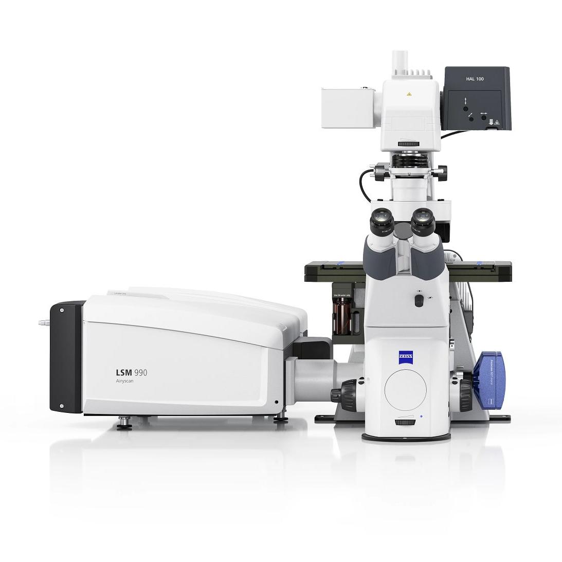
Instant volumetric high-speed imaging of living organisms- Instant volumetric imaging with one-snap 3D capture of fast biological processes up to 80 volumes per second.
- Capture long-term live imaging experiments with reduced phototoxicity.
- Combine Lightfield 4D with the flexibility of a fully featured LSM platform.


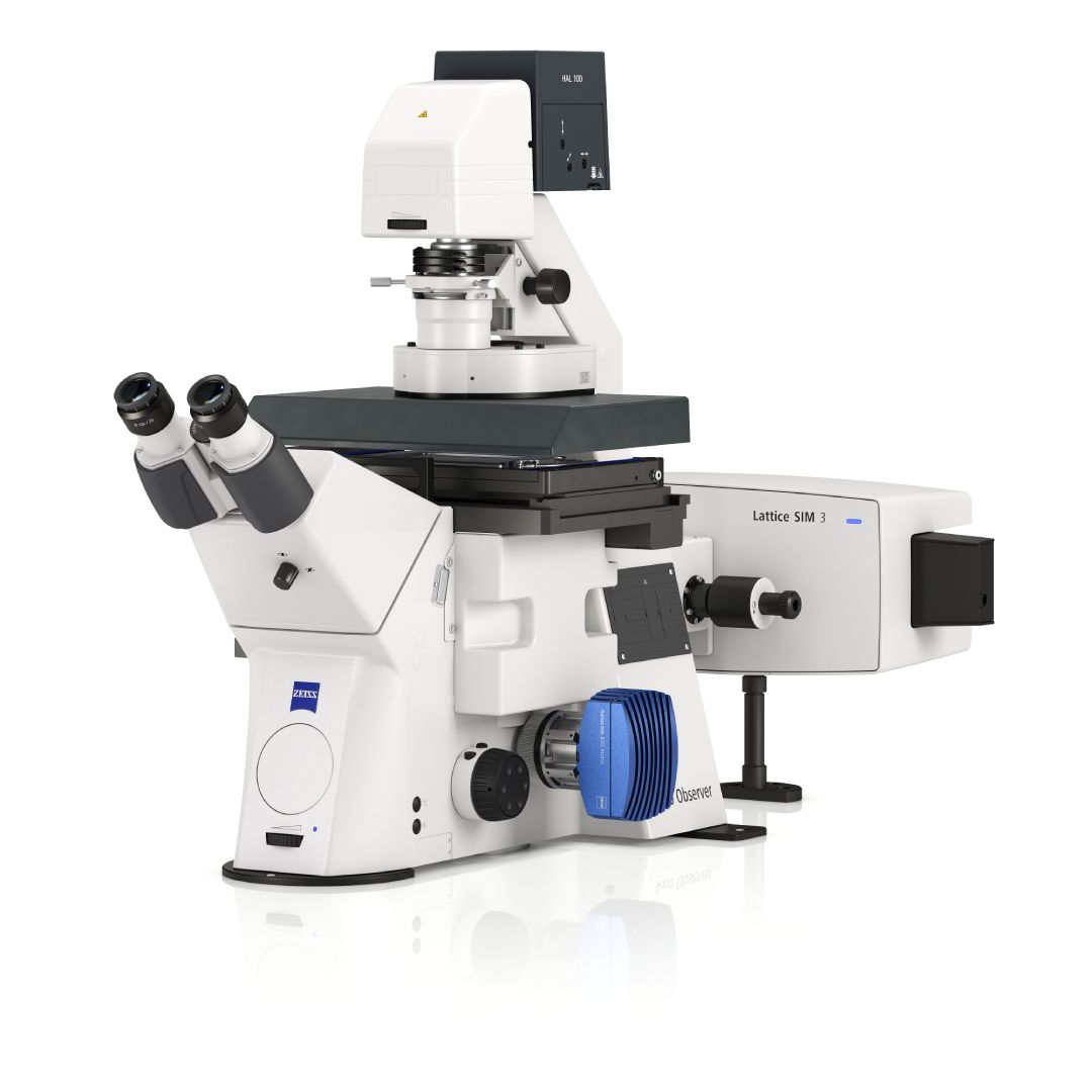
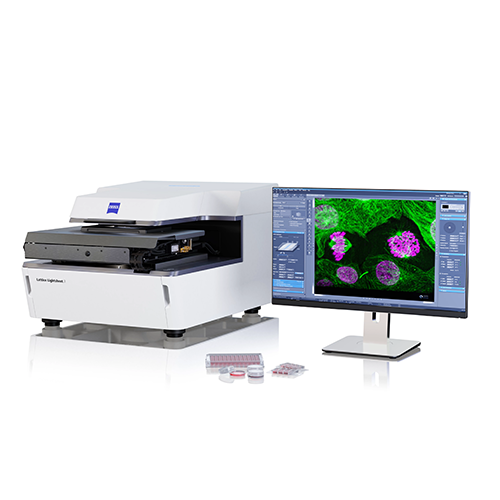
ZEISS Lattice Lightsheet 7
New functionality and capabilities- Now with single-molecule localization microscopy (SMLM). Track cellular dynamics for hours or days, then resolve structures at nanometer-scale precision.
- New sample screening capabilities. Increase experimental throughput with multiwell plate imaging & integrated analysis.
Software and Image Analysis
Improve the speed and efficiency of your workflows- ZEISS Microscopy Copilot: Optimize your imaging and analysis with our AI powered assistant for microscopy workflows on the LSM and widefield systems and within arivis Pro.
- ZEN Workflow: easily build and automate flexible experiments.
- AI assisted one-click measurements. Quickly detect objects without manual tracing.


MyZEISS
Your ZEISS knowledge ecosystem- Get started faster with guided onboarding, training, and learning resources.
- Track quotes, orders, trials, and installed systems in one place.
- Submit and manage support requests for quicker issue resolution.
ZEISS in the Cell Bio Program
Hear about our latest research from ZEISS experts and partners-
Lunch & Learn: Capturing biology across space, time, and scale From Cellular Dynamics to Human-Relevant 3D Biological Systems
Location: Exhibition Hall Theater 2
Presented by: Alma Arnold, Product Application Sales Specialist Team Lead, ZEISS Life Sciences Solutions
Abstract: As cell biology shifts toward human-relevant models, organoids, 3D cultures, and complex tissues, researchers are gaining new opportunities to study biology in more physiologically relevant contexts. At the same time, these systems challenge us to capture dynamic behavior, preserve 3D context, resolve subcellular organization, and transform increasingly complex datasets into meaningful biological insight.
Join ZEISS experts for a look at complementary imaging strategies, including live-cell, volumetric, high-resolution, automated, and AI-enabled workflows. Attendees will get an early look at new capabilities designed to extend what is possible across the cell biology workflow, from high-speed live-cell and volumetric imaging to larger-volume studies, automated multi-well experiments, advanced 3D biological models, and whole-cell molecular investigations.Join us to explore how imaging strategies can be matched to the biological question, balancing context, speed and resolution while moving beyond representative images toward quantitative measurements across cells, populations, and 3D models. -
Poster: Single-snapshot volumetric imaging for quantitative 3D drug-response screening of tumor co-culture organoids in 384-well format.
Author Block: Peter F. Favreau, PhD, Jason Fung, Nicolai Urban, PhD, Olivier Frey, PhD, Tamara Haefeli, Vimal Gangadharan, PhD
Carl Zeiss Microscopy, LLC; InSphero, AG, Schlieren, Zürich, Switzerland
Abstract:
Three-dimensional (3D) tumor models such as organoids recapitulate tissue architecture, cell-cell interactions, and drug penetration more faithfully than two-dimensional (2D) monolayers, making them increasingly central to oncology drug discovery and to immuno-oncology co-culture assays. Extracting quantitative 3D data at screening scale remains a bottleneck: conventional confocal imaging reconstructs each organoid from a serial stack of optical sections, which is slow across multiwell plates. A volumetric method that captures an entire organoid in a single acquisition would remove this constraint while preserving 3D morphology.Green fluorescent protein (GFP)-labeled HCT-116 colorectal carcinoma cells were co-cultured with red fluorescent protein (RFP)-labeled human dermal fibroblasts in a 384-well organoid plate at two tumor-to-stroma seeding ratios. Organoids were grown for 6 days, treated on day 4 with the topoisomerase I inhibitor SN-38 across four concentrations (0.5-500 nM) alongside vehicle and media controls, then cleared and counterstained. Whole organoids (200-325 µm) were imaged volumetrically on a ZEISS LSM 990 with Lightfield 4D, which captures one complete volume per well in a single snapshot without z-sectioning; selected wells were re-imaged at higher resolution on the same instrument. 3D analysis in ZEISS arivis Pro used deep-learning segmentation to extract per-well, per-channel cell counts.
Of 384 wells, 322 were analyzed; tissue loss during clearing and handling, primarily from 500 nM-treated spheroids, accounted for the remainder. SN-38 produced a dose-dependent reduction in GFP-positive tumor cells, reaching approximately 57% at 500 nM relative to media controls. RFP-positive fibroblast counts remained comparatively stable, declining modestly at the highest dose, consistent with a tumor-selective effect and demonstrating compartment-resolved readouts from dual-labeled co-cultures. Analyzing the same volumetric data as a 2D maximum-intensity projection (MIP) systematically undercounted cells: 3D counting recovered roughly 2.3-fold more tumor cells and 2.5-fold more nuclei than the MIP, because overlapping cells collapse along the projection axis.
Single-snapshot volumetric imaging enables quantitative 3D drug-response screening of tumor co-culture organoids in 384-well format, removing the throughput penalty of serial z-sectioning while delivering compartment-resolved, full-volume cell counts. Because the complete volume is captured in one acquisition, the more accurate 3D readout incurs no additional throughput cost. The platform is directly extensible to tumor-immune co-culture screening, offering immuno-oncology researchers a scalable route to 3D-resolved phenotyping and biomarker quantification.
-
Round Table Discussion: Are We Seeing Enough? A Discussion on 3D and Live-Cell Imaging
Understanding cell biology often requires capturing cellular structure in 3D and behavior over time. Join fellow researchers to exchange ideas, discuss common challenges, and learn how new microscopy approaches are helping scientists study increasingly complex biological systems.