You are on our international English website. This site features our entire product portfolio worldwide. The products featured may not be available in the US. If you are a citizen from the US, please visit your country website for local information and contacts.
You are on our international English website. This site features our entire product portfolio worldwide. The products featured may not be available in the US. If you are a citizen from the US, please visit your country website for local information and contacts.
Explore how ZEISS SL 800 supports everyday anterior segment imaging through a curated collection of clinical cases.
This selection showcases the clarity, detail, and contrast delivered by ZEISS SL 800 with TrueView Optics and VarioLight. Captured using the ZEISS SL Imaging Solution, the cases demonstrate the visualization capabilities of the system across a variety of anterior segment presentations.
Through high-quality imaging and documentation, the collection highlights the value of ZEISS SL 800 in supporting the observation and capture of anterior segment findings in clinical practice.
ZEISS SL 800 gallery
Cases contributed by Dr. Carceller
Conjunctival lymphangiectasia with localized conjunctival thickening
Dilated conjunctival lymphatic vessels may cause ocular irritation, foreign body sensation, and epiphora.
The microcysts can be visualized using both direct and indirect illumination but due to the small translucent nature of the microcysts indirect illumination can provide enhanced visualization of the medication side effect.
Multiple iris Lisch nodules associated with neurofibromatosis type 1
Lisch nodules are benign melanocytic hamartomas of the iris and are a characteristic ocular finding in patients with neurofibromatosis type 1.
Pterygium extending onto the cornea
Pterygia can alter corneal curvature and induce astigmatism, particularly as they extend further onto the cornea.
Persistent pupillary membrane (PPM) with prominent iris strands
PPM is a congenital remnant of the anterior vasculosa lentis and is usually asymptomatic when the visual axis remains unobstructed.
Diffractive intraocular lens visualized with retroillumination
Well-centered diffractive intraocular lens (IOL) visualized with retroillumination. This visualization technique allows for assessment of the IOL and posterior capsule.
White cataract with prominent tortuous episcleral vessels in Sturge-Weber syndrome
This vascular abnormality results in elevated episcleral venous pressure, poor drainage of the episcleral vessels and can lead to glaucoma in patients with Sturge-Weber syndrome.
Gonioscopic view of two iStent inject (Glaukos) devices implanted in the trabecular meshwork
These trabecular micro-bypass stents are designed to enhance aqueous outflow and reduce intraocular pressure in open-angle glaucoma.
Exposure keratitis with large epithelial erosion secondary to facial nerve palsy
Fluorescein dye with a cobalt blue filter enhance the visualization of both punctate erosions and very large central erosion as a result of incomplete lid closer after neurological paralysis.
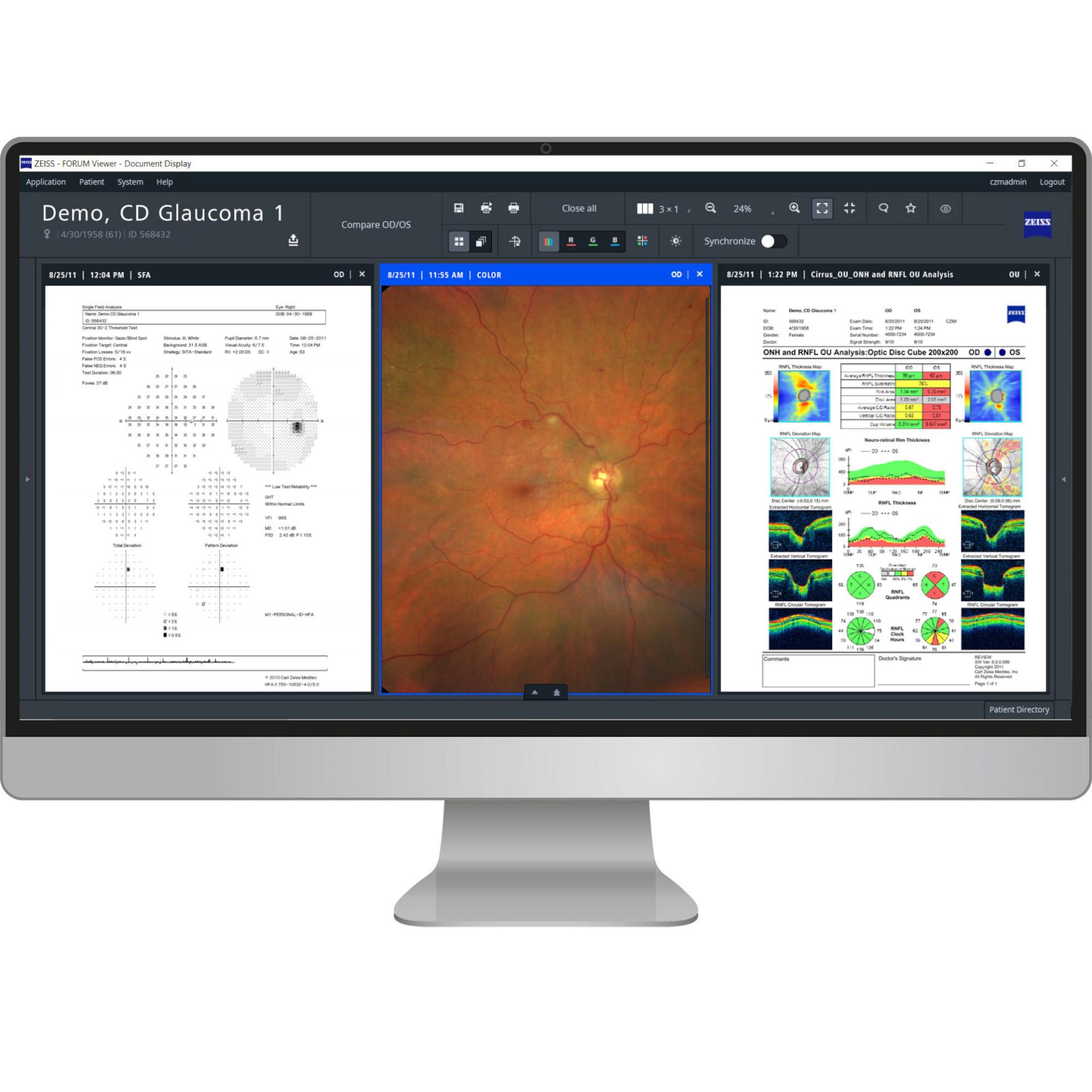
The ZEISS SL 800 has the best optics I have ever used. In combination with the ZEISS SL Imaging Solution, you get good quality images without being an expert photographer. I especially like to spend an extra minute making a report out of 6 images with the most important photos. Those will be easily available at FORUM from ZEISS whenever I see the patient again.
Learn more
ZEISS SL 800 and ZEISS SL Imaging Solution
ZEISS SL 800
With perfectly balanced ZEISS optics, extensive illumination options and a user-friendly operator concept directly at your fingertips, the SL 800 from ZEISS reveals details for diagnosis and optimizes your workflow.
The intuitive SL Imaging Solution from ZEISS takes your everyday slit lamp exams to the next level by adding the integration of high-quality image and video capture to exam reports, offering you the ability to document cases, to include in patient education, teaching, or publishing.