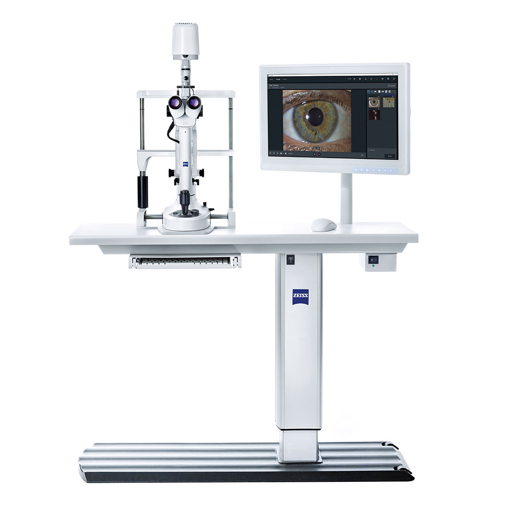

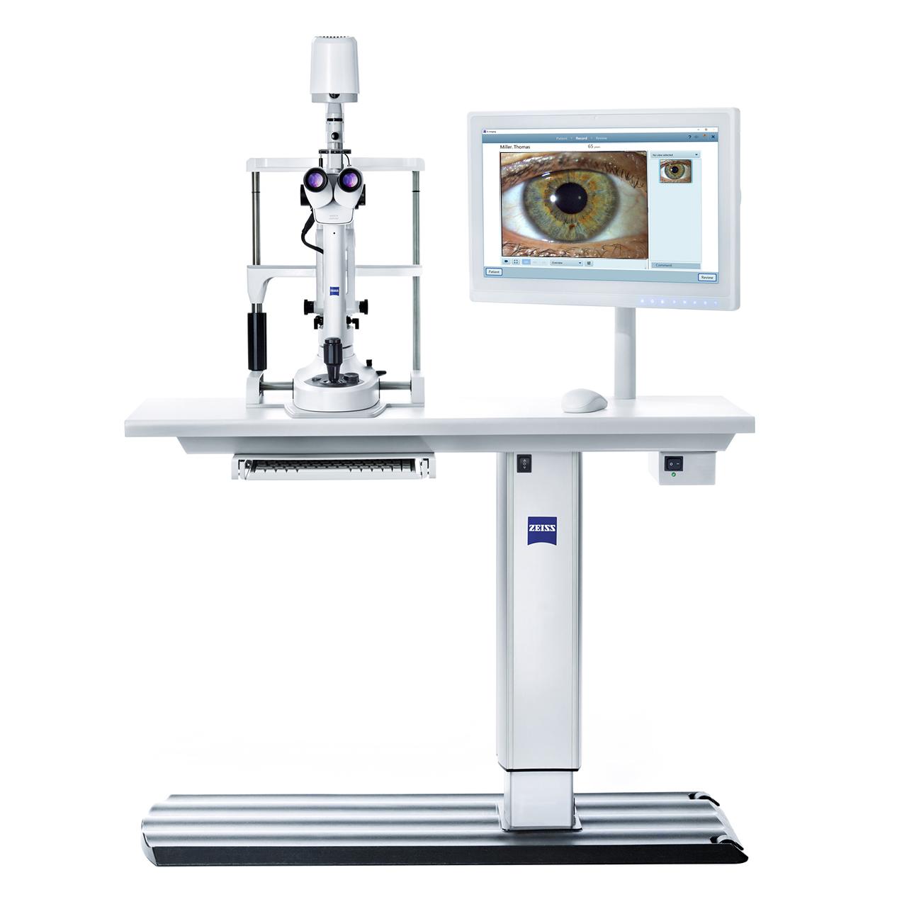
ZEISS SL Imaging Solution Seamless image and data capture for slit lamps
The intuitive SL Imaging Solution from ZEISS takes your everyday slit lamp exams to the next level by adding the integration of high-quality image and video capture to exam reports, offering you the ability to document cases, to include in patient education, teaching, or publishing. This all-round imaging solution features a modular concept, giving you the option to add slit lamp imaging seamlessly into your workflow on a preferential basis.
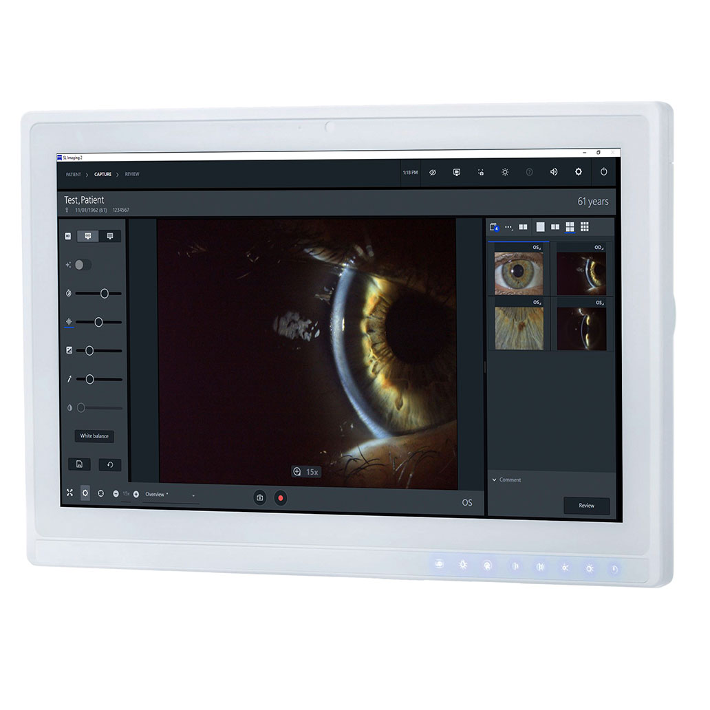

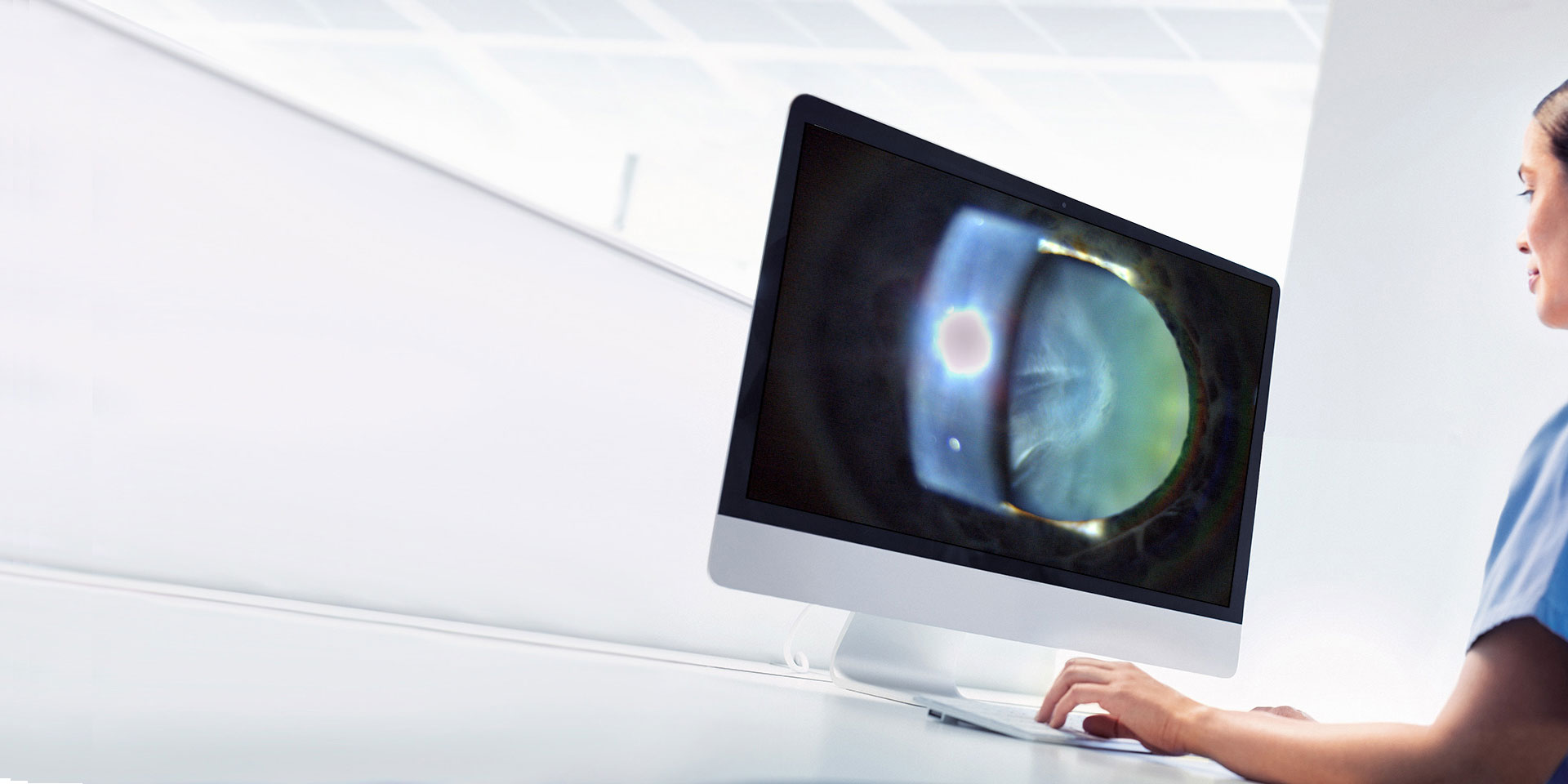
High-definition image and video capture
The smooth integration of camera and slit lamp allows to capture images and videos with clarity, magnifying tiny details. With a frame rate of up to 40 fps, broadcasting in real time without the worry of latency or lag is now possible with LiveView, enabling true-to-life color imaging — convenient tool for education or teaching.
Technical details:
- Camera with up to 4k resolution (4320 x 3240 pixels)
- True-to-life color: capture as seen through the slit lamp
- LiveView without latency: broadcast a live image on screen at up to 40 fps
Capture the details at up to 4k
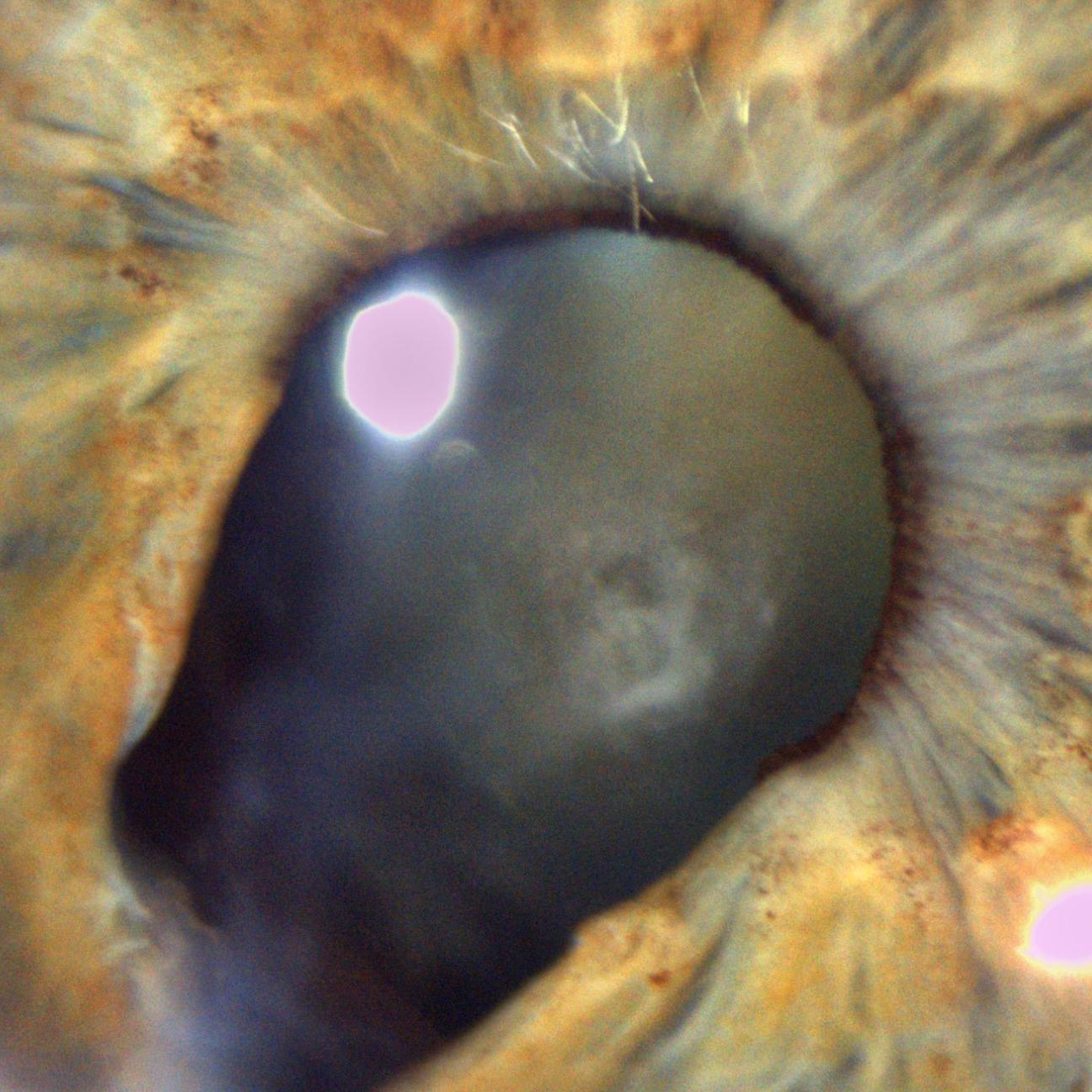

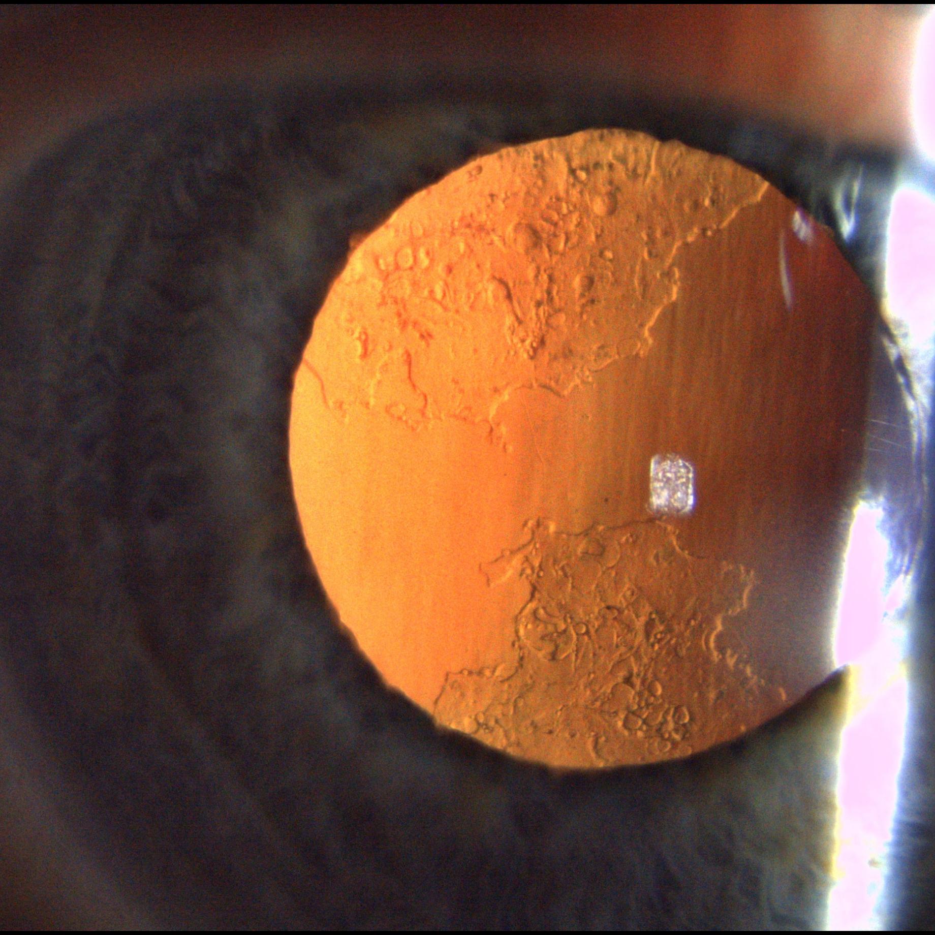
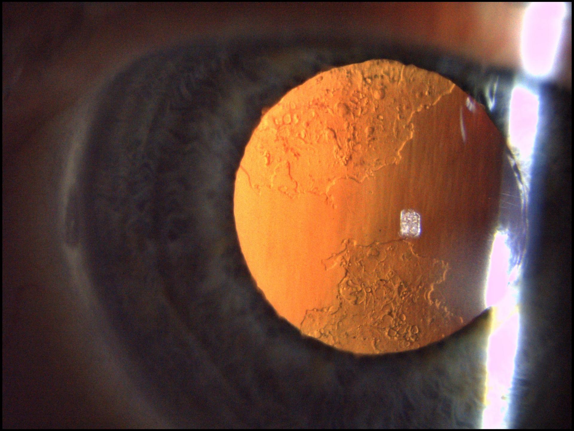
Traumatic coloboma of the iris and cataract as sequelae of penetrating injury
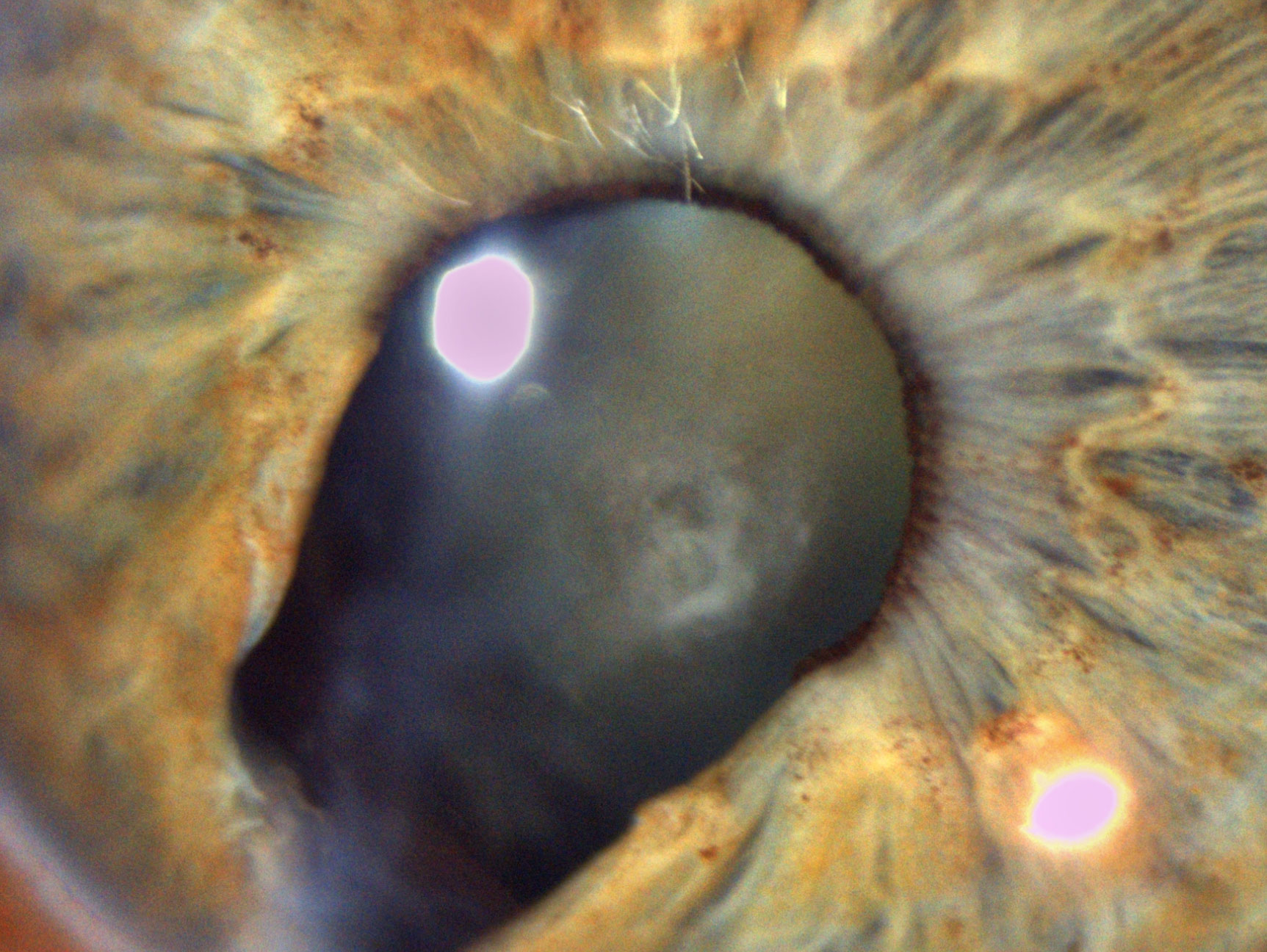

Traumatic coloboma of the iris and cataract as sequelae of penetrating injury

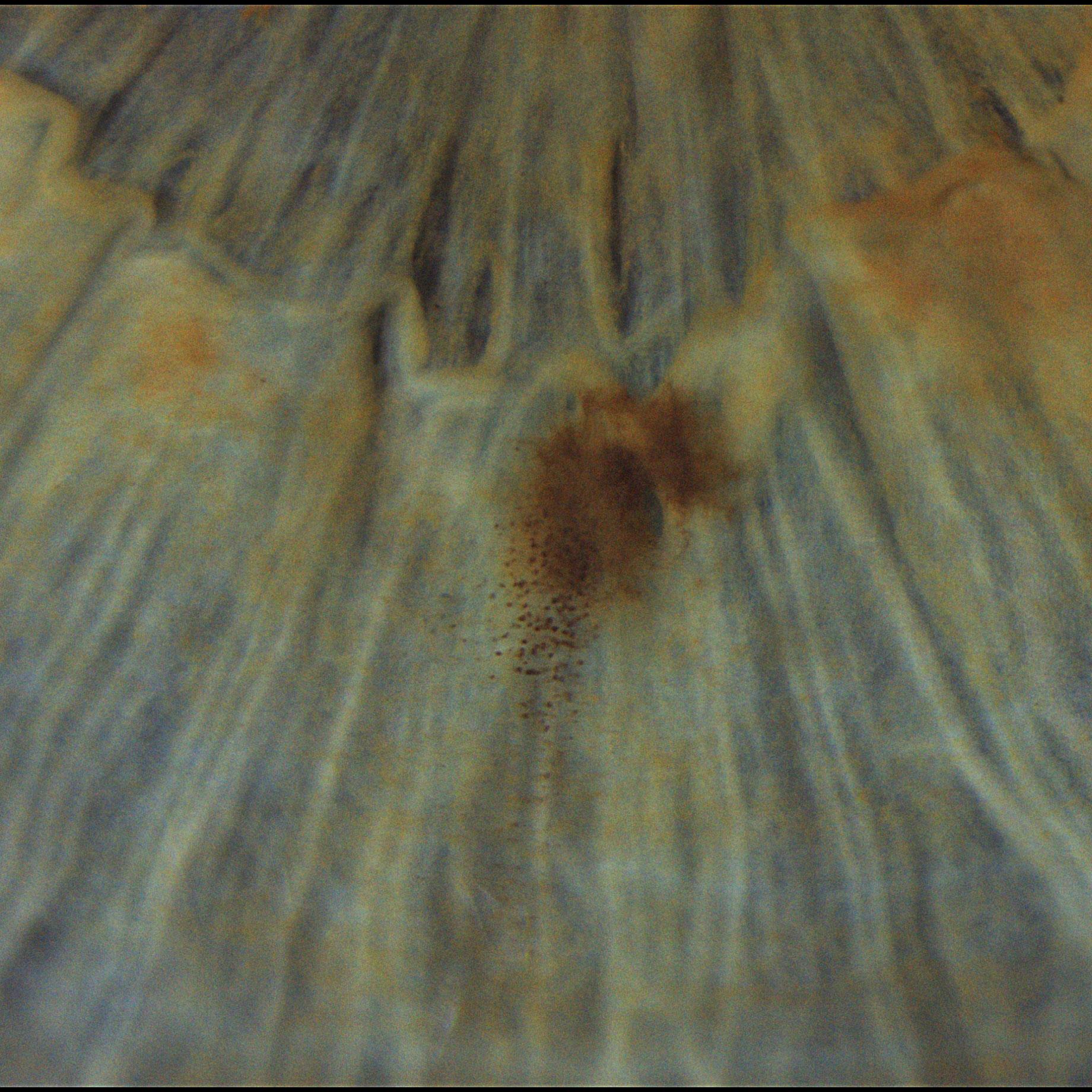
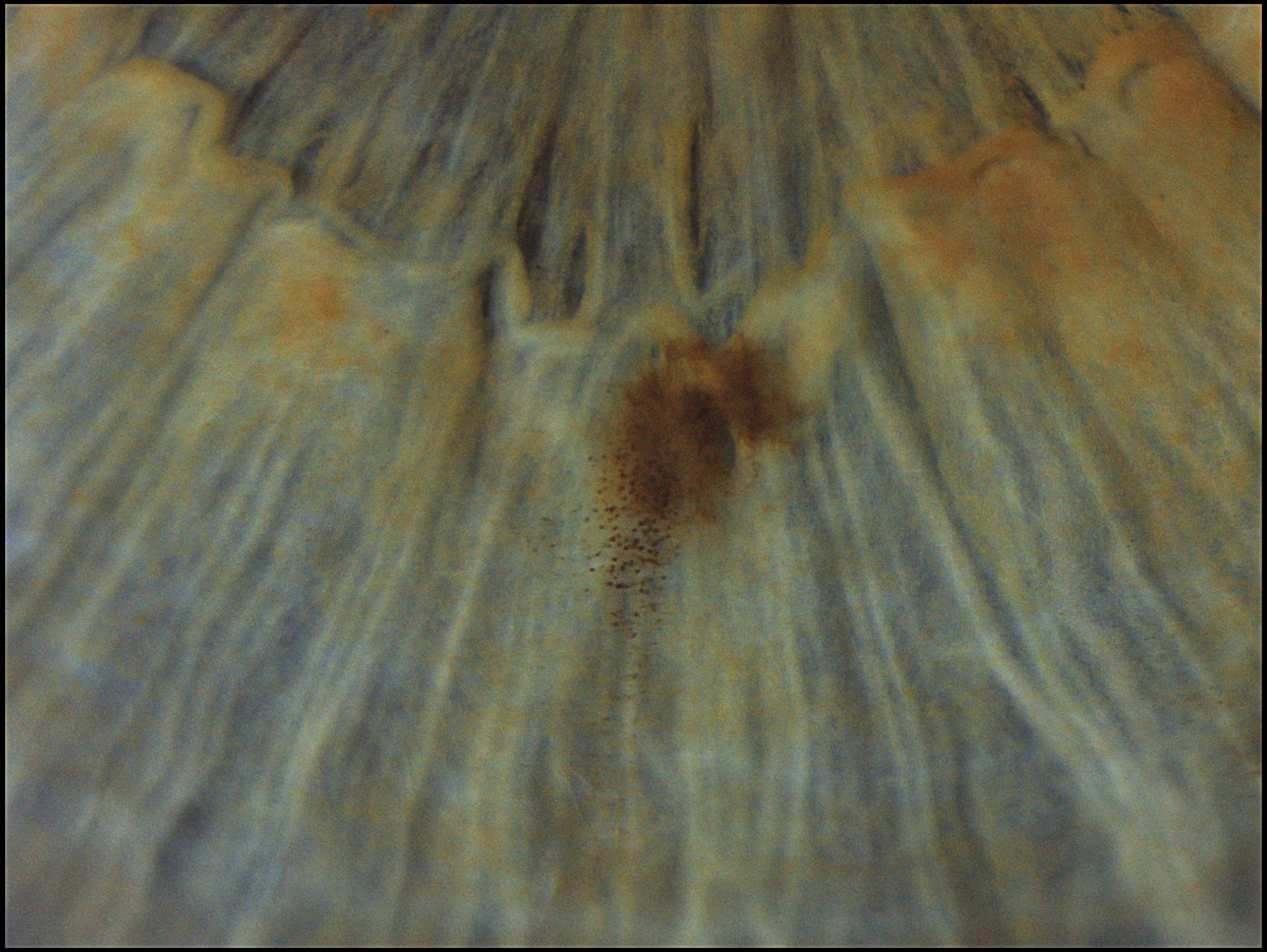
Faint corneal scars at high magnification after Lasik therapy

Faint corneal scars at high magnification after Lasik therapy
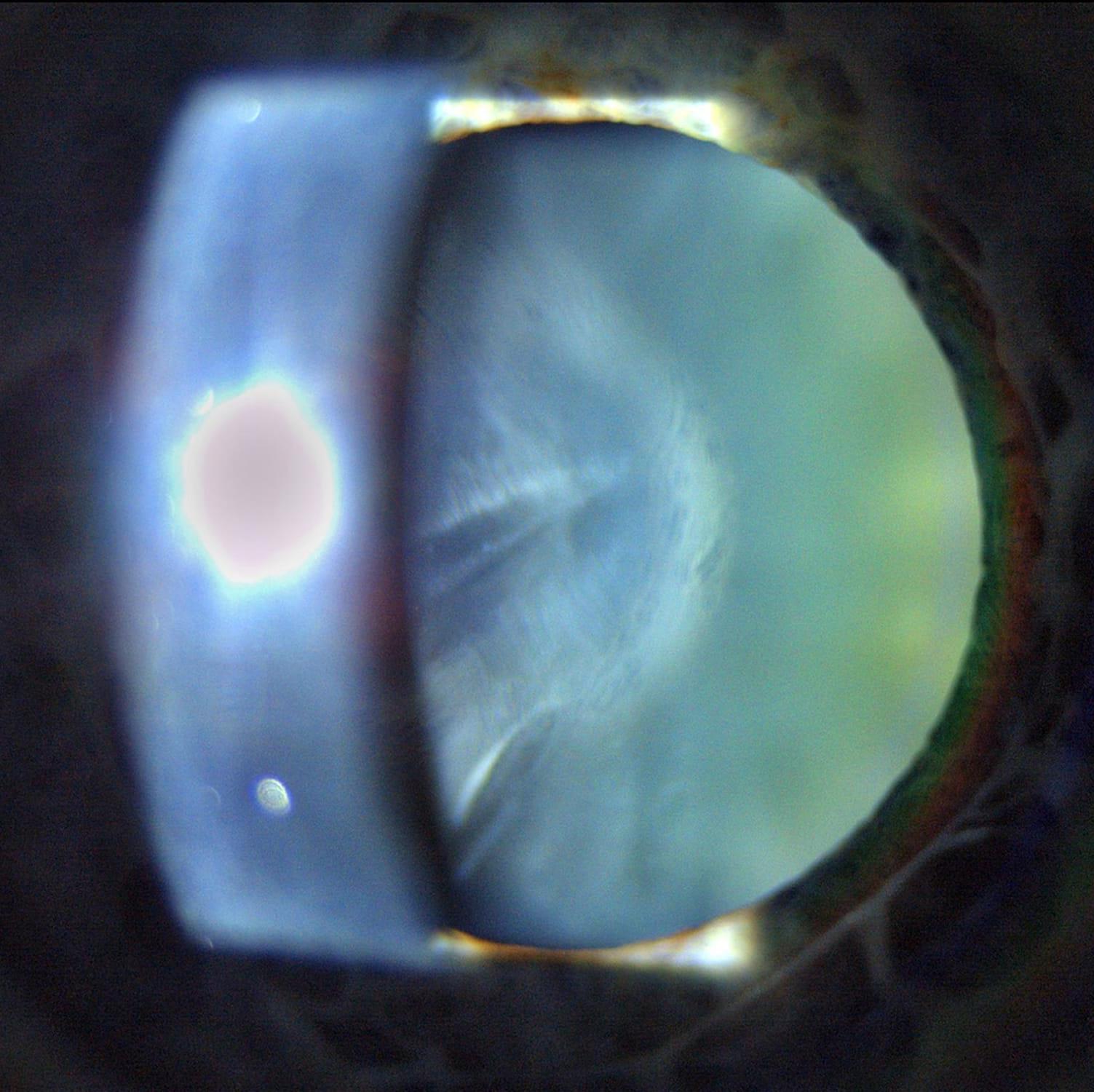

Cortical cataract

Cortical cataract

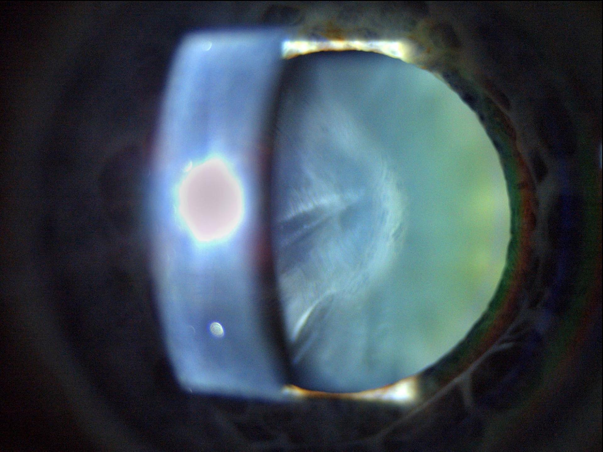
Posterior capsule opacification

Posterior capsule opacification

Small irisnevus with scattered nevus cells around

Small iris nevus with scattered nevus cells around
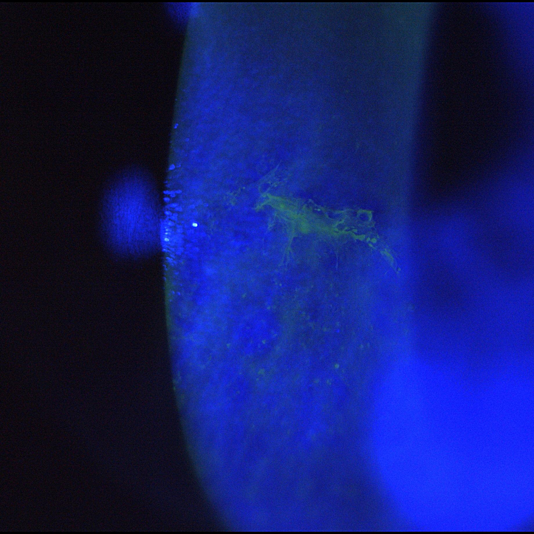

Epithelial ridge after corneal injury with fluorescein dye and blue light
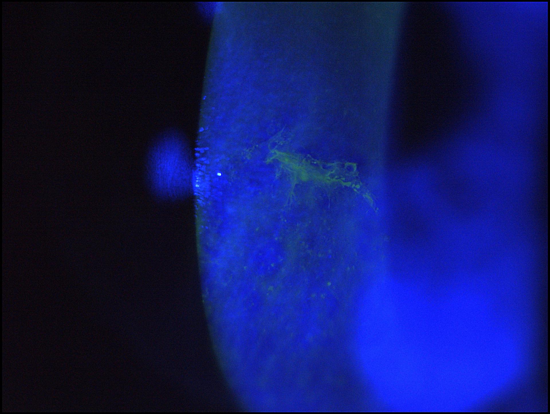

Epithelial ridge after corneal injury with fluorescein dye and blue light

Transillumination of the iris in albinism and pseudophakia

Transillumination of the iris in albinism and pseudophakia
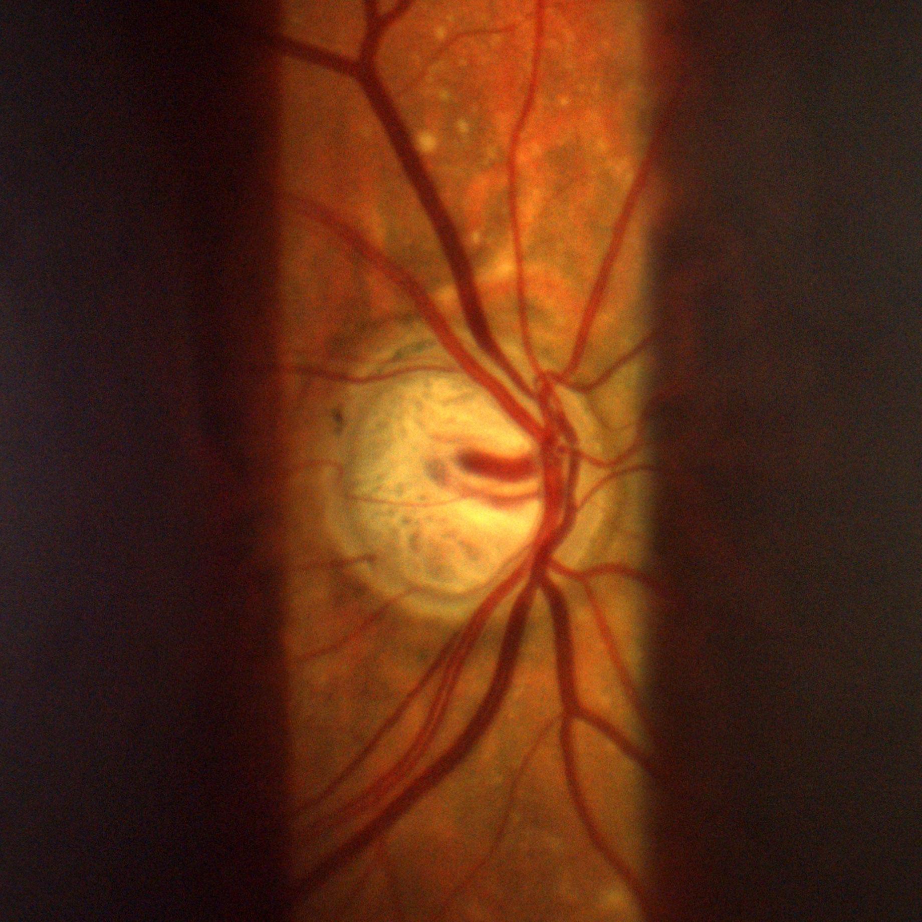

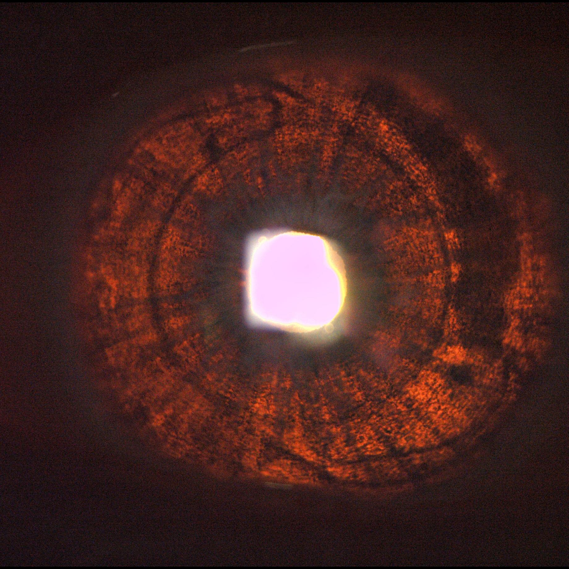
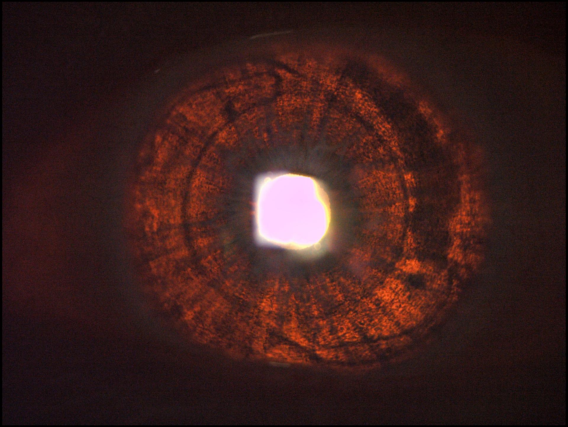
Deep excavation of optic nerve head in advanced glaucoma
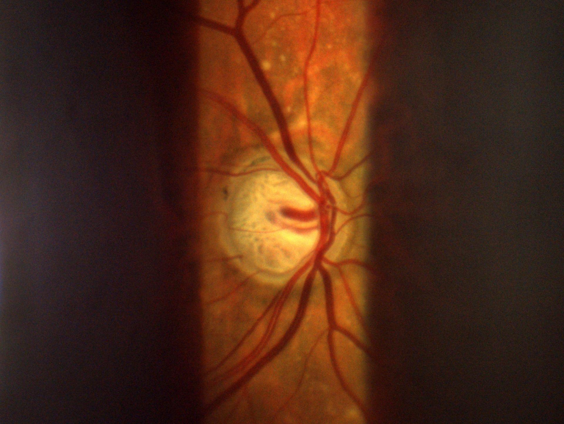

Deep excavation of optic nerve head in advanced glaucoma

Workflow-optimized design
The workflow-optimized software and hardware design1 of the SL Imaging Solution allows for both the actual exam and documentation to take place in parallel. With no need to take extra effort for creating images and videos, you can fully focus on your patients.
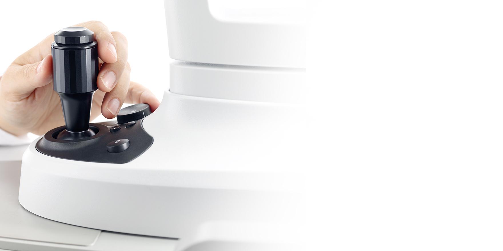
- Capture live exam images with the simple click of a joystick trigger or button1
- Capture images while simultaneously recording a video
- Compare recordings with the Flicker feature and add a second screen and let others watch the examination
Key benefits of the SL Imaging Solution:
- Automatic eye detection1: Automatically detects the eye position laterally.
- Live parameter1: Monitor the magnification level and defined camera parameters
- Camera profiles: Select a predefined settings profile, such as for slit images, or define your own individual profiles
- Quick edit: Edit and adjust camera settings, including white-balance, in live-mode and edit stored images and videos
- Instant preview: Review image quality immediately after capture
- Dark theme: In combination with the illumination of the panel PC, working in an dark environment is convenient


-
1
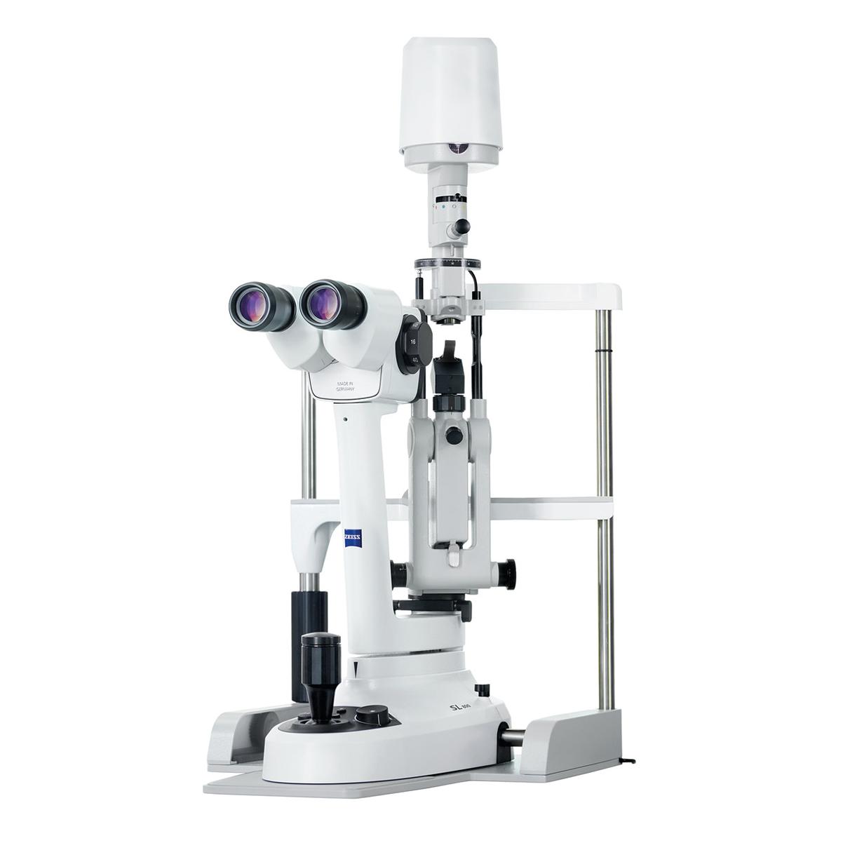
In combination with ZEISS SL 800 Slit lamp

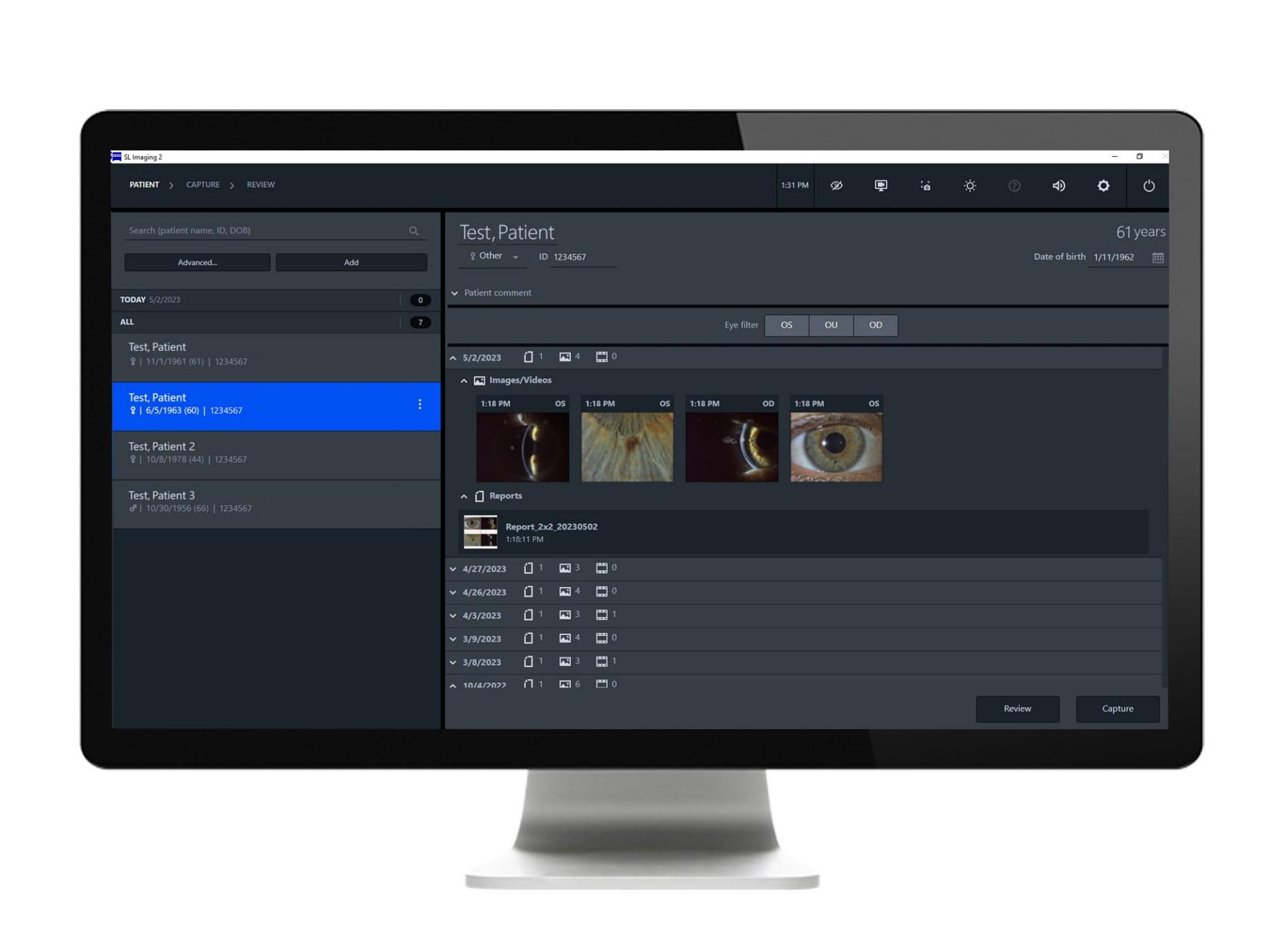
Digital documentation
One-click data export makes it convenient to add reports, images and videos to the digital patient record, streamlining your daily workflow. Share records with patients and publications, with students for education or with the ophthalmologic community for academics or to facilitate second opinions. Other benefits include:
- Individual report layouts
- Auto-fill reports with images, including a direct comment function
- Export images to FORUM and DICOM systems
- JPG and MP4 export to hard drive


Custom capabilities
Complete your setup with optional components like the SL Workstation (Panel PC with pre-installed software) and widefield illumination available from ZEISS for a comfortable practice integration.
First impressions
Downloads


Specifications
ZEISS SL Imaging Solution*For compatibility with other operating systems please contact your service contact.
Get in touch with us!
Receive more information about the product and availability in your country!Related products


-
1
In combination with ZEISS SL 800 slit lamp