Webinaire à la demande
Biopsies cérébrales virtuelles : corréler l'endomicroscopie confocale au laser (CLE) peropératoire et l'histopathologie virtuelle
Enregistré lors du 2e colloque ZEISS CONVIVO
23 juin 2021
· 20 MIN LECTURE
![]({"xsmall":"https://www.zeiss.com/content/dam/med-hcp/reference-master/myzeiss/peer-insights/free/pathology/509-neurosurgical-workflow-for-patients-with-brain-tumors/jennifer-eschbacher.jpg/_jcr_content/renditions/original.image_file.100.100.0,0,350,350.file/jennifer-eschbacher.jpg","small":"https://www.zeiss.com/content/dam/med-hcp/reference-master/myzeiss/peer-insights/free/pathology/509-neurosurgical-workflow-for-patients-with-brain-tumors/jennifer-eschbacher.jpg/_jcr_content/renditions/original.image_file.350.350.0,0,350,350.file/jennifer-eschbacher.jpg","medium":"https://www.zeiss.com/content/dam/med-hcp/reference-master/myzeiss/peer-insights/free/pathology/509-neurosurgical-workflow-for-patients-with-brain-tumors/jennifer-eschbacher.jpg/_jcr_content/renditions/original.image_file.350.350.0,0,350,350.file/jennifer-eschbacher.jpg","large":"https://www.zeiss.com/content/dam/med-hcp/reference-master/myzeiss/peer-insights/free/pathology/509-neurosurgical-workflow-for-patients-with-brain-tumors/jennifer-eschbacher.jpg/_jcr_content/renditions/original.image_file.350.350.0,0,350,350.file/jennifer-eschbacher.jpg","xlarge":"https://www.zeiss.com/content/dam/med-hcp/reference-master/myzeiss/peer-insights/free/pathology/509-neurosurgical-workflow-for-patients-with-brain-tumors/jennifer-eschbacher.jpg/_jcr_content/renditions/original.image_file.350.350.0,0,350,350.file/jennifer-eschbacher.jpg","xxlarge":"https://www.zeiss.com/content/dam/med-hcp/reference-master/myzeiss/peer-insights/free/pathology/509-neurosurgical-workflow-for-patients-with-brain-tumors/jennifer-eschbacher.jpg/_jcr_content/renditions/original.image_file.350.350.0,0,350,350.file/jennifer-eschbacher.jpg","max":"https://www.zeiss.com/content/dam/med-hcp/reference-master/myzeiss/peer-insights/free/pathology/509-neurosurgical-workflow-for-patients-with-brain-tumors/jennifer-eschbacher.jpg/_jcr_content/renditions/original.image_file.350.350.0,0,350,350.file/jennifer-eschbacher.jpg"})
Auteur
Dr Jennifer Eschbacher
Neuropathologiste au Barrow Neurological Institute, Phoenix, Arizona, État-Unis
RÉSUMÉ
Partage d'expérience entre pairs sur la corrélation entre les images par CLE peropératoire et l'histopathologie conventionnelle
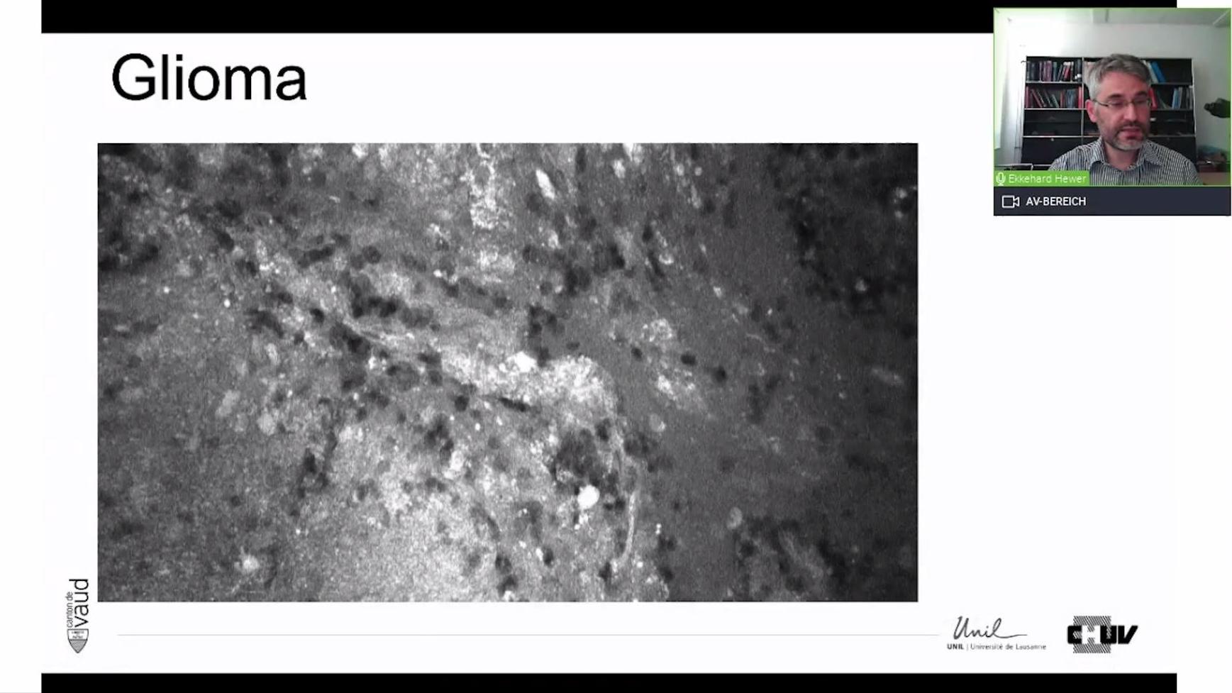
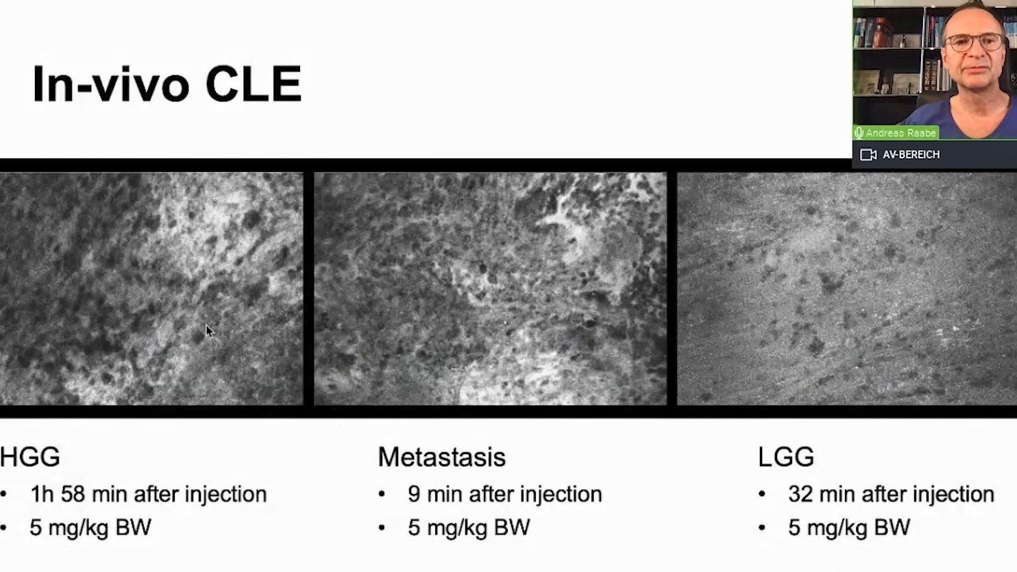
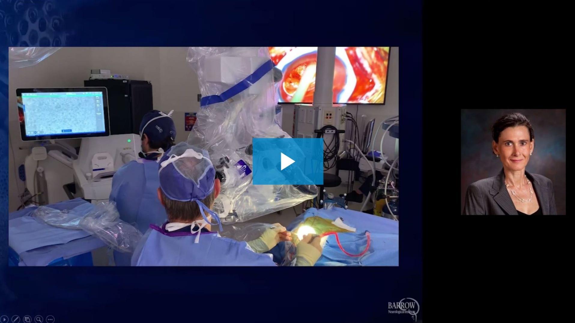
Le Dr Jennifer Eschbacher fait part de son expérience avec des biopsies virtuelles de tumeurs cérébrales au niveau cellulaire obtenues à l'aide de la technologie d'endomicroscopie confocale laser (CLE). Lors de cette présentation, elle parle des résultats de l'étude CLE en cours. Elle donne également des informations sur un cas enregistré de pathologie in vivo, suivi à distance depuis le laboratoire de pathologie, et démontre l'interaction de la pathologie neurochirurgicale.
![]({"xsmall":"https://www.zeiss.com/content/dam/med-hcp/reference-master/myzeiss/peer-insights/free/pathology/642-correlation-of-intraoperative-cle-images-and-conventional-histopathology/642--correlation-of-intraoperative-cle-images-and-conventional-h.jpg/_jcr_content/renditions/original.image_file.100.56.0,2,1916,1079.file/642--correlation-of-intraoperative-cle-images-and-conventional-h.jpg","small":"https://www.zeiss.com/content/dam/med-hcp/reference-master/myzeiss/peer-insights/free/pathology/642-correlation-of-intraoperative-cle-images-and-conventional-histopathology/642--correlation-of-intraoperative-cle-images-and-conventional-h.jpg/_jcr_content/renditions/original.image_file.360.203.0,2,1916,1079.file/642--correlation-of-intraoperative-cle-images-and-conventional-h.jpg","medium":"https://www.zeiss.com/content/dam/med-hcp/reference-master/myzeiss/peer-insights/free/pathology/642-correlation-of-intraoperative-cle-images-and-conventional-histopathology/642--correlation-of-intraoperative-cle-images-and-conventional-h.jpg/_jcr_content/renditions/original.image_file.768.432.0,2,1916,1079.file/642--correlation-of-intraoperative-cle-images-and-conventional-h.jpg","large":"https://www.zeiss.com/content/dam/med-hcp/reference-master/myzeiss/peer-insights/free/pathology/642-correlation-of-intraoperative-cle-images-and-conventional-histopathology/642--correlation-of-intraoperative-cle-images-and-conventional-h.jpg/_jcr_content/renditions/original.image_file.1024.576.0,2,1916,1079.file/642--correlation-of-intraoperative-cle-images-and-conventional-h.jpg","xlarge":"https://www.zeiss.com/content/dam/med-hcp/reference-master/myzeiss/peer-insights/free/pathology/642-correlation-of-intraoperative-cle-images-and-conventional-histopathology/642--correlation-of-intraoperative-cle-images-and-conventional-h.jpg/_jcr_content/renditions/original.image_file.1280.720.0,2,1916,1079.file/642--correlation-of-intraoperative-cle-images-and-conventional-h.jpg","xxlarge":"https://www.zeiss.com/content/dam/med-hcp/reference-master/myzeiss/peer-insights/free/pathology/642-correlation-of-intraoperative-cle-images-and-conventional-histopathology/642--correlation-of-intraoperative-cle-images-and-conventional-h.jpg/_jcr_content/renditions/original.image_file.1440.810.0,2,1916,1079.file/642--correlation-of-intraoperative-cle-images-and-conventional-h.jpg","max":"https://www.zeiss.com/content/dam/med-hcp/reference-master/myzeiss/peer-insights/free/pathology/642-correlation-of-intraoperative-cle-images-and-conventional-histopathology/642--correlation-of-intraoperative-cle-images-and-conventional-h.jpg/_jcr_content/renditions/original.image_file.1915.1077.0,2,1916,1079.file/642--correlation-of-intraoperative-cle-images-and-conventional-h.jpg"})