

Symposium "From 3D Light to 3D Electron Microscopy“
Hosted at the Francis Crick Institute | London, UKWelcome to the 8th Edition of the Symposium
From 3D Light to 3D Electron Microscopy

We are thrilled to announce that the 8th edition of the symposium “From 3D Light to 3D Electron Microscopy” is hosted by the Electron Microscopy Science Technology Platform at the prestigious Francis Crick Institute in London, UK. This event is a collaborative effort between the Francis Crick Institute, EMBL Heidelberg, VIB Ghent, and ZEISS, bringing together leading experts and enthusiasts in the field.
The symposium will feature a rich program of scientific sessions that span a wide spectrum of correlative workflows, addressing various imaging modalities, sample preparation techniques, and data analysis strategies. Participants will also have the unique opportunity to engage in instrument workshops and dynamic round table discussions. You will discover just how accessible correlative microscopy has become and explore its applications in clinical research environments.
We also warmly invite young scientists and those utilizing different imaging modalities beyond volume electron microscopy to join this event and learn more about the benefits from their peers. Knowledge and experience can be shared with your friends and other researchers in an inspiring and collaborative atmosphere.
Additionally, participants are encouraged to showcase their own scientific contributions through presentations selected from submitted abstracts or in short flash talks.
Don't miss this exciting opportunity to connect with fellow researchers and industry professionals, as we delve into the latest and emerging technologies in correlative microscopy, volume electron microscopy, and X-ray microscopy within the life sciences. Join us for an inspiring and collaborative experience that promises to enhance your understanding and application of these groundbreaking techniques!
Speakers

Yoshiyuki Kubota
Project researcher, EM team leader,
Division of Multisensory Integration Systems, National Institute for Physiological SciencesOkazaki, Japan

Candice Roufosse
Clinical Reader Renal Pathology
Imperial College London, Department of Immunology and Inflammation
London, UK

Ana Laura Vinagre Costa de Sousa
Electron Microscopy Facility Head
Gulbenkian Institute for Molecular Medicine
Lisbon, Portugal

Asandile Mangali
Asandile Mangali (PhD candidate),
Neuro Research Group (Prof. Ben Loos's lab), Physiological Sciences, Stellenbosch University, South Africa

Tanmay Bharat
Programme Leader Structural Studies Division MRC Laboratory of Molecular Biology
Cambridge, UK

Sven Klumpe
MBA/IMP, Fellow Research Institute of Molecular Pathology (IMP) and Institute of Molecular Biotechnology Austria (IMBA), Vienna BioCenter
Vienna, Austria


Stephan Handschuh
Staff Scientist,
VetCore Facility for Research, Imaging Unit,
University of Veterinary Medicine Vienna, Vienna, Austria

Nicolas Gueninchault
Head of Field of Business, X-ray Microscopy
ZEISS Microscopy
Dublin, USA

Edoardo D'Imprima
Head of CLEM Core facility
Humanitas Research Hospital
and Humanitas University
Milano, Italy

Patricia Goggin
Clinical Scientist and Head of the
Biomedical Imaging Unit, University Hospital Southampton and
University of Southampton
Southampton, UK

Alexandra Kerbl
Postdoc, Department of Evolutionary Neurobiology, Centre for Organismal Studies (COS), Heidelberg University
Heidelberg, Germany


Dominik Kutra
Senior Research Software Engineer,
EMBL Heidelberg, CBB, Kreshuk Group,
Heidelberg, Germany

Harald Hess
Senior Group Leader HHMI
Janelia Research Campus
Ashburn, USA
Program at a glance
Agenda overview and session detailsProgram overview
-
Program Item / SpeakerTitle
15:30 – 16:30 GMT
Registration & Coffee & Snacks
16:30 – 17:00 GMT
Welcome
Keynote Session, Chair: Lucy Collinson, The Francis Crick Institute, London
17:00 – 17:45 GMT
Keynote 1: Yoshiyuki Kubota
Large Volume Electron Microscopy Reveals Synaptic Architecture of Cortical Circuits in the Primate Prefrontal Cortex
17:45 – 18:30 GMT
Keynote 2: Candice Roufosse
A nanopathology platform for the prediction and early detection of kidney transplant rejection
18:30 – 20:00 GMT
Fingerfood & drinks @ The Crick
-
Program Item / SpeakerTitle
08:30 – 08:50 GMT
Welcome Coffee
09:00 – 09:15 GMT
Welcome & Housekeeping
09:15 – 09:45 GMT
Ana Laura Vinagre Costa e Sousa
Simple Tools, Big Insights: The Histo-CLEM Approach
09:45 – 10:00 GMT
Nicolas Brouilly
(3D)-CLEM in invertebrates: how worms and flies can shed light on ultrastructural Information
10:00 – 10:15 GMT
Hélène Roberge
Towards a scalable and reproducible open-source image processing workflow for 3D CLEM images
10:15 – 10:45 GMT
Coffee Break
10:45 – 11:15 GMT
Asandile Mangali
Increased environmental exposure to heavy metals in South African mining regions: A recipe for metal-induced neurotoxicity and mitochondrial dysfunction?
11:15 – 11:30 GMT
Flash Talks
11:30 – 13:00 GMT
Workshop 1 – 4 / Panel Discussion 1
13:00 – 14:00 GMT
Lunch
14:00 – 15:30 GMT
Workshop 1 – 4 / Panel Discussion 2
15:30 – 16:00 GMT
Coffee Break
16:00 – 16:30 GMT
Tanmay Bharat
Multi-scale, correlated imaging of multicellular specimens: from bacterial biofilms to eukaryotic infection
16:30 – 17:00 GMT
Sven Klumpe
Cryo-(P)FIB development from unicellular to multicellular organisms
17:00 – 17:15 GMT
Marketa Dalecka
Visualization of T-cell microvilli and their role in the immunological synapse by combined cryo-fluorescence microscopy and FIB-SEM tomography
17:15 – 17:30 GMT
Hannah Ochner
Sub-cellular chemical mapping using correlated cryogenic electron and mass spectrometry imaging
18:30 – 21:00 GMT
Conference Dinner
-
Program Item / SpeakerTitle
08:45 – 09:15 GMT
Welcome Coffee
09:15 – 09:45 GMT
Stephan Handschuh
X-ray microscopy for imaging of biomedical tissue samples
09:45 – 10:00 GMT
Cheng Xing
Multiscale volume electron microscopy mapping of the human liver reveals vascular-cellular organization and mitochondria-ER contacts
10:00 – 10:15 GMT
Aya Zucca
Precision Targeting for Volume Electron Microscopy Using Correlative Light and X-ray Imaging in the mito-APEX2 Expressing Larval Zebrafish Brain
10:15 – 10:45 GMT
Coffee Break
10:45 – 11:15 GMT
Nicolas Gueninchault
Elevating Life Sciences Imaging through X-ray Microscopy
11:15 – 11:30 GMT
Flash Talks
11:30 – 13:00 GMT
Workshop 1 – 4 / Panel Discussion 3
13:00 – 14:00 GMT
Lunch
14:00 – 15:30 GMT
Workshop 1 – 4 / Panel Discussion 4
15:30 – 16:00 GMT
Coffee Break
16:00 – 16:30 GMT
Edoardo D'Imprima
The potential and challenges of Volume Electron Microscopy for clinical research
16:30 – 17:00 GMT
Patricia Goggin
Bridging the Gap: Collaborative EM for Research and Clinical Impact
17:00 – 17:15 GMT
Chang Guo
Multiscale correlative imaging reveals neurovascular and membrane disruption by environmentally relevant particles
17:15 – 17:30 GMT
Abigail Lytton-Jean
ToCLEM: A Robust and Accessible Method for Correlative Light and Electron Microscopy
17:30 – 18:30 GMT
Drinks @ The Crick
-
Program Item / SpeakerTitle
08:45 – 09:15 GMT
Welcome Coffee
09:15 – 09:45 GMT
Alexandra Kerbl
Reconstructing organisms section by section - modular serial section electron microscopy to tackle whole-organism connectomics projects
09:45 – 10:15 GMT
Dominik Kutra
ilastik: dealing with bigger volumes
10:15 – 10:30 GMT
Stijn Karacoban
Optimizing iterative ion beam milling and multibeam scanning transmission electron microscopy for volume EM
10:30 – 10:45 GMT
Michaela Wilsch-Bräuninger
Correlative Analysis of Microorganism-Mineral-Interactions and their Impact on our Planet Earth
10:45 – 11:15 GMT
Coffee Break
Closing Keynote Session, Chair:Lucy Collinson, The Francis Crick Institute, London
11:15 – 12:00 GMT
Harald Hess
Larger Volume Imaging with Single and Multiple Electron Beams and Cryo-Super-Resolution Microscopy
12:00 – 12:15 GMT
Closing Words
Session Details
-
Erin Tranfield
-
Pippa Hawes
-
Elizabeth Duke
-
Yannick Schwab
-
Paolo Ronchi


Workshops
-
Volume electron microscopy (vEM) methods have the potential to reveal the underlying 3-dimensional ultrastructure of cells and tissues from a wide variety of sample types. However, large samples and the preparation methods necessary for optimal imaging often make it extremely difficult to identify and/or relocate a specific area for analysis.
Targeting to a specific region of interest can be achieved in several ways, ranging from simple visual landmarks for global positioning, through to the identification and localisation of groups of proteins within the greater cellular landscape. Each method has distinct strengths and weaknesses, and can ultimately be used successfully in isolation, or in multiple combinations as part of more complex imaging workflows.
In this workshop, participants will explore a correlative workflow using lab-based x-ray imaging to locate specific tissue types within a larger organism. The x-ray data will then be used to guide targeted block trimming with a semi-automated ultramicrotome, prior to vEM image acquisition using a serial blockface scanning electron microscope (SBF-SEM).
Workshop Leads:
Chris Peddie
The Francis Crick Institute, London, UK
Jenny Hounsome
The Francis Crick Institute, London, UK
Wei Guan
The Francis Crick Institute, London, UK
Sebastian M. Markert
Carl Zeiss Microscopy Oberkochen, Germany -
Array tomography is an accessible and non-destructive volume light and EM technique that allows considerable flexibility in sample preparation including correlative options. Series of ultrathin sections of a sample are collected onto either a solid support (e.g. coverslips using an ultramicrotome) or tape (e.g. using an ATUMtome type setup) before LM or SEM imaging. In correlative workflows, light microscopy can be performed prior to EM embedding, after embedding or after sectioning as appropriate, and these images can be imported into the Zeiss SEM Atlas acquisition software to locate areas of interest and enable easy targeting of automated, high resolution ultrastructural imaging in the SEM. Repeated imaging allows multiple ROI to be imaged at multiple resolutions to thoroughly interrogate the sample.
This SEM Array Tomography workshop will cover many aspects of the EM workflow, with key array collection techniques being explained and/or demonstrated live to attendees, with variations and alternatives also discussed. The Imaging workflow on a Zeiss SEM will also be explained and demonstrated live as well as previously obtained data shown.
Workshop Leads:

Jemima Burden
LMCB / UCL London, UK
Ian White
LMCB / UCL London, UK
Anne Weston
The Francis Crick Institute, London, UK -
This workshop will cover the principles of the VP-CLEM kit workflow, which is designed for high precision volume CLEM and built to be user-friendly, allowing deployment in light microscopy facilities without the need for specialised rooms and expert electron microscopists. We will discuss and demonstrate sample preparation of monolayers for in resin fluorescence (IRF) using easyIRF protocol, imaging of serial sections using a widefield fluorescence and single molecule localisation microscopy, and reconstruction of the resulting single molecule localisations using Picasso software. Furthermore, we will introduce and demonstrate the concept of post-staining and imaging of serial sections in an array tomography format using a benchtop scanning electron microscopy, enabling high-resolution volumetric CLEM overlays.
Workshop Leads:

Dumisile Lumkwana
The Francis Crick Institute, London, UK
Nicola Vahrmeijer
University of Stellenbosch, Stellenbosch, South Africa
Asandile Mangali
University of Stellenbosch, Stellenbosch, South Africa -
This 90-minute, demonstration-based workshop will introduce participants to the core ideas and practical possibilities of 3D image analysis for light and electron microscopy. Through a series of “show and tell” examples, we will outline typical analysis pipelines, touch on common data types and visualization approaches, and illustrate how structures can be segmented, quantified and combined across modalities. The session will also briefly highlight emerging approaches for handling larger, more complex datasets and building more reproducible analysis workflows.
Workshop Leads:
Helen Spiers
The Francis Crick Institute, London, UK
Martin Jones
The Francis Crick Institute, London, UK
Ya Zhou
The Francis Crick Institute, London, UK
Delisa Garcia
Carl Zeiss Ltd., Cambourne, UK


Panel Discussions
-
Paul Verkade
-
Ana Laura Vinagre Costa a Sousa
-
Anneke Kremer
-
Patricia Goggin


Download the Abstract Book


Location To Get There
The Crick is easily accessible by train, tube and bus.
Plan your route with the Transport for London journey planner
How to get to the Francis Crick Institute?
The venue is accessible through multiple transport options, ensuring a smooth and reliable journey regardless of how you choose to travel. Whether you arrive by tube, bus, car, train, or plane, each route offers clear connections that bring you directly to the area.
For more information in finding your way to the venue, click the link button:
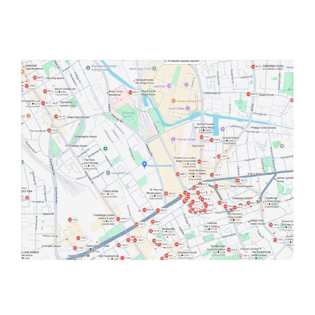

Hotels
Recommended Hotels in walking distance to the Francis Crick Institute, however, there are numerous other hotels in this regionClick on the reccomended hotels below to view more details:
- Radisson Blue Edwardian, Grafton Hotel
- Hub by Premier Inn London Kings‘s Cross hotel
- Travelodge (Bar Café) London Euston Hotel
- Point A Hotel London Kings Cross (Tune Hotel Kings Cross)
Organizing Commitee

Lucy Collinson
Head of Electron Microscopy,
The Francis Crick Institute,
London, UK

Pippa Hawes
Deputy Lead EM STP,
The Francis Crick Institute
London, UK

Yannick Schwab
Team Leader and Head of Electron Microscopy Core Facility EMBL
Heidelberg, Germany

Paolo Ronchi
Scientist EMBL
Heidelberg, Germany

Erin Tranfield
Head of the VIB BioImaging Core - Ghent VIB-UGent Center for Inflammation Research,
Ghent, Belgium

Anneke Kremer
Microscopy Specialist VIB Bioimaging Core Ghent VIB-UGent Center for Inflammation Research, Ghent, Belgium

Alexandra Elli
Solution Manager EM and XRM in Life Sciences Carl Zeiss Microscopy,
Jena, Germany