XRM State of the Science

3D/4D Imaging and Analysis with X-rays

An Educational Miniseries

  • What are some of the latest ways that scientists in my field are using X-ray microscopy to gain unique perspective inside their samples?
  • How does X-ray imaging fit in the landscape of other microscopy techniques, like optical and electron microscopy? When is a 3D and nondestructive approach valuable and necessary?
  • What are some of the recent technology innovations that are continuing to expand the scope of what XRM can offer the research community?

High resolution X-ray microscopy (XRM) has grown out of the synchrotron community, to provide a critical capability in today’s suite of lab-based characterization equipment. As the field continues to evolve rapidly and new innovations expand the reach of the technique, tune in to this miniseries to gain quick updates on how XRM is making an impact in your field of work.    

  • Short 15-minute episodes provide the critical information quickly and easily
  • Sign up for just the topics you are interested in
  • Emails will alert you when your chosen episode(s) become available
Topics & Schedule
Learn more about each episode
Ep.1 : Welcome and Introduction Dr. Will Harris | Available Immediately

Intended audience: Anyone registering for other episodes in the series, microscopy lab managers and directors

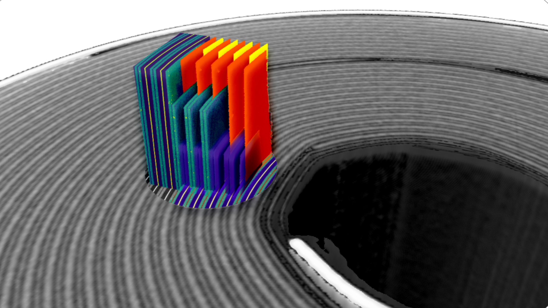
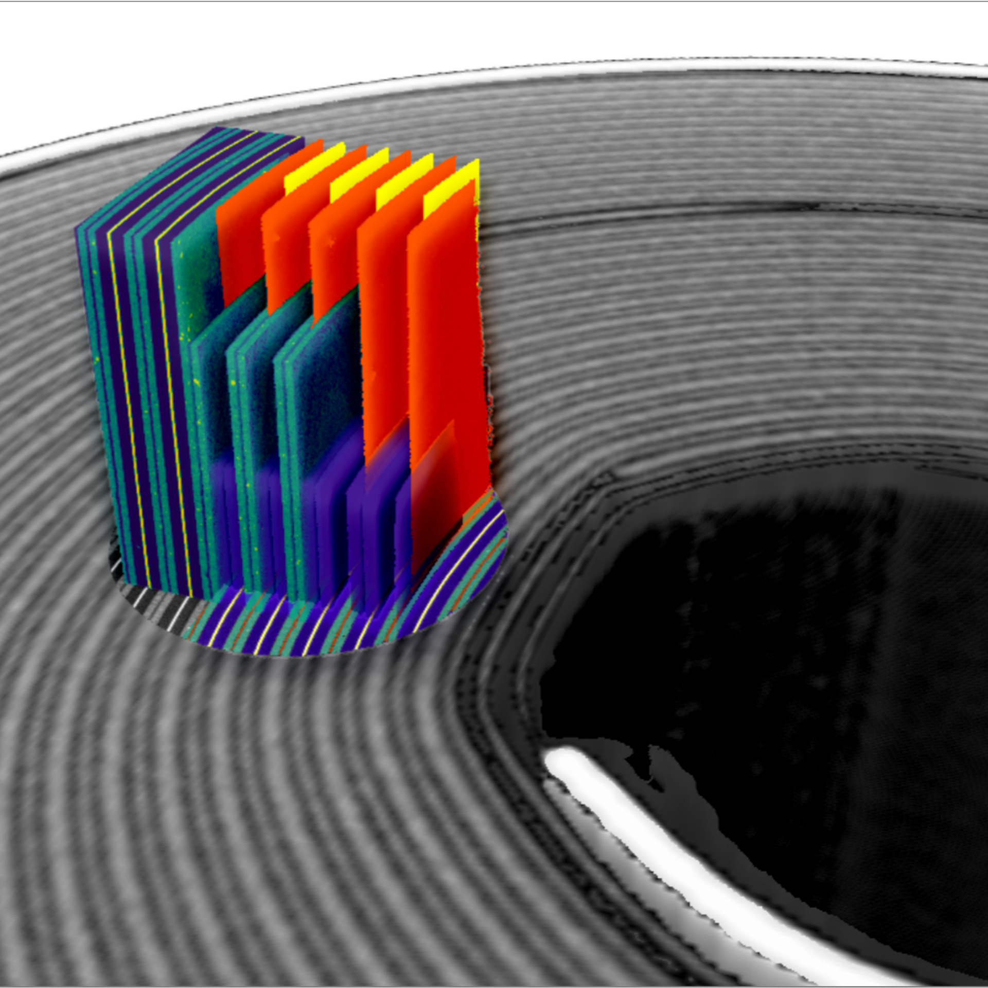
  • Gain an overview of 3D X-ray imaging
  • How does 3D X-ray complement other common microscopy techniques?​
  • Understand the main differences between different instrument types (microCT vs sub-micron XRM vs nanoscale XRM)​
  • See a brief survey of recent technology innovations supporting research​
  • Introduction to the remainder of the series
Ep.2: Energy Conversion & Storage Dr. Steve Kelly | Releases on May 30

Intended audience: Researchers in battery and fuel cell materials, electrochemists, chemical engineers

  • Using XRM to gain a nondestructive view inside intact cells
  • How XRM can help understand 3D electrode morphology, transport processes, defect distributions
  • Study degradation and cycling behavior using 4D imaging
Ep.3 : Life Sciences & Biomaterials Aubrey Funke MS | Releases on June 6

Intended audience: Core imaging facility personnel, multimodal imaging specialists, bone and mineralized tissue researchers, plant scientists, soft tissue researchers

  • Explore the power of non-destructive 3D imaging solutions in life science applications
  • Understand capabilities for structural imaging in soft and mineralized tissue without complex staining
  • Observe 3D cellular detail of plant structure without sectioning
  • Discover how XRM initiates multimodal, multi-scale workflows
  • Learn how to streamline your synchrotron or volume electron microscopy workflowsIntended audience: Anyone registering for other episodes in the series, microscopy lab managers and directors
Ep.4: Geoscience & Natural Ressources Dr. Rich Taylor | Releases on June 13

Intended audience: Geologists, Earth Scientists, Petroleum Engineers, Mining Technologists

  • See the latest advances in quantitative microCT for Geoscience
  • Understand quantitative material classification for 3D automated mineralogy
  • Use diffraction contrast tomography for non-destructive crystal orientation analysis
Ep.5 : Engineering Materials Andy Holwell | Releases on June 20

Intended audience: Materials Scientists, Engineers, Physicists, Chemists

  • See how to use 3D X-ray imaging to understand processing-structure-property relationships
  • Interrogate internal morphology in metals, composites, ceramics, and cementitious materials
  • Map cracks and failures in bulk or multi-materials systems non-destructively
  • See how diffraction contrast tomography reveals 3D crystallographic grain structures
Ep.6: Semiconductors & Electronics Cheryl Hartfield, MS | Releases on June 27

Intended audience: Semiconductor packaging R&D and failure analysts

  • Learn about packaging trends and the pressure on non-destructive package analysis
  • Understand 3D X-ray microscopy compared to microCT and its application in the package FA workflow
  • Gain insights into new technology for 4x faster 3D X-ray scans at submicron resolution on intact packages

Register

Please complete the form below and select the episodes that interest you. You will be alerted by email when those episodes become available.

Form is loading...

Name

If you want to have more information on data processing at ZEISS please refer to our data privacy notice.