Optical Sectioning

Explore advanced 3D imaging

Confocal, Lightfield & Structured Illumination Imaging

Experience ZEISS LSM 910 Lightfield 4D and Apotome 3 live

Three-dimensional fluorescence imaging has become essential for revealing complex biological structures and dynamic processes.
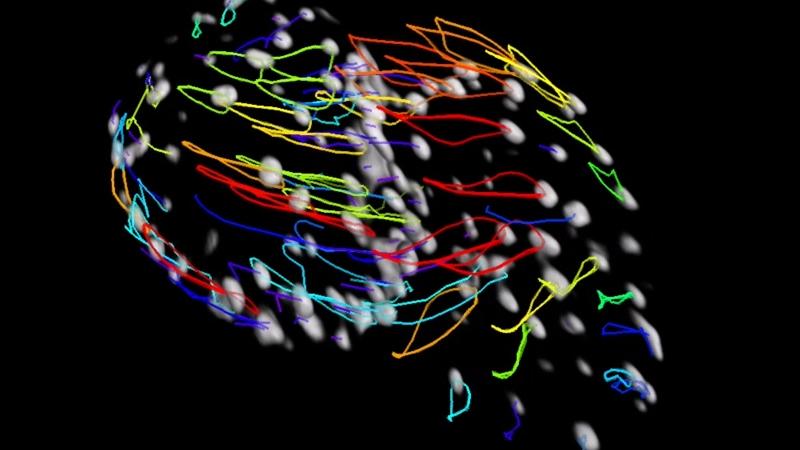
At this local workshop, experience a modern confocal laser scanning microscope equipped with Lightfield 4D imaging and structured illumination-based optical sectioning.

🎯 What you will learn

  • One Snap. One Volume. Capture real-time biological dynamics with Lightfield 4D
  • Experience the simplicity of optical sectioning with Apotome 3
  • Learn how to quantitatively analyze your data in 3D
  • Achieve high-contrast 3D imaging in thick specimens with confocal LSM
  • Capture instant volumetric information in a single acquisition
  • Enhance optical sectioning and resolution for challenging Life Science samples


  • 📅 Workshop | July 8, 2026

    Join us for a focused session combining:

    • A concise overview of key imaging principles
    • Live demonstrations on relevant biological samples
    • Practical guidance on:
      • Sample preparation
      • Parameter optimization
      • Data handling

    Application examples will cover workflows from fixed tissue to live-cell imaging.

    👉 Ideal for researchers aiming to expand their 3D imaging toolbox and identify the most suitable optical sectioning strategy for their work.

  • 🔍 Hands-On Experience | July 9–15, 2026

    Individual One-to-One System Demonstrations

    • Dedicated 2-hour demo session with your own samples
    • Direct interaction with ZEISS imaging experts
    • Compare imaging approaches on your specific application

    👉 Register your personal time slot during the workshop on July 8.

  • 🔬 Sample Preparation Guidelines (Lightfield 4D)

    Please bring:

    • Slides with coverslips
    • Ibidi chamber slides with coverslip bottoms
    • Well plates with coverslip bottoms

Agenda

  • 9.30 AM

    Registration & Coffee

    10 AM

    Innovative imaging workflows for biological dynamics and optical sectioning
    Jan Erik Heil, Andreas Bolzer, ZEISS

    10.45 AM

    Coffee break

    11 AM

    Live system demonstrations

    • Capturing biological dynamics in real time with LSM Lightfield 4D
    • Optical sectioning with Apotome 3- Quantitative 3D data analysis

    Including Lunch.

    2.45 PM

    End

  • Individual one-to-one system demonstrations

    Dedicated 2-hour demo session with your own samples

    👉 Register your personal time slot during the workshop on July 8.

Location

University of Regensburg

Faculty of Biology and Preclinical Medicine Universitätsstraße 31 , Room: VKL4.1.29 93053 Regensburg Deutschland
Registration

Form is loading...

If you want to have more information on data processing at ZEISS please refer to our data privacy notice