

New Benchmarks for Live Imaging
An overview of recent technologies for core facilitiesThe drive to image live specimens has revolutionized our understating of modern biology, and the careful balance of speed, gentleness and resolution is a prerequisite for optimal results.
This balance has recently been redefined and is paving the way for exciting new opportunities in the exploration of living specimens.
Here, we present the latest technology advancements from ZEISS that provide these new opportunities to complement your existing live imaging solutions.
Live Imaging in the Core Facility
Since the routine use of fluorescent proteins began in the 1990s, the imaging of living specimens has exponentially increased in popularity as an approach for sample exploration. The ever-increasing number of publications invoking live sample imaging means this is now one of the cornerstones of the light microscopy core facility.
The trade-offs between resolution, acquisition speed and gentleness for live experiments are well known. Indeed, aligning expectations of facility users in this regard is a critical step to avoid the misconception that many frames per second can be teamed with week-long acquisitions in multiple colors, with sub 100 nm resolution and in samples 200 µm thick.
Considerations for Live Imaging
A large proportion of live specimens are mounted on coverslips or in media filled dishes. Sealed dishes minimize the risk of contamination and provide the best means of keeping samples hydrated over long time periods.
For these dish-mounted samples, the length of acquisition or the number of frames within each acquisition needs to be adjusted to prevent any negative impact of light interaction1. This can result in the capture of datasets that miss or have insufficient resolution to visualize the key events of interest, or that run for insufficient time to capture the datapoints necessary for robust statistical analyses.
Ideally, providing a range of approaches in the facility that enable 3D acquisitions for longer and/or at higher resolution and in multiple colors would go a long way to shortening this list of compromises. With lattice lightsheet and lattice structured illumination microscopy, the latest technology developments move quickly towards this aspiration.
Lattice Lightsheet Microscopy
New possibilities for fast, volumetric imaging of cells
Lattice lightsheet technology hit the headlines following the landmark paper in 20142 and has since proved itself to be a powerful acquisition approach for long term study of a wide range of specimens.
Until recently, this technology has only been available in an upright configuration, and this has limited the scope and usability of the approach for a very wide user base. This has recently changed with the release of the ZEISS Lattice Lightsheet 7 platform which provides lattice lightsheet imaging but in a convenient, inverted configuration that is compatible with the same samples you would image using a confocal or spinning disc, for example.


ZEISS Lattice Lightsheet 7
The automatic alignments provided by Lattice Lightsheet 7 mean that even the most inexperienced of facility users can access this cutting-edge acquisition approach and capture 3D data of their classically mounted samples over hours and days at a time. Lattice lightsheet acquisition is already making its mark with homebuilt instruments generating numerous high impact publications.
With the launch of Lattice Lightsheet 7 this approach is set to become the new standard in 3D long term time-lapse imaging of petri dish or multi-well plate mounted specimens in the core facility. Learn how the team at the Centre for Dynamic Imaging (CDI) at the Walter and Eliza Hall Institute of Medical Research (WEHI) in Parkville, Victoria (Australia) are using Lattice Lightsheet technology to enable new discoveries (read the blog article).
Lattice Structured Illumination Microscopy
Sub-100 nm super resolution in living specimens
From the resolution perspective, the push for live super-resolution acquisition is clear from a doubling in the number of publications in the last 5 years. However, until recently restrictions in technology have prevented live, 3D multicolor acquisitions at resolutions below 100 nm.


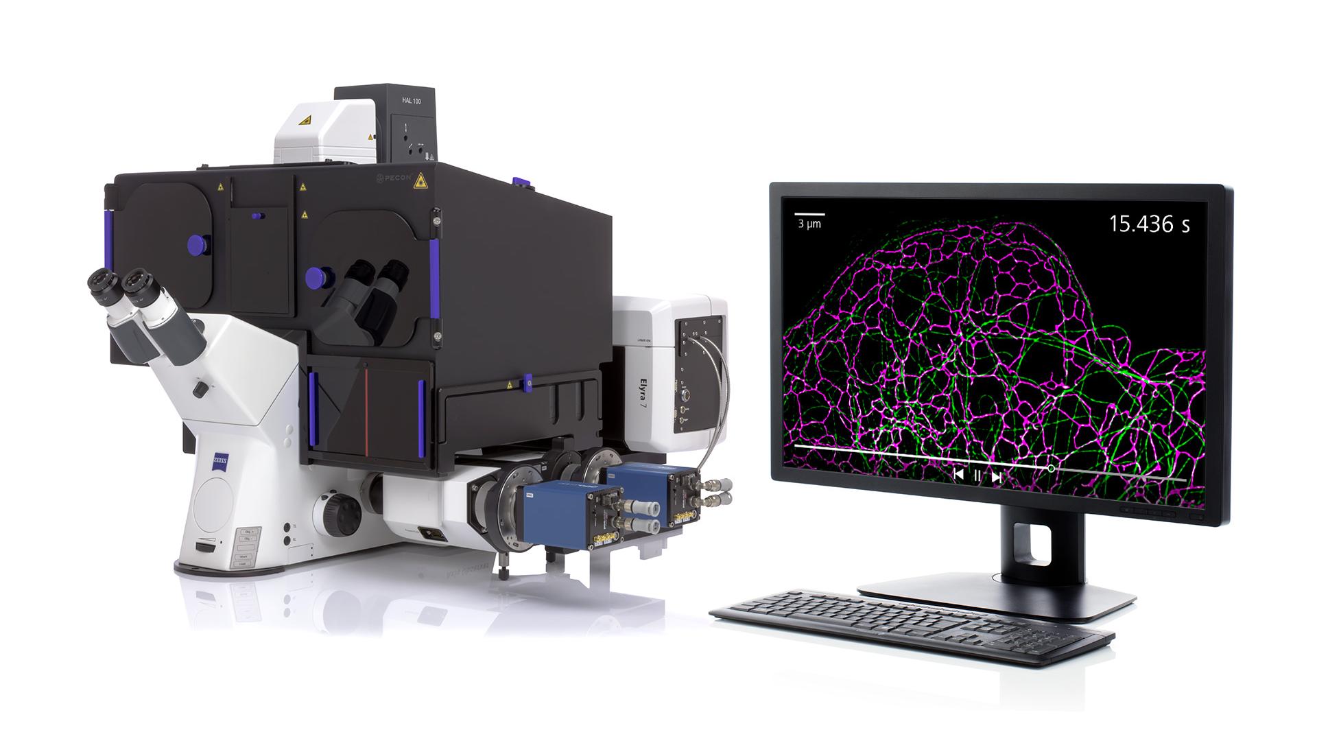
ZEISS Elyra 7 with Lattice SIM²
ZEISS Elyra 7 with SIM2 completely changes this and uniquely provides sub-organelle resolution down to 60 nm at live imaging speeds.
See how Elyra 7 with SIM² is changing the live imaging possibilities at the University of York across a wide range of application spaces (video)
Using standard dyes and fluorescent proteins, SIM2 is the ideal solution for core facility users seeking an easy imaging approach with the speed and light dose of widefield imaging, but with super-resolution results.
Find the Best Balance of Speed, Gentleness and Resolution for Every User
New benchmarks in live imaging
Selecting the right range of live imaging capability in the core facility can be challenging with so many alternative options available. Particularly for coverslip-based experiments, restrictions in terms of acquisition longevity or resolution have previously limited outcomes and experimental set ups.
The new benchmarks in live imaging set by the recent introduction of both Lattice Lightsheet 7 and Lattice SIM2 on inverted platforms promise to pave the way for new and exciting science as the drive to explore subcellular dynamics for longer and longer time durations continues apace.


Get in Contact with Us
Get in contact with a ZEISS imaging specialist for more information on technologies for live imaging and to learn more about ZEISS solutions for core facilities.
-
1
Bioessays: News and Reviews in Molecular, Cellular and Developmental Biology (Aug 2017); 39(8). Icha J, Weber M, Waters JC, Norden C. Phototoxicity in live fluorescence microscopy, and how to avoid it. DOI:10.1002/bies.201700003
-
2
Science (Oct 2014); 346(6208):1257998. Bi-Chang Chen et al, Lattice light-sheet microscopy: imaging molecules to embryos at high spatiotemporal resolution. DOI:10.1126/science.1257998