In archaeology, nails are useful markers of technological advancement. In most excavation sites, nails are the only remaining record of wooden structures such as ships, houses, coffins, sledges, and many other structures. Therefore, nails have a pivotal role in archaeological discovery.
Flavio Cognigni works at the Research Center on Nanotechnology Applied to Engineering (CNIS) - Sapienza University of Rome. This center is part of the Advanced Tomography and Microscopies (ATOM) project that is co-funded by Regione Lazio (“Infrastrutture Aperte per la Ricerca”). In his industrial PhD project in collaboration with ZEISS, the investigation of corrosion mechanisms in iron nail artifacts using non-destructive X-ray microscopy is a significant part of his research.
Flavio Cognigni (left) with Dr. Martina Bernabale (right) in front of the ZEISS Xradia Versa microscope they used in their studies.
Investigating an Archeological Iron Nail with Non-Destructive, Multi-Scale Microscopy
Mr. Cognigni and his colleague Dr. Martina Bernabale, a post-doctoral researcher in the Earth Sciences Department, created a comprehensive correlative microscopy workflow built on advanced multi-scale X-ray microscopy to characterize corrosion mechanisms in iron-base artifacts. This strategy provided the most information from the specimen with minimum sampling.
As seen in their publication M. Bernabale et al., they explored an iron nail from the 4th century BCE which had been unearthed during the excavations carried out by the Sapienza Archaeological Expedition in 2003 at the Motya archaeological site in Sicily, Italy.
Third-party Content Blocked
The video player is blocked due to your cookie preferences. To change the settings and play the video, please click the button below and consent to use of "Functional" tracking technologies.
An example of a multi-scale analysis using the Scout-and-Zoom procedure for the high-resolution scanning of a selected volume of the nail. This 3D reconstruction and segmentation of the nail shows finer details of the region of the interest (ROI) revealing hidden inclusions (green) in the metal core (blue). Soil and corrosion layers are reported respectively in yellow and red.
Multiscale Imaging of Iron Nail Artifact with X-ray Microscopy
In additional to studies with optical microscopy, micro-Raman spectroscopy and electron microscopy, X-ray microscopy was used to explore corrosion propagation and verify both the presence and the thickness of a metal core inside the nail while also monitoring the variations in density between different phases of the artifact.
ZEISS Xradia Versa 610 was key in these investigations as it overcomes the limits of X-ray computed tomography (CT) and offers non-destructive, multi-length scale visualization with an imaging field-of-view range from tens of millimeters down to tens of micrometers with a true spatial resolution of 500 nm.
Image A
Image B
Image C
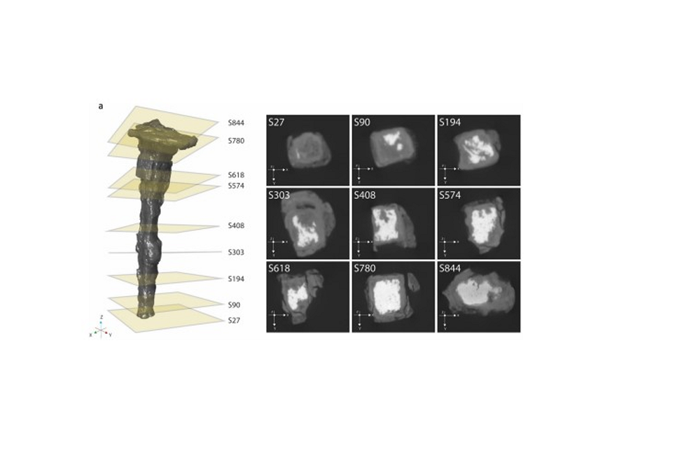
Image A: Overview image with virtual cross sections
Image A: Overview image with virtual cross sections
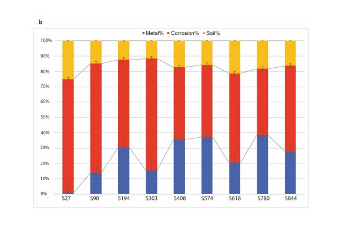
Image B: Histogram showing percentage of metal, corrosion and soil for each cross section.
Image B: Histogram showing percentage of metal, corrosion and soil for each cross section.
Image C: 3D reconstruction fo the nail.
Image C: 3D reconstruction fo the nail.
Low Resolution X-ray Microscopy for 3D Compositional Analysis
Low resolution imaging with ZEISS Xradia Versa 610 was used for non-destructive, 3D compositional analysis of the iron nail artifact.
Slide Show (right):
Image A shows an overview of the nail with the exact position of each virtual cross section acquired.
Image B quantifies the percentages of metal, corrosion and soil for each cross section in a histogram.
Image C illustrates the 3D reconstruction of the nail and segmentation of different layers: soil (yellow), corrosion layer (red), and metal (blue). The pie chart indicates the total percentage composition calculated by the imaging segmentation. A sagittal section of the 3D reconstruction showing the internal region of one half of the nail is also shown.
High Resolution X-ray Microscopy Unveils Microstructural Detail
A portion of the nail was selected for higher resolution imaging and analysis with X-ray microscopy.
(A) 3D rendering of analyzed section
(B) A sagittal cut of the 3D reconstruction reveals parallel structural discontinuities in the iron core.
(C) The structural discontinuities of the iron core, such as impurities and voids, are highlighted in this 3D reconstruction (green).
(D) A single cross-section showing the microstructure detail and corrosion stratigraphy in grayscale. The same cross-section is shown in (E) now with labels for metal (blue), goethite (Fe+3O(OH)) in the dense product layer (DPL) (pink), and iron magnetite (Fe3O4) (turquoise) and the transformed medium (TM) (red).
X-ray microscopy is a powerful tool to investigate a huge variety of samples non-destructively and provides quantitative information on chemical phase composition. It offers the possibility for a multi-scale and multi-modal 2D and 3D investigation of the sample.